18 minute read
Can the knowledge of eccentric training in achilles tendinopathy be used to treat superficial digital flexor tendinopathies in the equine?
Denise Kesson MCSP MSc Veterinary Physiotherapy Student
Introduction
Advertisement
Physiotherapy has been a valued component of health care in humans for over a century. The recognition of the benefits of Veterinary Physiotherapy in the injured animal has been relatively recent. The profile of the Veterinary Physiotherapist is rising in both the Veterinary world and with the general public. To increase the credibility of the profession it is important that interventions used are evidenced based.
The Equine Veterinary Journal published a review article by Buchner and Schildboeck (2006) on physiotherapy applied to the horse. This review systematically analysed published research on the effect of the various physiotherapeutic modalities used in practice applying the evidence-based medicine approach (Mair and Cohen 2003). Their findings were that there were insufficient good quality studies investigating the use of various physiotherapy treatments on horses to enable physiotherapists to work with an evidence based medicine approach to treatment.
The following year, in the same journal, McGowan et al (2007) built on this review stating that Buchner and Schildboeck had limited their review of evidence to veterinary literature. The feeling was that there are good quality studies performed in human literature that has led on from previous research performed on animals that can be applied to veterinary practice. McGowan et al emphasised how physiotherapy uses a range of treatment modalities for each individual patient after a formal assessment that will have led to a functional diagnosis. They therefore believed that it was more beneficial to discuss both the science and evidence base behind current issues in key areas of physiotherapy and to look at how they can be applied to the performance horse, rather than reviewing the research done concerning each treatment modality.
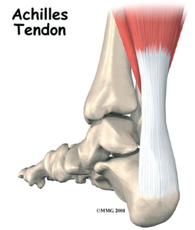
Achilles tendon
The aim of this literature review is to follow on from McGowan et al’s article and to examine the relevance and application of eccentric exercise as an evidenced-based physiotherapeutic approach to treat superficial digital flexor tendinopathy in the equine. Currently tendon problems affect both the human and equine athletes (Williams et al (2001), Maffulli et al (1999), Horshian et al (1998) and Moller et al (1996)). Management of Achilles tendinopathy in humans is predominantly conservative and physiotherapist led whereas the equivalent tendon in equines, the superficial digital flexor tendon, is managed by more invasive methods by the veterinary surgeon (Dyson (2004)).
Current literature exploring the definition of tendinopathy, its aetiology and the evidence surrounding the efficacy of eccentric exercises will be explored. The author will aim to draw from the literature evidence to justify the use of eccentric exercises in the management of superficial digital flexor tendinopathy in the equine.
Tendiniopathy Definition
The term tendinopathy is used to describe tendon pathology in the sheath and or body itself arising from overuse (Benazzo and Maffulli (2000)). It has replaced the diagnosis of “tendonitis” and “tendinosis” which are only used if there is evidence of inflammation found after histopathological examination (Sharma and Maffulli, 2006). It has replaced the diagnosis of “tendonitis” and “tendinosis” which are only used if there is evidence of inflammation found after histopathological examination (Sharma and Maffulli, 2006).
Tendon structure

In both human and the equine, tendinopathies can occur from the influence of both intrinsic, for example poor foot conformation or biomechanics (Eliashar et al (2004) and Nigg (1994)), and extrinsic factors, such as excessive repetitive
overloading of the tendon during vigorous training (Kvist (1994) and Kasashima (2004)). Selvanattie et al (1997) reports degeneration in the tendon is encouraged by the overloading of it during vigorous exercise. If the tendon is not able to regenerate as quickly as it is degenerating the end result is a tendinopathy. If intrinsic factors affect the overused tendon the risk of this process occurring increases.
Although it is widely recognised that tendon injuries are the most common in both human and equine athletes, little is understood about the aetiology of the conditions. At present tendinopathies are diagnosed once they become symptomatic and the ‘damage has been done’. Arnoczky (2008) believes that if we understood the underlying causes of tendon injuries we would be able to identify the athletes at risk and begin injury prevention.
Current Theories in Causes of Tendinopathy
The cause of tendinopathy is based mainly around several aetiological theories such as hypoxic or ischemic changes (vigorous exercise could lead to a reduction in oxygen delivery to the tendon and if this is prolonged, eventual tenocyte death), excessive apoptosis (normal programmed cell death is increased within the tendon), oxidative stress (ischemia occurs if the tendon is under increased load) and hyperthermia (when tendons are loaded heat is created. If they are repeatedly loaded and the heat cannot escape, cells within the tendon can become damaged) (Sharma and Maffulli (2005), Yuan et al (2003), Bestwick and Maffulli (2000), Birch et al (1997) and Goodship et al (1994)). All of these theories are yet to have significant scientific backing (Fredburg and Stengaard-Pedersen, 2008). The predominant theory is that tendinopathies are caused by overuse which causes microtrauma and the tendon is not given enough time during training to recover (Kjaer et al (2006)). Following on from this, early degeneration occurs and the tendon is vulnerable to injury (Tallon et al (2001)). It is known that, to have and maintain homeostasis within a tendon the tendon needs to be exposed to mechanical loading (Banes,et al (1995) and Wang and Ingber (1994)). The magnitude, regularity and length of time the tendon should be loaded for to maintain homeostasis is unknown (Arnoczky (2007) and (2002)). There have been several studies performed looking into the effects of over stimulating tendons, in vitro, either by repetitive loading and stretching (Wang et al (2003), Archambault et al (2002) and (2001), Skutek et al (2001) and Banes et al (1999)). The studies showed that the mechanical loading they exposed to the tendons led to the release of inflammatory cytokines and degenerative enzymes. However the mechanical strains used to produce these responses where excessive of normal physiological movement in either the length of time the stress was applied, the magnitude of stress applied or the amount of repetitions applied. It is therefore not appropriate to base clinical reasoning for treating tendon degeneration solely on these studies.
Both human and equine athletes present with tendon problems once they become symptomatic. By this time there is already a histological appearance of a failed healing response and the clinical diagnosis is tendinopathy. Therefore it is not the individual fibril damage caused by overloading of the tendons but the continued loading (or continued training) of the now inferior replacement collagen fibrils that leads to the progression of the pathological process.
Eccentric Exercises – the Evidence
As the understanding behind tendinopathies increases, the aims of treatment of this condition has been seen to change within the human field and we are moving away from addressing the inflammatory process to become more exercise focused (Allison and Purdam, 2009). Over the last 10 years there has been a growing interest in the use of eccentric exercises to treat tendinopathies. Eccentric exercise involves the muscle-tendon unit lengthening while it is loaded (Rees, 2008). This is the opposite of concentric exercises where the muscle-tendon unit shortens while it is loaded.
In 2005 Peers and Lysens proposed that eccentric exercises counteract the failed healing response of the tendon, encouraging the formation of cross links of the collagen fibres and facilitating the remodelling stage in healing. This theory was supported by Jeffery et al (2005). Allison and Purdam (2009) progressed this theory further in their paper hypothesising that there may be several mechanisms at work that lead to the efficacy of eccentric exercises such as;
1) Correcting the heterogeneity of the visco-elastic properties of the tendon caused by tendinopathies and potentially affecting the tendons aponeurosis, elastic recoil and spinal reflex feedback loops improving lower limb function.
2) Correcting the imbalance of the elastic recoil timing and spinal reflex feedback in both short and long reflex loops and thus changing optimal functional ranges of the passive and active components of the muscle-tendon unit.
3) Reducing the vascularity of the tendon by causing shearing forces between the tendon and the paratendon. The increase in vascularity associated with tendinopathies is thought to be a source for the pain (Ohberg and Alfredson (2004).
4) Adapting the mechanotransduction signalling (the ability of living cells to sense mechanical stress and respond by remodelling) in the passive structures of the muscletendon unit.
A trial by Stanish et al (1986) found that in 200 patients diagnosed with Achilles tendonitis 44% became completely pain free and 43% had a “marked improvement” in their pain levels after performing an eccentric loading programme once a day for 6 weeks. Although these results were
very promising there was no control group and, due to other weaknesses in the methodology, the results were grossly ignored by clinicians and eccentric exercises were not accepted as best practice.
It wasn’t until 1998 when Alfredson et al’s prospective randomised controlled trial confirmed the efficacy of eccentric exercises in Achilles tendinopathy. Although Alfredson et al’s methodology was considerably stronger than Stanish et al’s previous work; his prescribed dosage of eccentric exercises (15 repetitions of heel drops into discomfort twice a day for 12 weeks) was based on his clinical experience rather than any scientific reasoning.
Since Alfredson et al’s randomised controlled study, a lot more research has been dedicated to the efficacy of eccentric exercises using the exercise programme prescribed in Alfredson’s paper for Achilles tendinopathy. Between 2001 and 2004, three randomised controlled trials took place (Roos et al (2004), Mafi et al (2001), Silbernagel et al (2001)). They all used similar sample sizes of around 40 participants. Both Mafi et al and Silbernagel et al compared eccentric training with concentric training. Mafi used the eccentric overload training programme designed by Alfredson; Silberangel et al used a training method containing eccentric exercises as well as alongside other interventions (eg, stretching and applying ice). Roos et al also used Alfredson’s eccentric training programme along with night splints. By using two interventions the evidence of eccentric training as a standalone treatment is devalued. The other variability noted between the trials was the use of outcome measures. Roos et al used the Foot and Ankle Outcome Measure (Roos et al (2001)) compared to the other two who used the Visual Analogue Scale (Wewers and Lowe (1990).
Aside from Alfredson et al (1998) and Stanish et al (1986) there are three controlled trials (Shalabi et al (2004), Alfredson et al (2003) and Fahlstrom et al (2003). All of these trials followed the prescribed eccentric exercise routine of Alfredson et al (1998). Fahlstrom et al divided the participants into two groups, one for insertional Achilles pain and one for mid-portion Achilles pain finding eccentric exercises to be significantly more beneficial for mid-portion pain. None of these three controlled trials used control groups.
All studies conducted so far have showed a reduction in pre-reported pain levels with eccentric training for Achilles tendinopathy and all except Mafi et al (2001) had a greater reduction in pain in the eccentric training compared with the control group. Unfortunately due to the various outcome measures used and the heterogeneity of the populations used it is not possible to pool the results to support a conclusion that there is a statistically significant effect of eccentric training. There is enough evidence to justify the use of eccentric exercises clinically for Achilles tendinopathy and to support further research into their affect on the tendon.
Discussion
After reviewing the current literature supporting conservative management of Achilles tendinopathy by eccentric training, it is clear that the published work is only relevant to treating human tendons. It is also clear that the majority of our knowledge about the aetiology of tendinopathy and the effect on the tendons with exercise is based on the results from animal studies rather than the human population. Both the Achilles tendons and superficial digital flexor tendons are energy storing tendons designed to ensure propulsion is energy efficient. The knowledge gained about Achilles tendinopathy from studying animal tendons is used to support our treatment of the human tendons. Using this same reasoning, our knowledge of efficacy for tendinopathy treatment in humans could be transferred to the equine.
There is evidence to support the use of eccentric exercises clinically in the management of Achilles tendinopathy and that there is a stronger efficacy for its use in midportion pain (Roos et al (2004) and Fahlstrom et al (2003)). Arnoczky (2008) reported that superficial digital flexor degenerative changes associated with tendinopathy occurs predominantly within the central core fibrils increasing the strength of justification for using eccentric exercises for rehabilitation. It is time to step away from the need of randomised controlled trials and to focus on the underlying science behind the interventions to justify our treatment.
Conclusion
When evaluating the current literature supporting eccentric exercises it is important to bear in mind that the author who led the first randomised controlled trial proving the efficacy of the intervention has been involved in most of the research projects since. This could introduce bias into the literature body. Current findings are promising but there is a need to gain further insight into the mechanobiological mechanisms that may play a role in the cause of tendinopathy in both Achilles and superficial digital flexor tendons. It is important also to clarify the changes that occur within the tendon as a response to eccentric exercise training to either prove or disprove the current theories in the literature rather than measuring changes in pain levels. Lawson et al (2007) discuss the development of a technique to measure the strains that occur in equine tendons using a three dimensional lower limb mode and therefore there may be a possibility of using this technology to measure strain through tendons during eccentric loading.
This literature review continued on from McGowen et al’s (2007) paper and reviews the current literature surrounding tendinopathies and eccentric training. In the light of the findings, there is enough evidence available to justify supporting a more conservative approach to managing superficial digital flexor tendinopathy in the equine compared to more invasive methods. The author recommends development of an
eccentric exercise programme suitable for the equine as well as exploring the possibility of using eccentric training to prevent tendinopathies in the future.
References
Alfredson, H and Lorentzon, R (2003). Intratendinous glutamate levels and eccentric training in chronic Achilles tendinosis: a prospective study using microdialysis technique. Knee Surgery in Sports Traunatological Arthroscopy. 11:196-499
Alfredson, H, Pietila, T, Jonsson, P and Larentzon, R (1998). Heavy-load eccentric calf muscle training for the treatment of chronic Achilles tendinosis. American Journal of Sports Medicine 26: 360-366
Allison, G and Purdam, C (2009). Eccentric loading for Achilles tendinopathy – strengthening or stretching. British Journal Sports Medicine 43: 276-279
Archambault, J, Tsuzaki, M and Herzog, W (2002). Stretch and interleukin -1beta induce matrix metalloproteinases in rabbit tendon cells in vitro. Journal of Orthopaedic Research 20 :36-39
Archambault, J, Tsuzaki, M and Herzog, W (2001). Response of rabbit Achilles tendon to chronic repetitive loading. Connective Tissue Research 42: 13-23
Arnoczky, S (2008). Role of mechanobiology in the pathogenesis of tendinopathy: Lessons learned from horses and humans. AAEP Proceedings 54: 470-474
Arnoczky, S, Lavagnino, M ,and Egerbacher, M (2007). The mechanobiological etiopathogenesis of tendinopathy: Is it the over-stimulation or the under-stimulation of tendon cells? International Journal of Experimental Pathology 88: 217-226
Arnoczky, S , Lavagnino, M, Whallon, J and Hoonjan (2002). In situ cell nucleus in cell deformation under tensile load: a morphologic analysis using confocal laser microscopy. Journal of Orthopaedic Research 20: 29-35
Banes, A, Horesovsky, G, Larson, Tsuzaki M, Judex S, Archambault J, Zernicke R, Herzog W, Kelley S and Miller L (1999). Mechanical load stimulates expression of novel genes in vivo and in vitro in avian flexor tendon cells. Osteoarthritis cartilage 7: 141-153
Banes, A, Tsuzaki, M Yamamoto, J, Fischer T, Brigman B, Brown T and Miller L (1995). Mechanoreception at the cellular level: the detection, interpretation and diversity of responses to mechanical signals. Biochemical Cell Biology. 3: 841-848
Benazzo F and Maffulli N (2000). An operative approach to Achilles tendinopathy. Sports Medicine Arthroscopy Review. 8: 96-101
Bestwick, C and Maffulli, N (2000). Reactive oxygen species and tendon problems: review and hypothesis. Sports Medicine Arthroscopy Review. 8: 6-16
Birch, H, Wilson, A and Goodship, A (1997). The effect of exercise induced localised hyperthermia on tendon cell survival. Journal of Exp Biology (Pt 11): 1703-1708
Buchner, H and Schildboeck (2006) Physiotherapy applied to the horse: a review. Equine Veterinary Journal. 38(6) 574-580
Dyson, S (2004). Medical management of superficial digital flexor tendonitis: a comparative study in 219 horses (1992-2000). Equine Veterinary Journal. 36(5): 415-419
Eliashar E, McGuigan M and Wilson A (2004). Relationship of foot conformation and force applied to the navicular bone of sound horses at the trot. Equine Veterinary Journal 36: 431-435
Fahlstrom, M, Jonsson, P, Larentzon, R and Alfredson, H (2003). Chronic Achilles tendon pain treated with eccentric calf muscle training. Knees Surgery, Sports Traumatology and Arthroscopy. 11: 327-333
Fredburg, U and Stengaard-Petersen, K (2008). Chronic tendonopathy tissue pathology, pain mechanisms and etiology with a specific focus on inflammation. Scandanavian Journal of Medical Science in Sports 18: 3-15
Goodship, A, Birch, H, and Wilson A (1994). The pathobiology and repair of tendon and ligament injury. Veterinary Clinic North American Equine Practice. 10: 323-349
Houshian S, Tscherning T, Riegels-Nielsen P (1998). The epidemiology of Achilles tendon rupture in a Danish county. Injury 29:651–4 Jeffery, R, Cronin, J and Bressel, E (2005). Eccentric strengthening: Clinical applications to Achilles tendinopathy. New Zealand Journal of Sports Medicine. 33: 22-30
Kasashima, y, Takahashi T, Smith RK, Goodship AE, Kuwano A, Ueno T, and Hirano S (2004). Prevelance of superficial digital flexor tendonitis and suspensory desmitis in Japanese Thoroughbred flat racehorses in 1999. Equine veterinary Journal 36(4): 346-350
Kivst, M (1994). Achilles tendon overuse injuries in athletes. Sports medicine 18: 173-208
Kjaer, M, Magnusson, P, Krogsgaard, M, Boysenm Moller, J, Olesen, J, Heinemeier, K, Hansen, M, Haraldsson, B, Koskinen, S, Esmarck. B and Langberg, H (2006). Extracellular matrix adaption of tendon and skeletal muscle to exercise. Journal of Anatomy 208: 445-450
Lawson S, Chateau H, Pourcelot P, Denoix J-M, Crevier-Denoix N (2007). Sensitivity of an equine distal limb model to perturbations in tendon paths, origins and insertions Journal of Biomechanics 40 2510–2516
Maffulli, N, Barrass, V and Ewen, S (2000). Light microscopic histology of Achilles tendon ruptures. A comparison with unruptured tendons. American Journal of Sports medicine. 28: 857-863
Maffulli N, Waterston SW, Squair J, et al (1999). Changing incidence of Achilles tendon rupture in Scotland: a 15-year study. Clinical Journal of Sport Medicine 9:157–60
Mafi, N, Lorentzon, R and Alfredson, H (2001). Superior short term results with eccentric calf muscle training compared to concentric training in a randomised prospective multicentre study in patients with chronic Achilles tendinosis. Knee Surgery, Sports Tramatology, Arthroscopy. 11: 196-199
Mair, T and Cohen, N (2003). A novel approach to epidemiological and evidence based medicine studies in equine practice. Equine Veterinary Journal. 35: 339-340
McGowan, C, Stubbs, N and Jull, G (2007) Equine physiotherapy: a comparative view of the science underlying the profession. Equine Veterinary Journal 39 (1) 90-94
Moller A, Astron M and Westlin N (1996). Increasing incidence of Achilles tendon rupture. Acta Orthop Scand 67:479–481

Nigg, BM (1994). The role of impact forces and foot pronation: a new paradigm. Clinicla Journal of Sports Medicine. 11: 2-9
Ohberg, L and Alfredson, H (2004). Effects on neovascularisation behind the good results with eccentric training in chronic mid-portion Achilles tendinosis. Knee Surgery Sports Traumatological and Arthroscopy. 12: 465-470
Peers, K and Lysens, R (2005). Patellar tendinopathy in atheletes: Current diagnosis and treatment recommendations. Sports medicine 35: 71-87
Rees, J, Litchtwark, G, Wolman, R and Wilson, A (2008). The mechanism for efficacy of eccentric loading in Achilles tendon injury; an in vivo study in humans. Rheumatology. 47(10): 1493-1497
Roos, E, Engstrom, M, Lagerquist, A and Soderbers,B (2004). Clinical improvement after 6 weeks of eccentric exercises in patients with mid-portion Achilles tendinopathy – a randomised trialwith 1 year follow-up. Scandanavian Journal of Medical Science in Sports. 14: 286-295
Roos, E, Brandsson, S and Karlsson, J (2001). Validation of the Foot and Ankle Outcome Score for Ankle Ligament Reconstruction. Journal of Foot & Ankle International 22(10):788-794 Selvanetti A, Cipolla M and Puddu G (1997). Overuse tendon injuries: basic science and classification. Operative Techniques in Sports Medicine 5: 110-117
Shalabi, A, Kristoffersen-Wilberg, M, Svensson, L, Aspelin P and Movin T (2004). Eccentric training of the gastrocnemius-soleus complex in chronic Achilles tendinopathy results in decreased tendon volume and intratendinous signal as evaluated by MRI. American Journal of Sports medicine. 11: 1286-1296
Sharma, P and Maffulli, N (2006). Biology of tendon injury: healing, modelling and remodelling. Journal of Musculoskeletal Neuronal Interactions 6 (2): 181-190
Sharma, P and Maffulli, N (2005). Tendon injury and tendinopathy: healing and repair. Journal of Bone and Joint Surgery in America. 87: 187-202
Silbernagel, K, Thomee, R, Thomee, P and Karlsson J (2001). Eccentric overload training for patients with Achilles tendon pain – a randomised controlled study with reliability testing of the evaluation methods. Scandanavian Journal of Medical Science in Sports. 11: 197-206
Skutek, M, van Griensven, M, Zeichen, J and Brauer N, Bosch U (2001). Cyclic mechanical stretching enhances secretion of interleukin6 in human tendon fibroblasts. Knee Surgery, Sports Traumatology, Arthroscopy 9: 322-326
Stanish, W Rubinovich, R and Curwin, S (1986). Eccentric exercise in chronic tendonitis. Clinical Orthopaedic Related Research. 208: 65-68 Tallon C, Maffulli N and Ewen S (2001). Ruptured Achilles tendons are significantly more degenerated than tendinopathic tendons. Medical Science in Sports Exercise 33: 1983-1990
Wang, N and Ingber, D (1994). Control of cytoskeletal mechanics by extracellular matrix, cell shape and mechanical tension. Biophysiological Journal 66: 2181-2189
Wang, J, Jia F, Yang, G et al (2003). Cyclic mechanical stretching of human tendon fibroblasts increases the production of prostaglandin E2 and levels of cyclooxygenase expression; a novel in vitro model study. Connective tissue research 44: 128-133
Wewers M.E. & Lowe N.K. (1990). A critical review of visual analogue scales in the measurement of clinical phenomena. Research in Nursing and Health 13, 227-236
Williams R, Harkins L and Hammond C (2001). Racehorse injuries, clinical problems and fatalities recorded on British racecourses from flat racing and National Hunt racing during 1996, 1997and 1998. Equine Veterinary Journal 33:478–86
Yuan, J, Wang, M and Murrell, G (2003). Cell death and tendinopathy. Clinical Sports Medicine 22: 693-701
Achilles Tendon http://www.eorthopod.com/eorthopodV2/ index.php?ID=dbb85cd4171ee690cf f1b8cc7b0a57c6&disp_type=topic_ detail&area=20&topic_id=d46c40fda45e0a25 27a1ba3f5aa53cdd
Tendon Structure http://mcr.coreconcepts.com.sg/tendondisorders-inflammation-and-degeneration/


