
Abstract Stanford type A aortic dissection is a rapidly evolving pathological process often with poor prognosis if no urgent surgical management. However, it may be asymptomatic in the acute phase with late symptomatic presentation or incidental diagnosis on chest imaging. We report the case of a 52 year old man in whom a painless aortic dissection was discovered incidentally following a pericardial effusion assessment. The patient's condition was successfully managed with uneventful open heart surgery. This case demonstrates an extremely rare presentation of accidental hemorrhagic pericardial effusion caused by painless aortic dissection.
(ISSN: 2831-7416) Open Access Journal of Case Reports and Medical History www.acquirepublications.org/JCRMH Painless Aortic Dissection Revealed After its Rupture to the Pericardium: About a Case and Review of the Literature Bendahou H*,1, Abouriche A1, Selmaoui M1, Haboub M2, Arous S2, Bennouna G2, Drighil S2, Azzouzi L2 and Habbal R2 1Doctor, Cardiology Department, Hospital university of Casablanca 2Professor, Department of Cardiology, Hospital university of Casablanca *Corresponding author: Bendahou H, Doctor, Cardiology Department, Hospital university of Casablanca Received date: 01 Aug, 2022 | Accepted date: 10 Aug, 2022 | Published date: 13 Aug, 2022 Citation:Bendahou H, Abouriche A, Selmaoui M, Haboub M, Arous S, et al. (2022) Painless Aortic Dissection Revealed Afterits Rupture to the Pericardium: About a Case and Review of the Literature. J Case Rep Med Hist 2(4): doi https://doi.org/10.54289/JCRMH2200116 Copyright: © 2022 Bendahou H, et al. This is an open access article distributed under the terms of the Creative Commons Attribution License, which permits unrestricted use, distribution, and reproduction in any medium, providedthe original authorand source are credited.
Introduction Acute aortic dissection is defined by a tear in the intima of the aortic wall, creating an entry point for blood at the level of the elastic limit, within the media. This breach extends along the aortic wall, forming what is called a “false channel” of dissection [1]
Case report This is Mr. S.B, 52 years old, chronic smoker, followed for balanced arterial hypertension for 5 years under medical treatment, and without any other particular pathological history, presenting for a mild dyspnea. On clinical examination, we note an asymptomatic patient, without chest pain, in good general condition, with a blood presure = 135/85 mmHg, and a heart rate = 82 bpm, without
Case Report Volume 2 Issue 3
Abbreviations: TEVAR: Thoracic Aortic Endovascular Repair
Data on incidence in the general population are limited; estimates range from 2.6 to 3.5 per 100,000 people [2] Aorticdissectioncanbeasymptomaticintheacutephasewith late symptomatic presentation or following complications or an incidental diagnosis on imaging. We report the case of a 52 year old man, followed for arterial hypertension, in whom the diagnosis of pericardial effusion was made following NYHA stage II dyspnea. Complementary examinations confirmed a hemorrhagic pericardial effusion secondary to a chronic aneurysm dissecting the ascending aorta. The patient underwent successful and uneventful open heart surgery. This case illustrates an extremely rare presentation of an incidentally discovered hemorrhagic pericardial effusion caused by aortic dissection type A Stanford classification.

A complete biological assessment was carried out returning normal, without notable anomaly. The patient was urgently sent to the operating room of the cardiovascularsurgerydepartment,wheretheascendingaorta wasreplacedbyaDACRONprostheticsegment.Thesurgical procedure was uneventful, with good postoperative evolution.
www.acquirepublications.org/JCRMH
signs of heart failure.
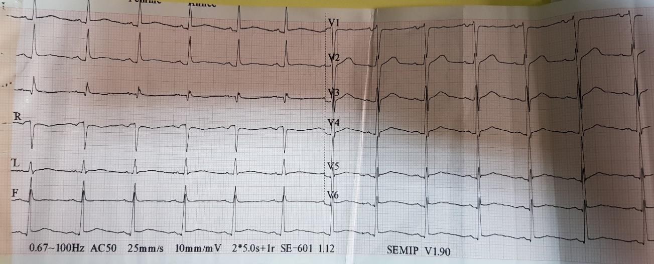
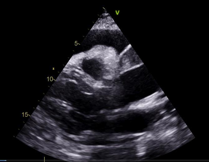
On the electrocardiogram, there is a regular sinus rhythm at 80 bpm, with no conduction or rhythm abnormality (Figure 1 An). echocardiography performed on admission shows moderately abundant pericardial effusion, predominantly posterior with: 14 mm next to the OD, 12 mm next to the RV, and 30 mm next to the LV, with good biventricular function, LVEF at 60%, with no variation in inspiratory flow, and a slightly dilated inferior vena cava at 21mm and not very compliant (Figure 2).
Journal of Case Reports and Medical History
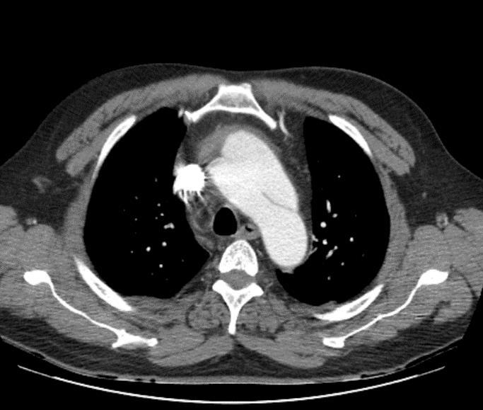
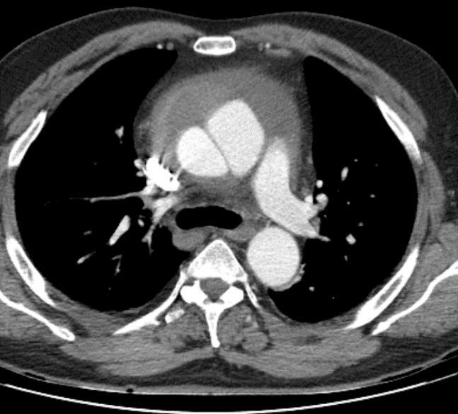
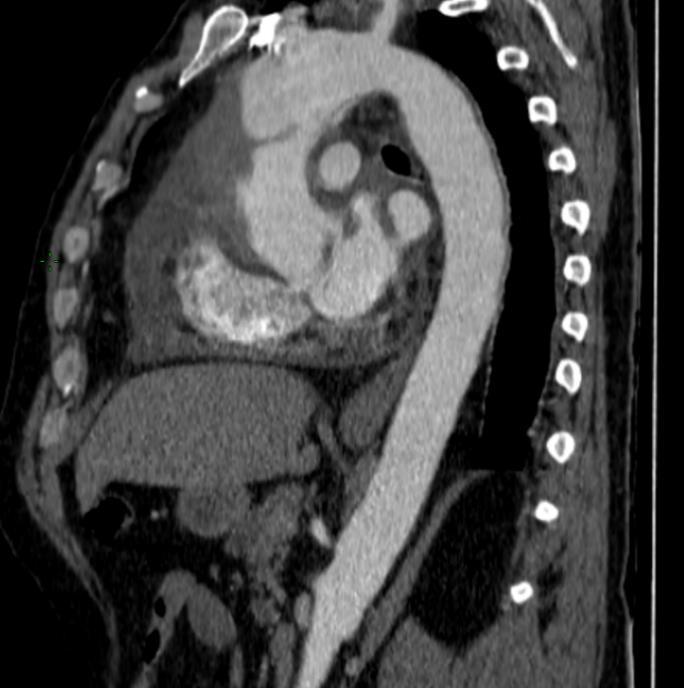
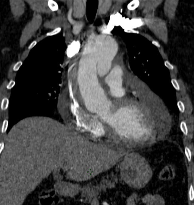
Also note a dilatation of the ascending thoracic aorta to 51 mm, with strong suspicion of aortic dissection. The aortic valve was normal and without leakage or stenosis. Forthis,athoracicCTangiographywasrequestedconfirming a type A aortic dissection of the STANFORD classification, of the ascending aorta extending to the aortic arch, associated with a parietal hematoma (Figures 3).
2 2
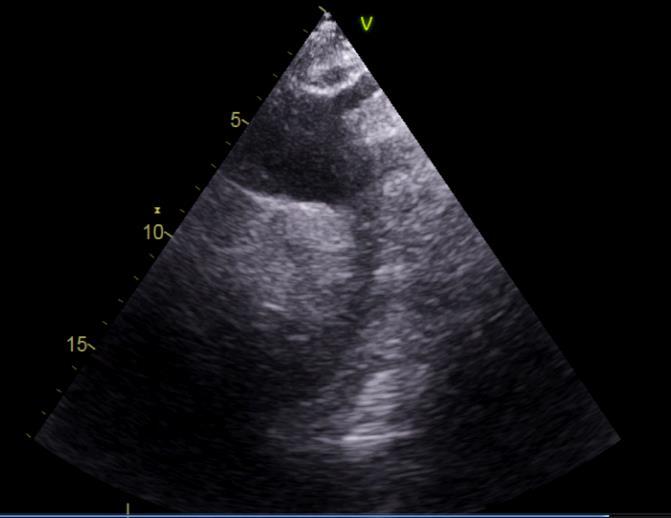
Figure 1: Normal ECG A B Figure 2: dissection of the ascending aorta (yellow arrow) on the major axis parasternal view (A) and the suprasternal view (B) Aspect of the coagulated pericardial effusion, on the long axis parasternal section (A)

Discussion
Figure 3: Appearance of aortic dissection type A of the STANFORD classification (blue arrow), extending from the ascending aorta to the aortic arch, associated with a parietal hematoma.
Journal of Case Reports and Medical History www.acquirepublications.org/JCRMH 3 3
The most important predisposing factor for acute aortic dissection is systemic hypertension. But there are other predisposing factors including collagen disorders such as Marfan syndrome, Ehlers Danlos syndrome, and annuloaortic ectasia. Bicuspid aortic valve, aortic coarctation, Turner syndrome, coronary artery bypass surgery, previous aortic valve replacement may very rarely be predisposing factors [2,4]
An aortic aneurysm is a localized dilation of the aorta. It can affect the different segments of the aorta: the aortic root, the ascending aorta, the aortic arch or the descending aorta.
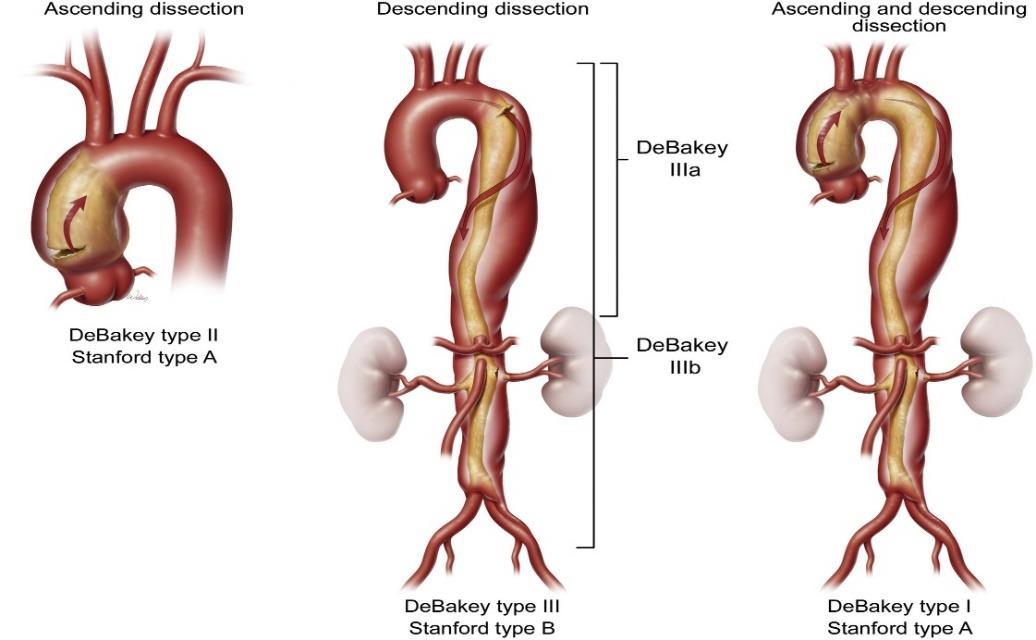
There are two classifications of dissections: Stanford (Table 1) and DeBakey (Table 2).
The natural course of thoracic aneurysms in particular is a progressive extension, with an increase in pressure on the wall of the aneurysm and thus an eventual rupture, which is called a dissection [2,3].
Stanford Type A includes dissections that involve the ascending thoracic aorta, while Type B dissections do not involve the ascending thoracic aorta. The DeBakey classification of type 1 concerns the entire
In a study [3], 60% of thoracic aneurysms involve the aortic root or the ascending aorta, and 40% involve the descending 10%aorta.involvethe arch and10%involvethethoraco abdominal aorta, with some involving more than one segment [2].
The diagnosis of aortic dissection is usually suspected in the presence of severe back pain, abrupt onset, tearing if it is a distal dissection of the descending artery or anterior chest pain if it is an ascending aortic dissection [2]
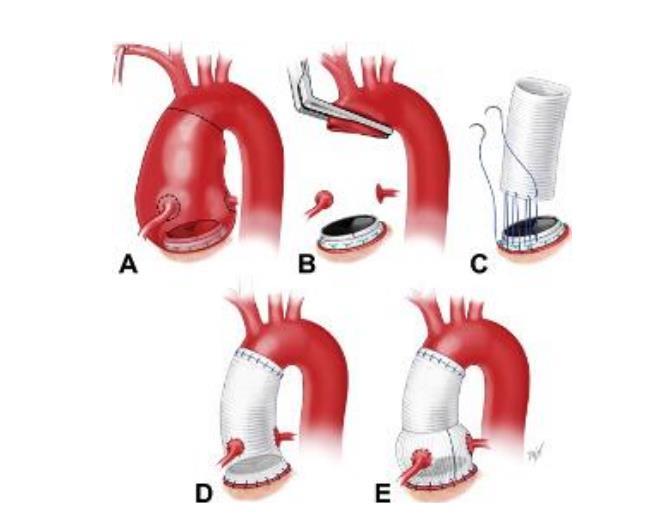
Table 1: Stanford classification Type Characteristic Type A Dissection involving the ascending aorta, regardless of the site of the primary tear Type B Dissection of the descending aorta Table 2: DeBakey classification Type Characteristic Type 1 Dissection of the ascending and descending thoracic aorta Type 2 Dissection of the ascending aorta Type 3 Dissection of the descending aorta Figure 4: Stanford and DeBakey classifications of aortic dissection [5]
aorta, type 2 dissections concern the ascending aorta and type 3 dissections concern the descending aorta. Thus, Stanford Type A dissection includes DeBakey Types 1 and 2, and Stanford Type B is equivalent to DeBakey Type 3 (Figure 4) [2].
Painless dissection has been reported, but is relatively rare. In an analysis of 977 patients from the International Acute Aortic Dissection Registry 3, only 63 patients (6.4%) had no pain [2]
Journal of Case Reports and Medical History www.acquirepublications.org/JCRMH 4 4
However, so called chronic type A aortic dissections represent a different category of patients presenting with atypical symptoms related to enlargement of the dissection, malperfusion (decreased visual acuity, pain in the extremities of the limbs or syncope), to valvular disease (dyspnea, edema of the lower limbs), or even asymptomatic at the start of the dissection, which prevents an early diagnosis [6]. As is the case of our patient. For this, a careful history is necessary to determine the symptoms announcing the beginning of a dissection [6] Andifthereisanysuspicion,thediagnosismustbeconfirmed by other specific imaging modalities, including transesophageal echocardiography, chest CT angiography or magnetic resonance angiography. And among the most evocative signs of the chronic picture of the dissection we find: a thick and immobile intimal flap, an aneurysmal dilation of the thoracic aorta or the presence of a thrombus in the false channel [6,7]
Chronic lesions limited to the ascending aorta that do not involve the aortic arch, aortic valve, or coronary arteries, can be repaired with endovascular stents, as a variant of thoracic aortic endovascular repair (TEVAR) [10], but studies are still very limited in this context, and publications are limited to clinical cases.
Regardingthetherapeuticmanagement,fortypeAdissection, the aim of the treatment is essentially the repair of the dissected aorta, taking into account the comorbidities of the patients. Endovascular treatment can be performed in patients unsuitable for open heart open surgical repair [9].
Data on outcomes of surgery in patients with chronic type A dissection on dissected native ascending aorta are limited to case reports [9]. But more often than not, the results of these surgeries are favorable. In a study carried out on the operative risk, hospital mortality was lower in patients with stable or so called chronic aortic dissection. Of the 67 operated patients, no intraoperative deaths and 3 early perioperative deaths [9].
Conclusion Chronic type A aortic dissection is a difficult diagnosis with a wide range of clinical presentations, including asymptomatic presentation or discovered following accidental hemorrhagic pericardial effusion. Pericardial puncture in this context is prohibited [6]. Therefore, it is strongly recommended to obtain a computed tomography of the chest before any diagnostic pericardial puncture. Surgical repair is the treatment of choice for chronic complicated Stanford type A aortic dissections. Replacement of all dissected aortic tissue and repair or replacement of the aortic valve can be done safely in the majority of patients with chronic type A dissection [9]
Journal of Case Reports and Medical History www.acquirepublications.org/JCRMH 5 5
Pericardial effusion is a common complication of Acute Type A Aortic Dissection, and generally occurs by two mechanisms. The first and most common is the transudation of fluid through the thin wall of the false channel into the pericardial cavity. Second, and it is less common, the dissected aorta can rupture directly into the pericardial space with subsequent tamponade [8] The pericardial effusion can coagulate as it did in our patient.
Cardiac tamponade is diagnosed in 8 to 10% of patients with acute type A aortic dissection, often painless, and represents a predictive factor for poor prognosis, and the leading cause of death [8].
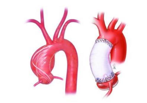
Depending on the extent of the dissection, open heart surgery remains the right alternative, and may include simple replacement of the ascending aorta with a supracoronary tube (DACRON tube, Figure 5, A) [9], or even the BENTALL technique (Figure 5, B) if severe aortic insufficiency or destruction of the aortic valve is associated. The technique of reimplantation of the aortic valve according to Tyrone David: is an alternative also can be carried out.
A B Figure 5: A, Replacement of the ascending aorta by supracoronary tube, DACRON tube. B, BENTALL technique.
The Task Force for the Diagnosis and Treatment of Aortic Diseases of the European Society of Cardiology (ESC) European Heart Journal. 35(41): 2873 2926.
8. Hiratzka LF, Bakris GL, Beckman JA, et al. (2010) theSurgeons,ofCardiovascularSocietyCollegeAmericanHeartAmericanpatientsVMACCF/AHA/AATS/ACR/ASA/SCA/SCAI/SIR/STS/S2010Guidelinesforthediagnosisandmanagementofwiththoracicaorticdisease.AReportoftheCollegeofCardiologyFoundation/AmericanAssociationTaskForceonPracticeGuidelines,AssociationforThoracicSurgery,AmericanofRadiology,AmericanStrokeAssociation,ofCardiovascularAnesthesiologists,SocietyforAngiographyandInterventions,SocietyInterventionalRadiology,SocietyofThoracicandSocietyforVascularMedicineJournalofAmericanCollegeofCardiology.121(13):e266369.
ACQUIRE PUBLICATIONS Volume 2 Issue 3
1. Nienaber CA. (2007) Pathophysiology of acute aortic syndromes. In: Baliga RR, Nienaber CA, Isselbacher EM, Eagle KA. Aortic Dissection and Related Syndromes. New York: Springer. 17 43.
9. Rylski B, et al. (2015) Outcomes of Surgery for Chronic Type A Aortic Dissection. Ann Thorac Surg. 99(1): 88 94. 10. Hynes MD, et al. (2016) Chronic Type A Aortic Dissection Two Cases and a Review of Current Management Strategies Conor F. AORTA. 4(1): 16 21.
6 6
Journal of Case Reports and Medical History
www.acquirepublications.org/JCRMH
3. Tsai TT, Evangelista A, Nienaber CA, et al. (2006) Long term survival in patients presenting with type A acute aortic dissection: Insights from the International Registry of Acute Aortic Dissection (IRAD). Circulation. 114(1 Suppl): I350 1356.
4. Larson EW, Edwards WD. (1984) Risk factors for aortic dissection: A necropsy study of 161 cases. Am J Cardiol. 53: 849 855.
5. Hong JC, Huu AL, Preventza O. (2021) Medical or endovascular management of acute type B aortic dissection The Journal of Thoracic and Cardiovascular Surgery. S0022 5223(21)00733 9.
Bibliographie
6. Abugroun A, et al. (2019) Chronic Type A Aortic Dissection: Rare Presentation of Incidental Pericardial Effusion. Hindawi. Case Reports in Cardiology. 7. Erbel R, Aboyans V, Boileau C, et al. (2014) 2014 ESC Guidelines on the diagnosis and treatment of aortic diseases: document covering acute and chronic aortic diseases of the thoracic and abdominal aorta of the adult
2. Conor F, Hynes, et al. (2016) Chronic Type A Aortic Dissection Two Cases and a Review of Current Management Strategies. AORTA. 4(1): 16 21.