2 minute read
NONINVASIVE OPTIONS


As mentioned previously, not all spots are biopsy worthy, but when there is doubt, some practitioners don’t have any other option but to cut the spot to test it. In reality, most spots fall along a continuum in terms of how suspicious they are (Figure 1). Some moles are so bland and normal looking that an experienced provider can immediately determine that they have no risk for melanoma and therefore are not biopsy worthy. At the other end of the spectrum, some spots are so clearly melanoma (e.g., the image marked “high risk” below that has all the ABCDE features), they will definitely require a biopsy. The challenging cases are those in the middle zone —they seem to be potentially suspicious to the eye, but it’s unclear how high the melanoma risk is. So providers often biopsy these out of an abundance of caution. However, if the provider has the right non-invasive tools, s/he can often get a better assessment of these middle-ground cases without cutting.
As mentioned in the below Figure, your provider may be able to use a range of non-invasive tools to evaluate your spot(s).
Spectrum of risk for melanoma based on visualization and genetic testing of spots. The diagram also addresses the use of non-invasive testing for spots that are suspicious but not clearly melanoma upon visual examination.
Some of the non-invasive tools your provider may use to evaluate your suspicious spots.
These tools can help your provider decide which of your spots are high risk enough that they warrant a biopsy. It’s helpful to ask your provider if they have these tools and whether they can be used for any of your spots.

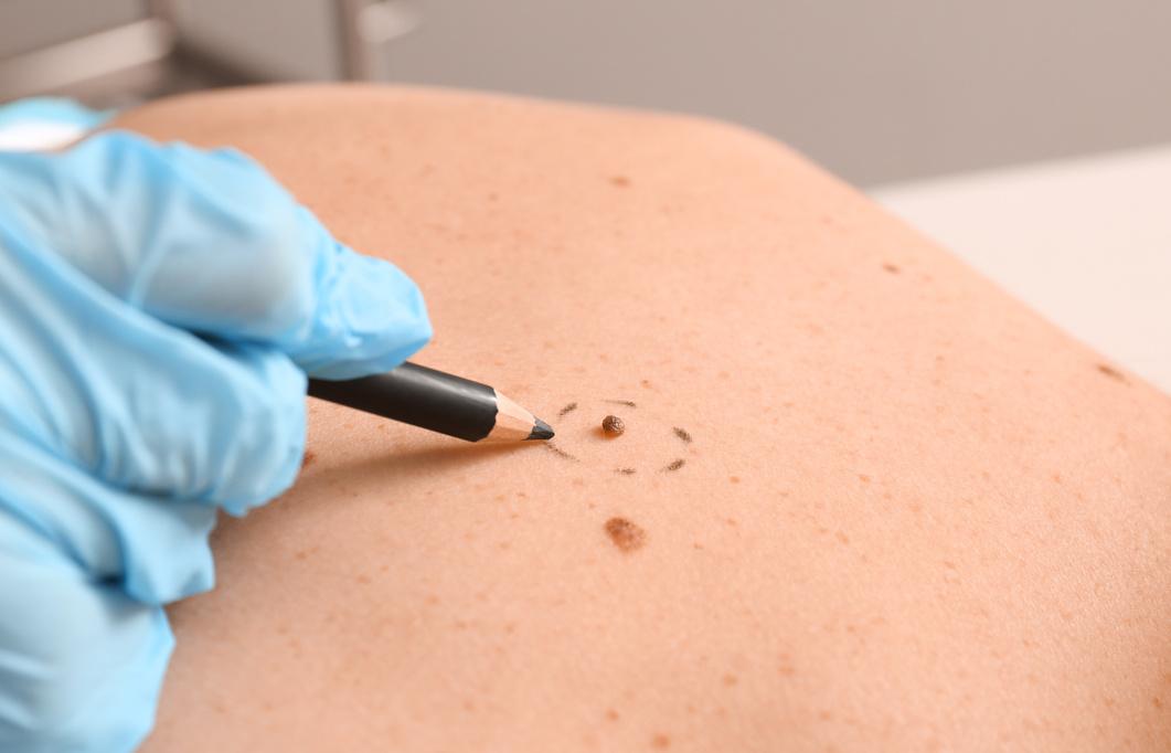
Dermoscopy is a technique in which the healthcare provider gently presses a handheld device called a dermatoscope against the suspicious lesion. Kind of like a specialized lighted magnifying glass, the dermatoscope allows the healthcare provider to look below the skin's surface for specific colors and patterns that would be consistent with melanoma or other skin cancers
Photography can be used to obtain close follow-up of a spot to see if it is changing or getting more suspicious. Photography can be used as part of dermoscopy as well, which provides a precise picture to examine over time
Mole mapping creates a comprehensive guide of all the moles or spots on someone's body. The spots are followed over time and photographed to look for subtle changes. This tool is often used for people with a lot of moles or who have a high risk of skin cancer


Reflectance confocal microscopy is a technique in which the provider uses laser light at an infrared wavelength to get a horizontal view of the layers of skin It produces an image similar to that observed from a biopsy
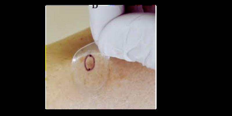
The non-invasive genetic sticker is a sticker applied to the skin that measures the presence of genes that are considered markers of melanoma (PRAME, LINC, and TERT)