17 minute read
RevivalUpdate Mike Perry surveys the news and research to report on new developments that bring us closer to the revival of cryonics patients
Revival Update
Scientific Developments Supporting Revival Technologies
Reported by R. Michael Perry
A Swarm of Slippery Micropropellers Penetrates the Vitreous Body of the Eye
Zhiguang Wu1, Jonas Troll, Hyeon-Ho Jeong, Qiang Wei, Marius Stang, Focke Ziemssen, Zegao Wang, Mingdong Dong, Sven Schnichels, Tian Qiu, and Peer Fischer
Science Advances 02Nov2018: Vol.4,no.11,eaat4388 DOI: 10.1126/sciadv.aat4388
Abstract
The intravitreal delivery of therapeutic agents promises major benefits in the field of ocular medicine. Traditional delivery methods rely on the random, passive diffusion of molecules, which do not allow for the rapid delivery of a concentrated cargo to a defined region at the posterior pole of the eye. The use of particles promises targeted delivery but faces the challenge that most tissues including the vitreous have a tight macromolecular matrix that acts as a barrier and prevents its penetration. Here, we demonstrate novel intravitreal delivery microvehicles—slippery micropropellers—that can be actively propelled through the vitreous humor to reach the retina. The propulsion is achieved by helical magnetic micropropellers that have a liquid layer coating to minimize adhesion to the surrounding biopolymeric network. The submicrometer diameter of the propellers enables the penetration of the biopolymeric network and the propulsion through the porcine vitreous body of the eye over centimeter distances. Clinical optical coherence tomography is used to monitor the movement of the propellers and confirm their arrival on the retina near the optic disc. Overcoming the adhesion forces and actively navigating a swarm of micropropellers in the dense vitreous humor promise practical applications in ophthalmology.
FromtheIntroduction
Ocular drug delivery plays an important role in ophthalmology and is used to treat diseases ranging from diabetic retinopathy, glaucoma, to diabetic macular edema. Although topical administration is currently available to treat diseases in the anterior of the eye including the cornea, ciliary body, and the lens, delivery to the posterior part of the eye via topical administration, systemic administration, and intravitreal injection is very ineffective and difficult because of the lacrimal fluid–eye barrier and the retina–blood barrier. To overcome these difficulties, nanoparticles have been injected into the eye, and their passive diffusion toward the retina has been investigated. Passive diffusion, however, suffers from long diffusion time and decreased activity of the biomedical agents. Moreover, it is systemic and therefore comes with an increased risk of side effects. It therefore still remains challenging to achieve targeted delivery with intravitreal administration.
Here, we report the first micropropellers that can penetrate the vitreous humor and that can reach the retina. The propellers are helical in shape, with the diameter that is comparable to the mesh size of the biopolymeric network of the vitreous and are functionalized with a perfluorocarbon surface coating that minimizes the interaction of the propellers with biopolymers, including collagen bundles that are present in the vitreous. The coating is inspired by a liquid layer found on the carnivorous Nepenthes pitcher plant, which presents a slippery surface on the peristome to catch insects. The nontoxic silicone oil and fluorocarbon coatings are also used as slippery surfaces in medical applications. Under the wireless actuation of an external magnetic field, the coated micropropellers not only show controllable propulsion but also can be driven as a large swarm over centimeter distances through the eyeball and can reach the retina within 30 min. The micropropellers are imaged with standard optical coherence tomography (OCT).
Source: http://advances.sciencemag.org/content/4/11/eaat4388, accessed 29 Dec. 2018.
Effective Wound Healing Enabled by Discrete Alternative Electric Fields from Wearable Nanogenerators
Yin Long, Hao Wei, Jun Li, Guang Yao, Bo Yu, Dalong, Angela LF Gibson, Xiaoli Lan, Yadong Jiang, Weibo Cai, and Xudong Wang
ACS Nano 2018, 12,12,12533-12540 DOI: 10.1021/acsnano.8b07038 PublicationDate(Web): November29,2018
Abstract
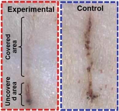
Skin wound healing is a major health care issue. While electric stimulations have been known for decades to be effective for facilitating skin wound recovery, practical applications are still largely limited by the clumsy electrical systems. Here, we report an efficient electrical bandage for accelerated skin wound healing. On the bandage, an alternating discrete electric field is generated by a wearable nanogenerator by converting mechanical displacement from skin movements into electricity. Rat studies demonstrated rapid closure of a full-thickness rectangular skin wound within 3 days as compared to 12 days of usual contraction-based healing processes in rodents. From in vitro studies, the accelerated skin wound healing was attributed to electric field-facilitated fibroblast migration, proliferation, and transdifferentiation. This self-powered electric-dressing modality could lead to a facile therapeutic strategy for nonhealing skin wound treatment.
FromtheReportofScienceDaily:
E-bandage Generates Electricity, Speeds Wound Healing in Rats Date: December19,2018
To power their electric bandage, or e-bandage, the researchers made a wearable nanogenerator by overlapping sheets of polytetrafluoroethylene (PTFE), copper foil and polyethylene terephthalate (PET). The nanogenerator converted skin movements, which occur during normal activity or even breathing, into small electrical pulses. This current flowed to two working electrodes that were placed on either side of the skin wound to produce a weak electric field. The team tested the device by placing it over wounds on rats’ backs. Wounds covered by e-bandages closed within 3 days, compared with 12 days for a control bandage with no electric field. The researchers attribute the faster wound healing to enhanced fibroblast migration, proliferation and differentiation induced by the electric field.
Sources: https://pubs.acs.org/doi/10.1021/acsnano.8b07038, https://www.sciencedaily.com/releases/2018/12/181219115519. htm, accessed 30 Dec. 2018.
A Host-Produced Quorum-Sensing Autoinducer Controls a Phage Lysis-Lysogeny Decision
Justin E. Silpe, Bonnie L. Bassler
Published: December13,2018DOI:https://doi.org/10.1016/j. cell.2018.10.059
Summary
A wound covered by an electric bandage on a rat’s skin (top left) healed faster than a wound under a control bandage (right). Credit: American Chemical Society
Skin has a remarkable ability to heal itself. But in some cases, wounds heal very slowly or not at all, putting a person at risk for chronic pain, infection and scarring. As early as the 1960s, researchers observed that electrical stimulation could help skin wounds heal. However, the equipment for generating the electric field is often large and may require patient hospitalization. Weibo Cai, Xudong Wang and colleagues wanted to develop a flexible, self-powered bandage that could convert skin movements into a therapeutic electric field. Now, they have developed a selfpowered bandage that generates an electric field over an injury, dramatically reducing the healing time for skin wounds in rats.
Vibrio cholerae uses a quorum-sensing (QS) system composed of the autoinducer 3,5-dimethylpyrazin-2-ol (DPO) and receptor VqmA (VqmA Vc ), which together repress genes for virulence and biofilm formation. vqmA genes exist in Vibrio and in one vibriophage, VP882. Phage-encoded VqmA (VqmA Phage ) binds to host-produced DPO, launching the phage lysis program via an antirepressor that inactivates the phage repressor by sequestration. The antirepressor interferes with repressors from related phages. Like phage VP882, these phages encode DNAbinding proteins and partner antirepressors, suggesting that they, too, integrate host-derived information into their lysislysogeny decisions. VqmA Phage activates the host VqmA Vc regulon, whereas VqmA Vc cannot induce phage-mediated lysis, suggesting an asymmetry whereby the phage influences host QS while enacting its own lytic-lysogeny program without interference. We reprogram phages to activate lysis in response to user-defined cues. Our work shows that a phage, causing bacterial infections, and V. cholerae, causing human infections, rely on the same signal molecule for pathogenesis.
Commentsfrom Science Daily:
Princeton molecular biologist Bonnie Bassler and graduate student Justin Silpe have identified a virus, VP882, that can listen in on bacterial conversations – and then, in a twist like something out of a spy novel, they found a way to use that to make it attack bacterial diseases like E. coli and cholera.
3D Nanofabrication by Volumetric Deposition and Controlled Shrinkage of Patterned Scaffolds
Daniel Oran, Samuel G. Rodriques, Ruixuan Gao, Shoh Asano, Mark A. Skylar-Scott, Fei Chen, Paul W. Tillberg, Adam H. Marblestone, Edward S. Boyden

Science 14Dec2018: Vol.362,Issue6420,pp.1281-1285 DOI: 10.1126/science.aau5119
Summary: ShrinkingProblemsin3DPrinting
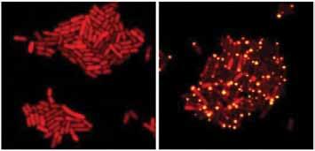
These E. coli bacteria harbor proteins from the eavesdropping virus. One of the viral proteins has been tagged with a red marker. When the virus is in the ‘stay’ mode (left), the bacteria grow and the red protein is spread throughout each cell. When the virus overhears that its hosts have achieved a quorum (right), the kill-stay decision protein is flipped to ‘kill’ mode. A second viral protein binds the red protein and sends it to the cell poles (yellow dots). All the cells in the right panel will soon die. Credit: Images courtesy of Bonnie Bassler and Justin Silpe,
Department of Molecular Biology, Princeton University
“The idea that a virus is detecting a molecule that bacteria use for communication – that is brand-new,” said Bassler, the Squibb Professor of Molecular Biology. “Justin found this first naturally occurring case, and then he re-engineered that virus so that he can provide any sensory input he chooses, rather than the communication molecule, and then the virus kills on demand.” Their paper will appear in the Jan. 10 issue of the journal Cell.
A virus can only ever make one decision, Bassler said: Stay in the host or kill the host. That is, either remain under the radar inside its host or activate the kill sequence that creates hundreds or thousands of offspring that burst out, killing the current host and launching themselves toward new hosts.
There’s an inherent risk in choosing the kill option: “If there are no other hosts nearby, then the virus and all its kin just died,” she said. VP882 has found a way to take the risk out of the decision. It listens for the bacteria to announce that they are in a crowd, upping the chances that when the virus kills, the released viruses immediately encounter new hosts. “It’s brilliant and insidious!” said Bassler.
Sources: https://www.cell.com/cell/fulltext/S0092-8674(18)31458- 2?_returnURL=https%3A%2F%2Flinkinghub.elsevier.com%2F retrieve%2Fpii%2FS0092867418314582%3Fshowall%3Dtrue, https://www.sciencedaily.com/releases/2018/12/181213142206.htm, accessed 29 Dec. 2018. Although a range of materials can now be fabricated using additive manufacturing techniques, these usually involve assembly of a series of stacked layers, which restricts threedimensional (3D) geometry. Oran et al. developed a method to print a range of materials, including metals and semiconductors, inside a gel scaffold. When the hydrogels were dehydrated, they shrunk 10-fold, which pushed the feature sizes down to the nanoscale.
Abstract
Lithographic nanofabrication is often limited to successive fabrication of two-dimensional (2D) layers. We present a strategy for the direct assembly of 3D nanomaterials consisting of metals, semiconductors, and biomolecules arranged in virtually any 3D geometry. We used hydrogels as scaffolds for volumetric deposition of materials at defined points in space. We then optically patterned these scaffolds in three dimensions, attached one or more functional materials, and then shrank and dehydrated them in a controlled way to achieve nanoscale feature sizes in a solid substrate. We demonstrate that our process, Implosion Fabrication (ImpFab), can directly write highly conductive, 3D silver nanostructures within an acrylic scaffold via volumetric silver deposition. Using ImpFab, we achieve resolutions in the tens of nanometers and complex, non–self-supporting 3D geometries of interest for optical metamaterials.
DiscussionadaptedfromAnneTrafton, MIT News:
Existing techniques for creating nanostructures are limited in what they can accomplish. Etching patterns onto a surface with light can produce 2-D nanostructures but doesn’t work for 3-D structures. It is possible to make 3-D nanostructures by gradually adding layers on top of each other, but this process is slow and challenging. And, while methods exist that can directly 3-D print nanoscale objects, they are restricted to specialized materials like polymers and plastics, which lack the functional properties necessary for many applications. Furthermore, they
can only generate self-supporting structures. (The technique can yield a solid pyramid, for example, but not a linked chain or a hollow sphere.)
To overcome these limitations, Edward S. Boyden and his students decided to adapt a technique that his lab developed a few years ago for high-resolution imaging of brain tissue. This technique, known as expansion microscopy, involves embedding tissue into a hydrogel and then expanding it, allowing for high resolution imaging with a regular microscope. Hundreds of research groups in biology and medicine are now using expansion microscopy, since it enables 3-D visualization of cells and tissues with ordinary hardware.
By reversing this process, the researchers found that they could create large-scale objects embedded in expanded hydrogels and then shrink them to the nanoscale, an approach that they call “implosion fabrication.” Currently, the researchers can create objects that are around 1 cubic millimeter, patterned with a resolution of 50 nanometers. (This is comparable to a twentieth of a millimeter resolution over a field of view of 1 meter, or 2,000 pixels.) There is a tradeoff between size and resolution: If the researchers want to make larger objects, about 1 cubic centimeter, they can achieve a resolution of about 500 nanometers. However, that resolution could be improved with further refinement of the process, the researchers say.
exception, but the restricted geometry of interactions across the heterodimer interface (primarily at the heptad a and d positions) limits the number of orthogonal pairs that can be created simply by varying side-chain interactions. Here we show that protein–protein interaction specificity can be achieved using extensive and modular side-chain hydrogen-bond networks. We used the Crick generating equations5 to produce millions of four-helix backbones with varying degrees of supercoiling around a central axis, identified those accommodating extensive hydrogen-bond networks, and used Rosetta to connect pairs of helices with short loops and to optimize the remainder of the sequence. Of 97 such designs expressed in Escherichia coli, 65 formed constitutive heterodimers, and the crystal structures of four designs were in close agreement with the computational models and confirmed the designed hydrogen-bond networks. In cells, six heterodimers were fully orthogonal, and in vitro—following mixing of 32 chains from 16 heterodimer designs, denaturation in 5 M guanidine hydrochloride and reannealing—almost all of the interactions observed by native mass spectrometry were between the designed cognate pairs. The ability to design orthogonal protein heterodimers should enable sophisticated proteinbased control logic for synthetic biology, and illustrates that nature has not fully explored the possibilities for programmable biomolecular interaction modalities.
Commentsfrom Science Daily:
Sources: http://science.sciencemag.org/content/362/6420/1281. long, http://news.mit.edu/2018/shrink-any-object-nanoscale1213, accessed 29 Dec. 2018.
Scientistsprogramproteinstopairexactly
Technique paves the way for the creation of protein nanomachines and for engineering of new cell functions
Zibo Chen, Scott E. Boyken, Mengxuan Jia, Florian Busch, David Flores-Solis, Matthew J. Bick, Peilong Lu, Zachary L. VanAernum, Aniruddha Sahasrabuddhe, Robert A. Langan, Sherry Bermeo, T. J. Brunette, Vikram Khipple Mulligan, Lauren P. Carter, Frank DiMaio, Nikolaos G. Sgourakis, Vicki H. Wysocki & David Baker
Date: December19,2018
Source: University of Washington Health Sciences/UW Medicine
Nature,2018; DOI: 10.1038/s41586-018-0802-y
Abstract
Specificity of interactions between two DNA strands, or between protein and DNA, is often achieved by varying bases or side chains coming off the DNA or protein backbone—for example, the bases participating in Watson–Crick pairing in the double helix, or the side chains contacting DNA in TALEN–DNA complexes. By contrast, specificity of protein–protein interactions usually involves backbone shape complementarity, which is less modular and hence harder to generalize. Coiled-coil heterodimers are an

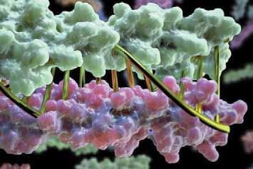
Proteins designed on computer and tested in the lab look a lot like DNA. Credit: Institute for Protein Design/ University of Washington Health Sciences/UW Medicine
Summary
Proteins designed in the lab can now zip together in much the same way that DNA molecules zip up to form a double helix. The technique could enable the design of protein nanomachines that can potentially help diagnose and treat disease, allow for the more exact engineering of cells and perform a wide variety of other tasks. This technique provides scientists a precise, programmable way to control how protein machines interact.
Sources: https://www.nature.com/articles/s41586-018-0802-y, https://www.sciencedaily.com/releases/2018/12/181219133219. htm, accessed 29 Dec. 2018.
A Roadmap to Revival
Successful revival of cryonics patients will require three distinct technologies: (1) A cure for the disease that put the patient in a critical condition prior to cryopreservation; (2) biological or mechanical cell repair technologies that can reverse any injury associated with the cryopreservation process and long-term care at low temperatures; (3) rejuvenation biotechnologies that restore the patient to good health prior to resuscitation. OR it will require some entirely new approach such as (1) mapping the ultrastructure of cryopreserved brain tissue using nanotechnology, and (2) using this information to deduce the original structure and repairing, replicating or simulating tissue or structure in some viable form so the person “comes back.”
The following is a list of landmark papers and books that reflect ongoing progress towards the revival of cryonics patients:
Jerome B. White, “Viral-Induced Repair of Damaged Neurons with Preservation of Long-Term Information Content,” Second Annual Conference of the Cryonics Societies of America, University of Michigan at Ann Arbor, April 11-12, 1969, by J. B. White. Reprinted in Cryonics 35(10) (October 2014): 8-17.
Brain,” in Brian Wowk, Michael Darwin, eds., Cryonics: Reaching for Tomorrow, Alcor Life Extension Foundation, 1991.
Ralph C. Merkle, “The Molecular Repair of the Brain,” Cryonics 15(1) (January 1994):16-31 (Part I) & Cryonics 15(2) (April 1994):20-32 (Part II).
Ralph C. Merkle, “Cryonics, Cryptography, and Maximum Likelihood Estimation,” First Extropy Institute Conference, Sunnyvale CA, 1994, updated version at http://www.merkle.com/cryo/cryptoCryo.html.
Aubrey de Grey & Michael Rae, “Ending Aging: The Rejuvenation Breakthroughs That Could Reverse Human Aging in Our Lifetime.” St. Martin’s Press, 2007.
Robert A. Freitas Jr., “Comprehensive Nanorobotic Control of Human Morbidity and Aging,” in Gregory M. Fahy, Michael D. West, L. Stephen Coles, and Steven B. Harris, eds, The Future of Aging: Pathways to Human Life Extension, Springer, New York, 2010, 685-805.
Chana Phaedra, “Reconstructive Connectomics,” Cryonics 34(7) (July 2013): 26-28.
Michael G. Darwin, “The Anabolocyte: A Biological Approach to Repairing Cryoinjury,” Life Extension Magazine (July-August 1977):80-83. Reprinted in Cryonics 29(4) (4th Quarter 2008):14-17.
Gregory M. Fahy, “A ‘Realistic’ Scenario for Nanotechnological Repair of the Frozen Human
Robert A. Freitas Jr., “The Alzheimer Protocols: A Nanorobotic Cure for Alzheimer’s Disease and Related Neurodegenerative Conditions,” IMM Report No. 48, June 2016.
Ralph C Merkle, “Revival of Alcor Patients,” Cryonics, 39(4) & 39(5) (May-June & July-August 2018): 10-19, 10-15.
Cryonics is an attempt to preserve and protect human life, not reverse death. It is the practice of using extreme cold to attempt to preserve the life of a person who can no longer be supported by today’s medicine. Will future medicine, including mature nanotechnology, have the ability to heal at the cellular and molecular levels? Can cryonics successfully carry the cryopreserved person forward through time, for however many decades or centuries might be necessary, until the cryopreservation process can be reversed and the person restored to full health? While cryonics may sound like science fiction, there is a basis for it in real science. The complete scientific story of cryonics is seldom told in media reports, leaving cryonics widely misunderstood. We invite you to reach your own conclusions.
How do I find out more?
The Alcor Life Extension Foundation is the world leader in cryonics research and technology. Alcor is a non-profit organization located in Scottsdale, Arizona, founded in 1972. Our website is one of the best sources of detailed introductory information about Alcor and cryopreservation (www.alcor.org). We also invite you to request our FREE information package on the “Free Information” section of our website. It includes:
A fully illustrated color brochure A sample of our magazine An application for membership and brochure explaining how to join And more!
Your free package should arrive in 1-2 weeks. (The complete package will be sent free in the U.S., Canada, and the United Kingdom.)
How do I enroll?
Signing up for cryopreservation is easy! Step 1: Fill out an application and submit it with your $90 application fee. Step 2: You will then be sent a set of contracts to review and sign. Step 3: Fund your cryopreservation. While most people use life insurance to fund their cryopreservation, other forms of prepayment are also accepted. Alcor’s Membership Coordinator can provide you with a list of insurance agents familiar with satisfying Alcor’s current funding requirements. Finally: After enrolling, you will wear emergency alert tags or carry a special card in your wallet. This is your confirmation that Alcor will respond immediately to an emergency call on your behalf.
Not ready to make full arrangements for cryopreservation? Then become an Associate Member for $5/month (or $15/quarter or $60 annually). Associate Members will receive:
Cryonics magazine by mail Discounts on Alcor conferences Access to post in the Alcor Member Forums A dollar-for-dollar credit toward full membership sign-up fees for any dues paid for Associate Membership
To become an Associate Member send a check or money order ($5/month or $15/quarter or $60 annually) to Alcor Life Extension Foundation, 7895 E. Acoma Dr., Suite 110, Scottsdale, Arizona 85260, or call Marji Klima at (480) 905-1906 ext. 101 with your credit card information. You can also pay using PayPal (and get the Declaration of Intent to Be Cryopreserved) here: http://www.alcor.org/BecomeMember/associate.html


