13 minute read
Biopsy of the skin


Linda Chan MBBS (2014), James Cook University Resident Medical Officer, Concord Repatriation General Hospital I am passionate about dermatology and medical education. As a combination of my two passions, I am particularly interested in increasing the amount of practical, high-quality dermatological resources pitched at medical students.
Abstract
Advertisement
Obtaining a skin biopsy is a relatively simple but important tool in the diagnosis and management of both skin neoplasms and inflammatory skin conditions. Many students are cautious about performing a skin biopsy. This may reflect a lack of proficiency or uncertainty regarding where to sample the skin, how to choose the biopsy method, and how to handle the sample. This article explores all the practical aspects of taking a skin biopsy, and explores how to choose the most appropriate site and technique for a biopsy.
Introduction
A skin biopsy is one of the most important tools in the diagnosis and management of skin diseases [1-7] . In principle, it involves removing a specimen of skin from the disease site for further evaluation under a microscope by a pathologist. It is a simple procedure that provides valuable additional evidence for the confirmation of skin malignancies and assists in confirming or excluding disease processes that share similar clinical presentations [1-7] .
Skin biopsies aid clinicians in arriving at an accurate diagnosis of cutaneous malignancies and in obtaining features for prognostication [1,7] . They are also helpful in understanding the zone of pathology in blistering skin conditions [1] and differentiating atypical presentations of inflammatory dermatosis [7] . Histological features that are obtained from skin biopsies allow clinicians to exclude specific diagnoses that exhibit overlapping clinical features [7] . Elizabeth Dawes-Higgs MBBS, MSc(Med), BE(Mech), DPD, PhD, FACD Dr Liz Dawes-Higgs is a highly educated and passionate dermatologist. She currently teaches and mentors students through the Australian College of Dermatologists and Concord Hospital. She is a state and national examiner for the Australasian College of Dermatologists. Dr Dawes-Higgs received the Schering-Plough Award for her laboratory-based research. She was awarded her Doctor of Philosophy from The University of Sydney for research on the biomechanical properties of skin.
What does having a skin biopsy involve?
Skin biopsies are usually performed in a medical practitioner’s office under local anaesthesia. The skin at the biopsy site is marked and cleaned [6] . Injection of the local anaesthetic produces transient stinging. After the skin specimen is collected, the clinician may close the wound with a suture if needed and a dressing may be applied.
Do patients have to stop any of their usual medications?
Warfarin does not need to be stopped for skin biopsies, but patients must inform his or her doctor so that an appropriate plan for the surgical technique and equipment for haemostasis is available. An international normalised ratio between 2.5–3.0 is generally accepted for simple skin surgery [2] .
Less is known about the use of antiplatelet therapy. It appears that the use of clopidogrel and aspirin increases the risk of complications during Moh’s surgery [2] . The cessation of dual antiplatelet is generally not necessary [2] .
Preparing the skin for biopsy
The chosen biopsy site will be marked with a surgical marker to avoid obliteration after the injection of local anaesthesia [1] . The area is then cleaned with a disinfectant.
Choosing a disinfectant
Alcohol reduces skin flora by 75% within one minute of application, but is mainly effective against Gram-positive microorganisms [2] .
Povidone-iodine solution and chlorhexidine have a broader antibacterial spectrum, including some Gram-negative microorganisms, and are commonly used in skin surgery [2] .
It is recommended to make a note of the direction of Langer’s lines (skin tension lines), which are generally parallel to the direction of collagen in the dermis [1] . Incisions made parallel to the Langer’s lines will close more easily and with better cosmetic results than those made perpendicularly [1,3,4] .
Local anaesthesia administration
Local anaesthesia is injected using a 29-gauge or 30-gauge needle. The needle is drawn back to check for blood, in order to ensure there is no risk of injecting the anaesthetic into the systemic circulation [4] . The initial injection is made perpendicular to the skin to minimise the sting. Deeper injections are less painful but do take longer to achieve anaesthesia [1] .
Usually, 1–2% lignocaine with 1:100 000 adrenaline is used. Other options include mepivacaine and bupivacaine [2] .
Topical agents such as EMLA cream (a mixture of prilocaine and lidocaine) or 4% lidocaine cream can be combined with injected local anaesthesia. Two hours of occlusion with EMLA will anaesthetise skin up to 5 mm deep [2,4] . This depth is sufficient for the chest, abdomen, face, and genitals but not for the palms, soles, and back, which have a thicker epidermis [4] .
Different types of skin biopsies commonly performed
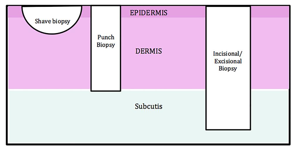
Choosing the best form of biopsy technique requires knowledge of the level of the lesion in the skin [2] . Figure 1 illustrates the levels of skin architecture reached by the common techniques.
Figure 1. Levels of skin architecture. Punch biopsy This is ideal for diagnostic purposes as it produces full thickness skin specimens [3] .
Advantages: It has a high ease of performance and produces uniformly shaped tissue [4] .
Disadvantages: The material can be inadequate and not include deeper tissue [4] .
Technique: It is performed using a circular blade attached to a pencil-like handle (Figure 2). The blade size ranges from 2–8 mm in diameter. A punch of 3–4 mm is enough for most conditions [3,8] . Small diameter biopsies, such as 2 mm, are rarely used and reserved for cosmetically sensitive sites such as the face.
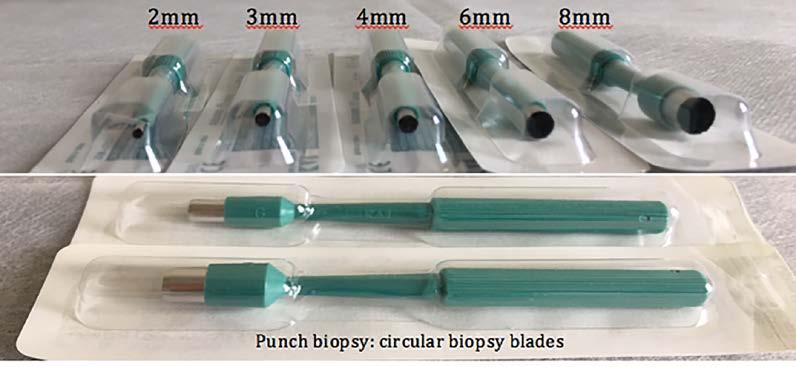
Figure 2. Punch biopsy blades.
After choosing the appropriate size of punch biopsy, the skin is stretched with the thumb and second finger perpendicular to the normal skin tension lines. The punch blade is placed perpendicular to the skin, with constant downward pressure in a circular motion. When the blade reaches the subcutaneous adipose tissue, there is a sensation of “give”, indicating that a full thickness cut has been made.
The blade is then removed and the specimen carefully extracted to avoid crushing.
If necessary, the wound is closed with a single layer of interrupted sutures. Generally, 4-0 or 5-0 monofilament nylon is used for the body and scalp and 6-0 nylon is used for the face [1,3] . Sutures on the face can be removed in 3–5 days. Sutures on the chest, abdomen, arms, or scalp can be removed in 7–10 days. Sutures in the back and legs can be removed in 12–20 days [1] .
Shave biopsy Shave biopsies are quick to perform and do not require suturing for closure. It is most suited for lesions elevated above the skin, such as seborrheic or actinic keratosis, skin tags, wards, superficial basal cell carcinomas, and squamous cell carcinomas [1] .
Advantages: They are quick to perform and provide large epidermal specimens [1,2] .
Disadvantages: This technique is not suitable for pigmented lesions and will usually leave a depressed scar at least the size of the initial lesion [1] .
Technique: The portion of the lesion that is above the level of the skin is shaved off using a blade [9] . The skin is left to heal via secondary intention with a dressing applied over the top. Saucerisation biopsy This is similar to a shave biopsy and ideal for vesiculobullous disorders and larger seborrheic keratoses.
Its advantages and disadvantages are similar to those of the shave biopsy technique [1,2] .
Technique: The biopsy blade is held between the thumb and index finger and bends to form an arc, which allows its intersection into the dermis (Figure 3). The plane of cleavage passes through the reticular dermis. It is performed using a shaving blade [4] . The wound is not sutured and is allowed to heal by secondary intention [4] .
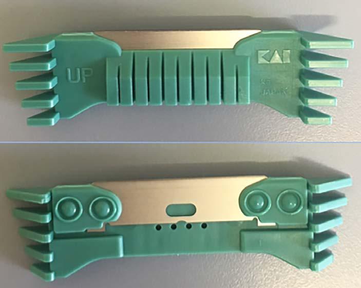
Figure 3. Saucerisation biopsy blades.
Incisional biopsy The main indication for an incisional biopsy is to obtain a sizable quantity of tissue to avoid diagnostic error.
Advantages: This is highly useful in diagnosing panniculitis, scarring alopecia, non-pigmented skin cancers with central necrosis, and larger vessel vasculitis as it provides a sizeable specimen that will allow the pathologist to review all features [2] .
Disadvantages: This is a time-consuming process and requires expertise [5,6] .
Technique: The technique is identical to that of an excisional biopsy [5,6] . The incision should also follow the skin tension lines [2] .
Excisional biopsy This is used for lesions that cannot be removed with a punch biopsy due to size, depth, or location, pigmented lesions suspicious for melanoma, and keratoacanthomas [5,6,10] .
Advantages: Excisional biopsy allows for the greatest diagnostic accuracy as the pathologist can examine the entire lesion [1] .
Disadvantages: This technique requires the greatest amount of expertise, training, and time to perform [1] .
Technique: A surgical scalpel is used. The entire lesion is completely removed up to the subcutaneous tissue plane. The wound is closed using two layers of sutures [4] .
How do you choose the site and type of skin biopsy?
Skin cancers A small punch biopsy from the centre of the lesion is recommended for suspected basal cell carcinoma (BCC) or squamous cell carcinoma (SCC) [6] . The area with the best diagnostic yield is the centre of the lesion, as it avoids confounding reactive changes around the periphery. If, however, there is central necrosis, an incisional biopsy that includes both the central portion and peripheral tissue is preferred [6] .
For SCCs, sufficient tissue needs to be biopsied for the pathologist to visualise the junction between the epidermis and dermis. One study found invasive SCCs in 20% of cases of actinic keratosis which were found by shave biopsy. The patients in these cases subsequently underwent re-excisions [6] .
BCCs often contain a mixture of growth patterns. Therefore, a single punch biopsy may not identify an aggressive component in 50% of cases.
Suspected melanomas should be excised with a 2 mm clinical margin and not partially biopsied [5] .
Inflammatory skin conditions Lesions with the most advanced inflammatory changes are preferred in most cases, but those that show secondary changes, such as excoriation, should be avoided [1,7,8] .
The sample should include maximal lesion area and minimal normal skin [1] .
Areas to be sampled should preferably be free from topical treatment for one month prior to the biopsy [7] .
If there is suspected vasculitis, lesions 48–72 hours old are ideal for histological analysis. For direct immunofluorescence, lesions less than 24 hours old should be sampled.
The incisional biopsy technique should be used for panniculitis, larger vessel vasculitis, and annular lesions, for example, granuloma annulare [7] . Ulcers, erosion, and blistering conditions A lesion formed within 48 hours is preferred as it provides the most specific histology features [1,7] .
Whenever possible, remove the vesicles intact during the biopsy. Bullae should be biopsied at the edge, keeping the blister roof attached [1] .
Immediate and long-term complications of skin biopsies
Anaphylaxis True anaphylaxis to local anaesthetic agents is very rare. However, reactions to additives may masquerade as a true allergy. In patients at risk of type I hypersensitivity, an intra-dermal test can be performed for local anaesthetics or a patch test for EMLA [4] . Bleeding This is the most common complication, especially on the scalp, face, genitals, and in elderly patients with atrophic skin [4] .
Other risk factors are a low platelet count, coagulopathy, haemophilia, and von Willebrand disease. It can also be drug-induced by substances such as aspirin, clopidogrel, heparin, and over-the-counter use of fish oil, garlic, and ginseng supplements [9] .
Majority of bleeding is due to the rupture of small venules. The application of pressure for 2-3 minutes stops the oozing. Swabs that are soaked with hydrogen peroxide, 20–40% aluminium chloride, or Monsel’s solution can also be helpful. If there is an identifiable bleeding vessel, this can be cauterised or ligated [4,9] . Damage to other structures Damage to nerves and vessels is rare. A biopsy in the pre-auricular areas can occasionally damage the facial nerve, as the branches are superficial [9] . Infection In one study, 22 out of 100 diagnostic biopsies showed signs of clinical infection. In the same study, two biopsy lesions had wound dehiscence alone and five showed signs of both infection and dehiscence. The most common bacterium on wound swab was Staphylococcus aureus [9] .
The incidence of secondary infection in the groin and axillae is higher, therefore biopsy in these areas should be avoided if possible [1] .
If the wound is frankly purulent or associated with cellulitis, oral antibiotics should be prescribed. Infected wounds in the hands, feet and, intertriginous areas are often infected with Candida and can respond to topical antifungal ointments [1] . Systemic risk factors for infection include an immunosuppressed state, particularly diabetes mellitus [9] .
Scarring Scarring with or without pigmentary changes is not uncommon after the biopsy site heals. Hypopigmentation is common after removing lesions from hyperpigmented lesions. Hypertrophic scarring tends to occur over the deltoid and chest areas. It is best to avoid these areas if possible [1] .
The incision should be made along the relaxed skin tension lines to provide a good cosmetic scar [9] .
Delayed healing Problematic healing tends to occur over the tibia, especially in diabetic patients or those with arterial and venous insufficiency [1] . This should be explained during the consenting process.
Sending the specimen to the pathologist
The skin specimen is placed in a jar containing the fixative formalin (Figure 4). It generally takes between one to two weeks for the final pathology report to be released. The report may be inconclusive, therefore correlation with the clinical findings is important. In certain conditions, repeated biopsies may be required.
There are several ancillary tests that can be used by the pathologist to aid in the diagnosis of skin diseases. The most common are direct immunofluorescence (DIF), microbiological culture, flow cytometry, and polymerase chain reaction analysis such as lymphocyte Figure 4. Specimen jar. clonality. Many of these additional tests will require non-formalin fixed specimens [7] .
Conclusion
Obtaining an appropriate skin biopsy remains one of the most fundamental clinical skills in the diagnosis and management of both inflammatory skin conditions and skin malignancies [1-7] . It is, therefore, crucial to understand the considerations involved in choosing an optimal biopsy site and a suitable biopsy technique to maximise the histological diagnostic features and minimise adverse clinical events.
Correspondence Linda Chan linda.chan@my.jcu.edu.au
[1]
[2]
[3]
[4]
[5]
[6]
[7]
[8]
[9]
[10] References
Alguire PC, Mathes BM. Skin biopsy techniques for the internist. J Gen Intern Med. 1998;13(1):46-54. Llamas-Velasco M, Paredes BE. Basic concepts in skin biopsy. Part I. Actas Dermosifiliogr. 2012;103(1):12-20. Zuber TJ. Punch biopsy of the skin. Am Fam Physician. 2002;65(6):1155-8. Nischal U, Nischal K, Khopkar U. Techniques of skin biopsy and practical considerations. J Cutan Aesthet Surg. 2008;1(2):107-11. Luk PP, Vilain R, Crainic O, McCarthy SW, Thompson JF, Scolyer RA. Punch biopsy of melanoma causing tumour cell implantation: another peril of utilising partial biopsies for melanocytic tumours. Australas J Dermatol. 2015;56(3):227-31. Harvey NT, Chan J, Wood BA. Skin biopsy in the diagnosis of neoplastic skin disease. Aust Fam Physician. 2017;46(5):289-94. Harvey NT, Chan J, Wood BA. Skin biopsy in the diagnosis of inflammatory skin disease. Aust Fam Physician. 2017;46(5):283-8. Keeling BH, Gavino AC, Gavino AC. Skin Biopsy, the allergists’ tool: how to interpret a report. Curr Allergy Asthma Rep. 2015;15(10):62. Abhishek K, Khunger N. Complications of skin biopsy. J Cutan Aesthet Surg. 2015;8(4):239-41. Llamas-Velasco M, Paredes BE. Basic concepts in skin biopsy. Part II. Actas Dermosifiliogr. 2012;103(2):100-10.


