8 minute read
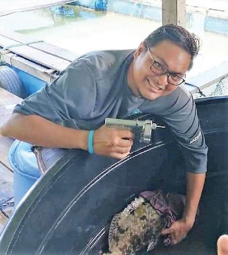
Leong Tak Seng surveyed ectoparasites in crimson snappers and hybrid groupers in a cage farm in Malaysia
Tilapia lake virus: Clinical signs, disease diagnosis and prevention
By Raanan Ariav and Natan Wajsbrot
Tilapia is the second most important finfish cultured worldwide. Among the secrets for tilapia’s success are its low production costs while maintaining high product quality, particularly its nutritional content. Additionally, tilapia tolerates high stocking densities and is highly resistant to diseases. Indeed, tilapia farmers were able to effectively deal with bacterial disease agents, such as Streptococcus spp. and Aeromonas spp., through different approaches, including the use of feed additives and antimicrobials, coupled with improved management and strict biosecurity strategies. However, the tilapia industry has recently witnessed the emergence of highly virulent infectious diseases such as the tilapia lake virus (TiLV) that has been affecting wild and farmed tilapia for over a decade.
TiLV was first officially identified and reported in 2014 at the Sea of Galilee, Israel. After learning more about the disease’s origin and progression, the sharp decline of wild-caught tilapia in the Sea of Galilee since 2009 was linked to an increasing presence of TiLV in the wild fish populations. Awareness on this novel virus spreads rapidly and by May 2017 the Food and Agriculture Organization of the United Nations (FAO) triggered a global alarm. The FAO warned that the virus can adversely impact global food security and nutrition and advised countries importing tilapia to take appropriate risk management strategies such as diagnostic testing, health certification, and imposition of quarantine measures and contingency plans to contain the outbreaks.
Transmission and risk factors
Tilapia is the main host of TiLV but other important warm water aquaculture species can also be infected. Some fish species such as snakeskin gourami (Trichogaster pectoralis), iridescent shark (Pangasianodon hypophthalmus), walking catfish (Clarias macrocephalus), striped snakehead fish (Channa striata), climbing perch (Anabas testudineus), common carp (Cyprinus carpio), silver barb (Barbodes gonionotus), Asian sea bass (Lates calcarifer) and Indian major carp (Labeo rohita) have been found to be resistant to this emerging aquatic disease. This apparent resistance to TiLV might be due to the lack of viral receptors or other mechanisms that enable replication in the hosts. Nevertheless, other factors, such as stress, co-infection and environmental conditions also play important roles in the vulnerability to TiLV.
TiLV is transmitted both horizontally (among and between individuals of the same generation) and vertically from broodstock to offspring. Under experimental conditions, the virus was detected in faeces and contaminated water after a successful intragastric infection, suggesting an oral–faecal route of transmission. The virus can thus spread horizontally amongst conspecifics inhabiting the same water body. old larvae). This also means that it is theoretically possible to test larvae for the presence of TiLV before shipping them to farms; this represents a significant opportunity to create TiLV pathogen-free larvae – a major biosecurity step. It is also hypothesised that molluscs, aquatic insects and invertebrates are potential carriers of TiLV and thus the virus could also be transmitted through these organisms. However, more research needs to be performed to confirm transmission pathways via these other vectors. Overall, these multiple transmission pathways make the containment of outbreaks particularly challenging.
Despite TiLV being a recently identified virus, some direct effects have already been identified as significant risk factors for its propagation, namely the presence and proximity of infected, wild or farmed populations, and water temperatures ranging from 25°C to 31°C. Other risk factors include any change that might affect the immune status of the fish and make them more vulnerable to the presence of the virus, either directly altering the fish immunological competence or disrupting their homeostasis. The latter, which forces the fish to energetically rebalance its physiological conditions, are referred to as immunosuppression drivers and include suboptimal environmental parameters, increased stocking densities and presence of secondary bacterial and/or parasitic infection in the background.
Clinical signs and diagnosis
Clinical signs of TiLV include lethargic movements and changes in fish behaviour, such as swimming near the water surface, swirling motion, reduced schooling, imbalanced movements and loss of appetite (Figure 1). Ocular and skin lesions, discolouration and abdominal swelling can also be present. However, TiLV infection is not necessarily limited to these symptoms and behavioural changes. Since these clinical signs are similar to those associated with various other tilapia diseases, the identification of the disease often relies on more specific diagnostic methods, such as histological observations or more recent molecular tools, involving polymerase chain reaction (PCR).

Unilateral or bilateral opacity of the lens and cataract due to TiLV greater understanding of the multiple factors involved in the disease process, fostering the knowledge of TiLV interaction with tilapia and the development of effective methods for controlling the virus.
TiLV control and prevention
Currently, biosecurity measures and good management practices are the best prevention and control measures for TiLV (Figure 2). All live tilapia shipments (including eggs) must be highly regulated and monitored for possible presence of TiLV. Since TiLV can be transmitted vertically, the establishment of a specific pathogenfree (SPF) stocking broodstock is essential to avoid the spread of the disease. Although this requires rigorously screening broodstock for this disease, it will ensure TiLV-free fingerlings, which is key to controlling the disease during early production.
Aquaculture facilities should apply strict biosecurity measures and guidelines to prevent the virus from spreading in the farm. The starting point must be a biosecurity plan, which outlines disease monitoring routines, strict regulations for quarantine of new fishes and specific protocols for disinfecting materials, water
Histological observations


Histopathological changes associated with TiLV are observed in the brain, liver, spleen, gills, eyes and kidney. The most common histopathological feature in infected tilapia is syncytial hepatitis, characterised by the evident development of multiple nuclei in a single hepatic cell. In addition, recurrent histopathological lesions found in TiLV infected fish were driven by the obstruction of the kidney and the brain. Ocular infections can also be recorded in fish over the course of the disease, which lead, in extreme cases, to endophthalmitis and cataracts.
Molecular analysis
The development of molecular detection techniques for TiLV, such as in situ hybridisation (ISH) and PCR, allow early detection of the disease. In addition, these molecular techniques provide a
Superficial ulceration and secondary Aeromonas infection due to TiLV


Contact Syndel


(800) 283-5292 / salesasia@syndel.com Anesthetics / Treatments / Spawning Biosecurity / Antimicrobials Nutrition / Transport


GLOBAL LEADER IN FISH HEALTH SOLUTIONS
Figure 2. Control and prevention measures for TiLV.
and vehicles. A vital routine task is also the immediate removal of moribund and dead fish from tanks to avoid the transmission of TiLV. The use of disinfectants should also be promoted, following several studies showing that common disinfectants, such as sodium hypochlorite (NaOCl), hydrogen peroxide (H2O2) or formalin, can reduce TiLV load to a minimum level.
Vaccination may also be an effective tool for preventing TiLV outbreaks. A vaccine for TiLV management is not yet available, but studies have shown that fish that survive the infection present effective protective immunity to TiLV. These findings suggest that tilapia can develop immunity to the virus and thus a TiLV vaccine may control the spread of the disease. TiLV vaccines (both attenuated and inactivated) based on cell culture preparations are currently being developed and their efficacy is being tested in laboratory conditions. Vaccination seems a promising solution, but further research is required to determine the best vaccine type (live-attenuated or inactivated virus) and the most effective vaccination method (immersion, injection or oral).
The way forward
The spread of TiLV to Latin America, Africa and Asia may cause a significant economic loss to tilapia producers. This will consequently lead to social and environmental challenges. Therefore, the FAO emphasises the need for an international monitoring program aimed at identifying populations of tilapia that are infected and implementing strict preventive measures among TiLV-free tilapia populations.
While the global trade of live fish is nowadays a common reality, the transport of live tilapia, from broodstock to eggs or juveniles, must be properly regulated to avoid the spread of TiLV. However, and since it is not clear yet if other aquatic animals can spread the virus or act as a reservoir, it is imperative that monitoring and research efforts are promoted to better understand the role of different transmission vectors and how to regulate them.

References are available on request
Dr Raanan Ariav, DVM, is VP Aquaculture and Executive Manager at Phibro Animal Health. Email: Raanan.ariav@pahc.com
Natan Wajsbrot, BSc is Fish pathologist and Microbiologist in the R&D team at Phibro Aqua.
FLEXIBLE DISSOLVED OXYGEN CONTROL

Protect your stock for less with the superior accuracy and reliability of OPTICAL RDO BLUE dissolved oxygen probes. Choose our convenient HANDHELD CONFIGURATION for easy spot checking. Or connect RDO Blue to your systems for FULL AUTOMATION and increased productivity. Learn more at in-situ.com/blue


