
4 minute read
project and restoration. CMR Center Materials Research
F. Rizzi R.Giorio B. D’Incau S. Franceschi A. Lazzari A. Sella S. Pozzacchio C. Rigoni F. Romano C. Scardellato
CMR Center Materials RESEARCH
Advertisement
beniculturali@cmr-lab.it www.cmr-lab.it
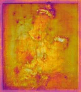
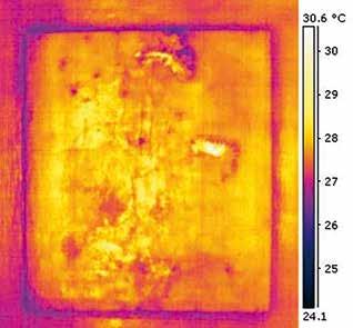
Fig 1. Visible light painting with mapping of the sampling points 8. Thermography 9. Digitally superimposed thermography on the image of the visible light painting
THE MADONNA OF ANCONETTA, DIAGNOSTICS, PROJECT AND RESTORATION
DIAGNOSTIC IMAGING: RADIOGRAPHIC IMAGING; IR REFLECTOGRAPHY, UV FLUORESCENCE INVESTIGATIONS, IR THERMOGRAPHIC IMAGES
Diagnostic imaging techniques, such as IR reflectographic observation, X-ray radiographic technique and IR thermographic mapping of the artwork, have allowed to identify areas with different electromagnetic response and different radiopacity in the icon. This distinc tion was useful for the collection of the micro samples that were distributed in the various identified sectors in order to characterize the different stratigraphic packages (Fig. 1). The combination of imaging techniques with chemical - stratigraphic characterization has made it possible to perform a digital mapping of the different types of stucco reintegration interventions, differentiating the various interventions from the original ground prepara tory layer. The following types of stucco painting preparation were therefore identified: Type 1: original preparation with plaster and animal glue, spread in two layers, the first with larger grain size plaster. Type 2: refers to a subsequent restoration intervention, consist ing of a primer based on calcium carbonate, drying oil added with barium white. The characteristic radiopacity of the latter made it possible to recognize its limits on the plate, identifying both the filling of the original ground layer and paint layer gaps (lacuna) and the overflowing additions. Type 3: referable to a second intervention after the original, based on calcite, plaster and drying oil. Also, in this case it was possible, thanks to the radiographic investigation, to map graph ically on digital support to report the area.
01
02


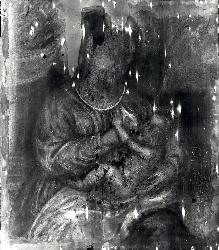
Fig 2. Image of the painting after removing the repaintings. X-ray painting image .X-ray painting image with mapping of ground layers and nails
03


Fig 3. IR infrared painting image. Image of the real painting digitally superimposed in infrared. X-ray painting image digitally superimposed in infrared
04

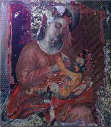

Fig 4. Ultraviolet image with the mapping of the different identified repaintings. Visible light painting image after the removal of warnish with mapping of the identified repaintings distinguished by removal and conservation

05
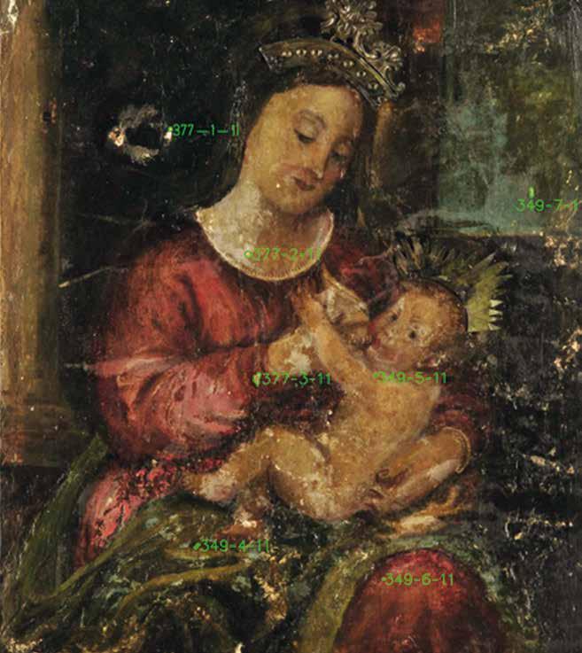

Fig 5. Visible light Painting with the sampling point 8th. Image of Crosssection with reflected light optical microscope 9. Scanning by electron microscope (SEM backscattered electron image)
X-ray analysis and thermographic images also showed the presence of metal elements as a support for the positioning of the ex-voto. These elements are not visible to the naked eyes as, being historicized, they are masked by layers of stucco and pictorial film. After removing the layers of varnish, other useful information was obtained through ob servation in UV fluorescence and grazing light. The non-coplanarity and the different fluorescence, -as a result of ultraviolet stimulation, -of pictorial materials, led to identify the coexistence of at least seven pictorial integration interventions carried out on the artwork.
MICRODESTRUCTIVE DIAGNOSTICS: STRATIGRAPHIC SECTIONS AND CHEMICAL – PHYSICAL ANALYSIS OF MICRO SAMPLES Stratigraphic investigations and microanalysis represent traditional techniques which, even with the limitation of taking samples from the artwork, provide certain references and fundamental chemical data that cannot always be extrapolated with certainty from diagnostic image techniques only, such as the composition of single overlapping layers. The chemical investigations on stratigraphic sections were performed by scanning elec tron microscope analysis accompanied by energy dispersed microscopy and Fourier transformed infrared. There were characterized three different types of ground layers that testify to the repeated retouching to which the artwork was subjected. The pigment analyzes also helped to pro vide indications regarding the possible dating of the repainting. In fact, above the oldest ground layer we have pigments certainly prior to 1700 while in subsequent retouching we also find pigments of eighteenth-century or later origin, such as barium white, zinc white and bice blue. The histological analysis of a sample taken from the wooden support (back of the slab), classified the essence of the wood as belonging to the species Picea excelsa Link (Spruce).
CONCLUSIONS The partnership between training bodies, protection bodies and specialists in the sector has made it possible to bring together professional figures who have different skills, who use different but at the same time complementary technologies. This has revealed numerous data on the construction techniques of the artwork and on past restoration methods. The subsequent digital processing allowed the operators to carry out the restoration in a conscious way, allowing, for example, to remove the overflowing parts of the non-original grouting with the scientific awareness that the oldest pictorial layer was present under it, thus managing to find the original portions of pictorial film. Finally, digitalization has allowed the comparison of the various diagnostic techniques, al lowing an immediate reading of the analytical data even in a highly complex artwork from a stratigraphic and material point of view.


