2 minute read
Cervical Spine Films
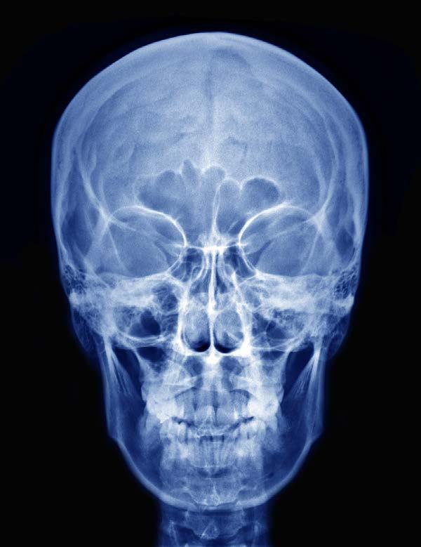
Figure 3.
The Waters view is an angled PA view of the skull that looks specifically for sinusitis or for facial fractures. The frontal and maxillary sinuses are what can most easily be seen with fractures visible on the inferior orbital rim, maxillae, zygoma, and zygomatic arch. The patient stands upright and faces the x-ray source.
Advertisement
A skull film using the Schuller method will detect problems with the base of the cranium. It cannot be used in suspected cervical spine fractures or when subluxation of the cervical spine is suspected. It can be used to detect multiple areas of the face, such as the hard palate, mandible, and both the sphenoid and ethmoid sinuses. The occipital bone can also be visualized. The patient is upright or supine for this x-ray. The neck must be hyperextended in order to see the structures of the base of the skull.
CERVICAL SPINE FILMS
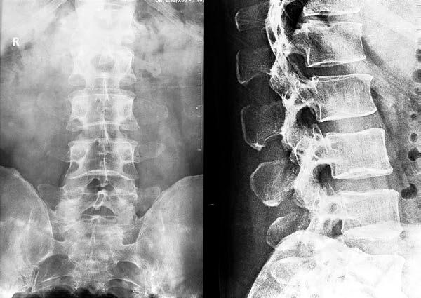
Spine films can be done on the cervical, thoracic, or lumbar spine. In a good cervical spine film, the x-ray must visualize all seven vertebrae plus the C7 to T1 junction. The cervical spine film can be done on the erect patient in either the AP or PA view. Figure 4 shows a cervical spine film (both AP and lateral):

Figure 4.
The lateral film is also done on the erect patient, but the side view of the patient is taken. The arms are held to the sides and the shoulders should be kept as low as possible in order to have an unobstructed view of all seven vertebrae and the first thoracic vertebra. An oblique view can also be done to see subtle fractures as well as the lateral foraminae. Patients with injuries may need to have this x-ray done while supine.
The cervical AP spine can be done using what’s called the “Fuchs Method”. This is an additional view that can detect the dens located in the foramen magnum. It cannot be used on the patient with degenerative diseases of the upper cervical spine or fractures in this area. The dens or odontoid process is a process or projection of the second cervical vertebra that sticks up through the first cervical vertebra.
The Judd method of obtaining the cervical spine film looks at the dens and the atlas as they can be visualized through the foramen magnum. As in the previous technique, it cannot be used for degenerative diseases or fractures of the upper cervical spine. The patient lies prone for this view with the neck extended and the tip of the chin located on the x-ray table.
The AP odontoid process view will specifically view the odontoid process or dens through the open mouth in order to see the first and second cervical vertebrae. The patient is supine during the film with a vertical beam that is slightly angled at 10 degrees.


