13 minute read
Tourniquet use in knee replacement – the why, the what and how to do without


Jonathan Phillips, Ben Waterson, Andrew Toms and Keith Eyres
Jonathan Phillips is a specialist knee arthroplasty, trauma and revision surgeon, appointed at the Royal Devon and Exeter Hospital in 2015. He is a member of the ODEP and Beyond Compliance committees, as well as both the Primary and Revision BASK Knee Arthroplasty Working Groups.
Advertisement
Ben Waterson undertook his specialist training in orthopaedic surgery in Edinburgh and undertook the adult lower limb reconstruction fellowship in Vancouver. He was awarded a Medical Doctorate (MD) from Edinburgh University for his research in the field of knee surgery and the impact of alignment on total knee replacements. He has been working as a consultant in the Exeter Knee Reconstruction Unit since 2018 and is an Honorary Senior Lecturer at Exeter University.
Andrew Toms is the Professor of Orthopaedics at the Princess Elizabeth Orthopaedic Centre, Exeter. He trained on the Stoke / Oswestry rotation and in Vancouver, Canada. He currently sits on the NJR Editorial Board, the Bone & Joint Infection Registry board, BASK Research Group and the RCS Robotic Advisory Group for Orthopaedics.
Keith Eyres is a Consultant Orthopaedic Surgeon practicing at the Nuffield Health Exeter Hospital and the Royal Devon and Exeter NHS Foundation Trust. He specialises in hip and knee replacement, knee revision and rheumatoid surgery.
A recent Cochrane review published in December 2020 re-opened the debate of tourniquet use during knee replacement and suggested we minimise its use. However, a 2016 survey of 547 consultant knee surgeons, members of the British Association of Surgery for the Knee (BASK), demonstrated that 90% used a tourniquet during total knee replacement surgery, the majority of whom did not wish to stop doing so1.
Why use a tourniquet?
Tourniquet advocates explain that the use of a tourniquet enables more accurate and quicker surgery due to the presence of a blood-free field2.
There is also an argument that cement penetration into the bone is improved if there this a dry bed into which cement can interdigitate. There is evidence for this in hip replacement surgery too where obviously without the use of a tourniquet; modern cementing techniques involve washing and drying the prepared bone surfaces, suction catheters and pressurisation of cement both before and after implantation3. There is a risk that poor cementation and pressurisation may lead to earlier implant loosening, although this link in knees is not as clear as in hips.
Surgical training is still largely apprentice-based. Therefore, if surgeons train to perform surgery using a tourniquet, it is likely that they will continue to use one throughout their career.
What is the argument not to use a tourniquet?
While tourniquets may provide a bloodfree field to allow for more accurate and/or efficient surgery, it does so at the compromise of producing an essentially avascular limb for the duration of the surgery.
When a limb is avascular, tissues are unable to undertake aerobic respiration, and as such this may lead to an increase in anaerobic metabolic products or rhabdomyolysis4. There are time limits suggested for the length of time for tourniquet use (a maximum of two hours is generally recommended). Beyond this time period, the use of a tourniquet may have a detrimental effect not only on the tissues of the limb, but it also increases the risk of reperfusion injury once the tourniquet is released. The release of anaerobic metabolites can cause an adverse effect to the cardiovascular system5.
Tourniquets are applied in the mid-thigh region and may not fit well for certain body types (such as overweight patients with significant adipose tissue and short legs). Frequently this is the typical knee replacement patient. A poorly fitted tourniquet may either reduce access to the more proximal end of the incision or moveduring the case leading to a compromise of sterility.
Tourniquets may fail intra-operatively, or the position of the tourniquet on the leg may change during the procedure such that the delivered pressure to the tissues drops below the systolic blood pressure but remains above the diastolic blood pressure, leading to a venous-tourniquet, where bleeding can appear worse and may only be resolved through deflating the tourniquet mid-procedure, while still having compromised exposure due to the presence of the tourniquet near the wound.
Tourniquet pain is well described in the literature and can be avoided by not using a tourniquet6.
A blood-free field may allow a clearer view during dissection or approach, however it may also risk masking iatrogenic injury of the vessels, a well-recognised risk in knee replacement surgery. Although this risk is very low, subtle vascular injuries may be missed intraoperatively if a tourniquet is used for the entire case, which may have devastating consequences for the patient. There are also patients with calcified vessels where compression may cause plaque fracture and thrombosis.
Advocates of tourniquet use have argued that blood loss is decreased. Indeed, there are publications demonstrating reduced blood loss intra-operatively, however the total blood loss (intra- and post-operatively) has been found to be similar with or without a tourniquet7,8. Modern anaesthesia techniques, with the use of tranexamic acid and improved pre-operative assessment to avoid performing surgery in patients with anaemia, have all meant that the concerns regarding blood loss post-knee replacement are far less of an issue, and blood transfusion after primary arthroplasty is now very uncommon.
Operating without a tourniquet means that small arterial bleeders (such as from the lateral geniculate vessels) are identified and cauterised intra-operatively, thereby reducing the risk of post-operative haematoma.
There is an additional risk of pressure injury and chemical injury from tourniquet usage. The pressure injury can cause subcutaneous fat necrosis, permanent soft tissue damage, nerve palsy and local pressure injury to the skin. Careful tourniquet application should limit this type of injury as long as the recommended time limitsare adhered to. The use of alcoholic preparations for pre-operative skin cleansing can lead to a chemical burn if left in contact with the skin beneath the tourniquet.
Many of these issues have led to some surgeons applying a tourniquet but only inflating it during the cementation process. Thereby, surgeons gain the benefit of using a tourniquet without the risks of a prolonged tourniquet time. As mentioned earlier, modern cementing techniques in total hip replacement do not rely on tourniquet use and in vitro lab testing shows that the same result is achievable in the knee using a cement gun and suction catheter to overcome the back bleeding pressure. Bleeding from bony surfaces at the time of cementation can be overcome through surgical technique described below3.
Are there any drawbacks to not using a tourniquet?
The main drawback to not using a tourniquet is that visibility can be poor, making careful dissection harder particularly in the presence of inflamed synovium or if the anaesthetist is unable to avoid hypertension.
An additional minor concern can be the intra-operative appearance, which can be unappealing to surgeons used to a strict bloodless field. There is also increased risk of a splash hazard particularly while performing aerosol generating procedures (AGPs) such as during power-tool use.
What does the evidence say?
A recent Cochrane review published in December 2020 based on 41 randomised controlled studies including 2,819 people concluded6:
“The evidence indicates that knee replacement surgery performed with a tourniquet increases the risk of serious complications needing additional healthcare, many of which will be avoided if a tourniquet is not used. Most people do very well after knee replacement, but like any major surgery, there are risks and use of a tourniquet may exacerbate these.”
The findings published in the review6 stated:
• Pain on the first day after surgery is probably worse with a tourniquet. On average, on a scale of 0 to 10 (higher scores = worse pain), people operated on with a tourniquet rated their pain as 5.81. People operated on without a tourniquet rated their pain as 4.56 (average difference: 1.25 points).
• Knee function one year after surgery is probably similar with or without a tourniquet. On average, on a scale of 0 to 100 (higher scores = better functioning), people operated on with a tourniquet rated their knee function as 89.74. People operated on without a tourniquet rated their knee function as 90.03 (average difference: 0.29 points).
• Satisfaction with treatment may be similar with or without a tourniquet. Six months after the operation, 94% of people operated on with or without a tourniquet were ‘extremely’ or ‘very’ satisfied with their treatment.
• There may be little or no difference in health-related quality of life with or without a tourniquet. On average, on a scale of 0 to 100 (higher scores = better quality of life), people operated on with a tourniquet rated their quality of life as 54.64. People who had surgery without a tourniquet rated their quality of life as 56.17 (average difference: 1.53 points).
• Serious adverse events such as blood clots in the leg or lung, infection, or reoperation other than to replace the artificial joint are probably more likely to occur with a tourniquet. Five per cent of people operated on with a tourniquet reported serious adverse events compared to 2.9% of people operated on without a tourniquet.
Although this is not compelling evidence for surgery without a tourniquet, it should be enough to make us question why we use a tourniquet. Our unit has performed tourniquet-free knee arthroplasty for over fifteen years, with low rates of revision surgery and high rates of patient satisfaction and many publications.
The Exeter cementation technique for tourniquet-free knee replacement
Where possible, spinal anaesthesia is used to provide a more hypotensive anaesthetic. Our experience has shown that the cut bone and incised soft tissues bleed less when sedation is added to reduce patient agitation or restlessness. Since 2012, we have used two grams of intravenous tranexamic acid on induction which has improved the surgical field. However, it is important to administer this at least 15 minutes before surgery starts. Diathermy should be used to cauterise bleeding vessels during the capsulotomy and applied to the meniscal beds. Another key step is to use a cannula with suction attached, inserted into the femur during bone preparation and placed close to the cut bone surfaces before cementation. Finally, infiltration of high-volume (usually 140-160ml) low dose local anaesthetic with adrenalin (adjusted to body weight) into the capsule and soft tissues throughout the knee, consolidates the advantage of being able to cauterise bleeding areas prior to wound closure.
Positioning of the patient, prepping and draping is much easier without a tourniquet in place especially in shorter obese limbs and this is an obvious advantage in revision surgery when the length of the procedure precludes tourniquet use. The leg is supported with a side thigh support and a foot bolster. After skin incision with the knee flexed, the capsule is opened and the patella everted or subluxed after knee extension. When the knee is flexed back into position again bleeding from the cut capsule stops.
The cannula with trocar (Biomet or Wolff needle) is inserted into the area of the medial femoral condyle to the depth of the venting holes two centimetres from the bone surface. The position is marked with a pen. When the trocar is removed, the femoral canal can be reamed allowing decompression of blood and fat from within the femoral canal (Figure 1). Careful positioning of the cannula avoids contact with fixation pin placement of the distal cutting block or the four in one cutting jig (Figure 2).

Figure 1: Femoral Condyle with intraosseous suction in place.

Figure 2: Intraoperative distal femoral cutting jig.
The suction attached to the cannula is set at a low suction pressure and aspirates minimal amounts of blood but dramatically reduces blood spray when the bone surfaces are cut. This drying effect is greater when the femur has not been perforated for example when navigation or robotic surgery is performed.
The tibia is cut and prepared in a standard way and the joint is balanced. Infiltration of 50ml of local anaesthetic into the posterior capsule is performed before cementing. Either the femur or tibia can be cut first depending upon surgeon preference and both techniques are routinely performed at our centre.
Prior to cementation, place the leg in extension and carefully observe for bleeding, particularly from the lateral meniscal bed or at the notch if the posterior cruciate ligament has been removed. The benefit of not using a tourniquet is that all bleeding points can be addressed intra-operatively using diathermy.
At the time of tibial cementation, the trocar and cannula is placed into the tibial metaphysis to allow bone washing and drying. The recommended point of needle insertion is medial to the tuberosity heading laterally and parallel to the cut bone surface approximately 15mm below the surface and angled to avoid the cavity for the tibial tray and keel. We have found that using the softer lateral metaphysis (in a varus knee in particular) is adequate for preparation of the whole of the tibia and avoids the risk of iatrogenic fracture in sclerotic bone (Figure 3). The suction cannula technique has been previously published and has demonstrated improved cement penetration in an in vitro setting (Bucher). It is postulated that an improved cement mantle may improve long term implant survival. In osteoporotic bone there is a risk of over penetration of bone cement, in which cases we either turn off the suction on tray insertion or do not use the suction needle technique.

Figure 3: Tibial suction catheter in situ.
The tibial surface is washed with pulsed lavage and dilute aqueous chlorhexidine (Chlorhexidine Acetate BP 0.02%; Baxter, Norfolk), and dried with the suction needle, a standard sucker and a dry swab prior to cementation.
We currently favour Simplex HV for cementation due to its longer working time and we apply the cement to the surface using a gun (particularly now as most cement can be mixed in a gun with vacuum suction) although it can be applied to the cut surface using spatula.
The key action is to get a swift seal of the cut surface with cement so that the suction effect is optimised, and cement can be seen being pulled down into the trabeculae. With cement pre-coating of the tibial component the implant is impacted into place. The cement and implant surfaces must be dry prior to implantation.
The cannula is removed after implant positioning and reinserted into the previously marked medial femoral aspiration hole. The femoral surface is washed and dried and a perfect surface for cementation is presented (Figure 4). The femoral component is pre-coated with cement and a horseshoe-shape of cement is applied manually or by gun to the dry cut bone surface and will appear to be pulled into the fixation holes and trabeculae. When the implant has been impacted the leg is extended fully and the patella is cemented. There is no need to aspirate the patella bone.

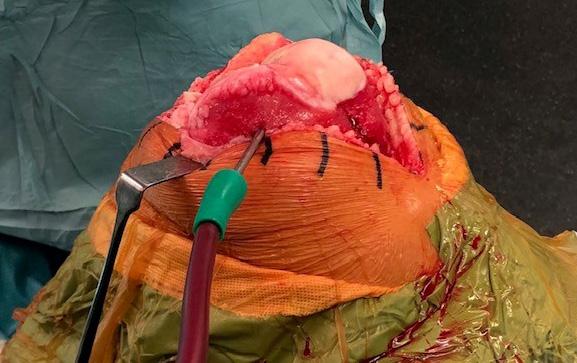
Figure 4: Femoral suction turned on showing rapid blanching of distal femur.
During the cementation phase the second 50ml syringe of LIA is injected into the medial and lateral gutters and capsule regions. Any residual bleeding should be addressed at this stage to minimise post-operative haematoma formation.
The final syringe of LIA is infiltrated into the subcutaneous areas and the wound is closed in a standard manner. Removal of the suction needle must be documented as part of the ‘count’ and the needle itself checked to confirm it is intact.
A similar technique is used for uni-condylar replacement noting that the cannula is inserted into the tibia away from the cut surface to prevent fracture propagation and then introduced into the respective femoral condyle to achieve dry bone surfaces. For patella-femoral replacement, the needle is only inserted into the medial femoral condyle to achieve the desired effect.
Conclusion
The latest evidence from a Cochrane review has demonstrated that tourniquet use during knee replacement surgery increases the risk of serious complications. This increase is modest, but we believe it is an unnecessary risk and that tourniquet use can be avoided by using a few simple techniques.
The risks and benefits of tourniquet use have been discussed and for those surgeons wishing to stop using a tourniquet, the method used at Exeter has been described.
References
References can be found online at: www.boa.ac.uk/publications/JTO.


