9 minute read
Evolution and current management of the paediatric supracondylar fracture


James Hunter and Anna Clarke
The supracondylar fracture of the humerus is still quoted as the case most trainees would like least on their first on-call consultant night. Mercer Rang prefaced his chapter on elbow fractures with the quote “pity the young surgeon whose first case is a fracture around the elbow”. He maintained it was an ancient saying, but searching for the reference is fruitless, leading to the possibility that he was quoting himself. This really does not need to be the case as we approach the end of the first quarter of the 21st century, and here we will try to summarise the tips and techniques that make the management of this fracture much easier.
As a registrar in the 1980s, the expectation was that one would manage a displaced supracondylar fracture by closed reduction and immobilisation, in either a cast or more traditionally a collar and cuff. This immediately exposed one to the conundrum of elbow flexion, whereby hyper flexion was required for stability, but was the very thing that endangered the vascular supply and could cause the compartment syndrome that resulted in Volkmann’s ischaemic contracture – the very worst results that could be obtained. Various alternative treatment methods were available; skin or skeletal traction, open reduction and K wiring (strongly recommended by several senior surgeons of that era) and closed reduction with percutaneous Kirschner wiring, which had been described a decade earlier. It is this latter method which is now the standard. Traction produces excellent results, but lengths of stay in hospital are between 10 and 25 days. Routine open reduction can lead to unacceptable levels of stiffness and modern equipment, especially image intensifiers, have facilitated the performance of close reduction and percutaneous pinning (CRPP).
Classification
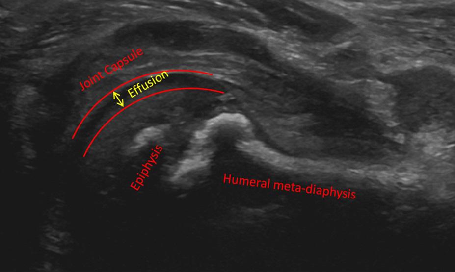
Five percent of supracondylar fractures are of the flexion type, which if displaced is particularly difficult to manage (Figure 1).
The classification of extension type supracondylar fracture has been made unnecessarily complicated by adherence to the method of Gartland, which requires considerable modification to work.
Gartland originally described mild, moderate and severe displacement of the supracondylar fracture, and this became modified to:
Undisplaced
Angulation with an intact posterior cortex
Complete displacement.
This version of the classification has been shown to have good reproducibility, but fails to distinguish between some type 2 fractures that can be managed non-operatively, and others that are less stable and require fixation. Most of the fractures that defy classification have an element of rotation at the fracture site. Von Laer introduced the concept of fractures that were rotated being distinct from those that were not and created a classification with four groups (Figure 1). There are other four group classifications, such as that of Lagrange and Rigault, and that of the AO group. Whichever one is used, the key is to distinguish unstable fractures requiring reduction and fixation from those that can be managed in a sling, possibly after a nudge straight.

What treatment when?
Undisplaced and angulated fractures can be managed in a collar and cuff or cast. The former is preferable, as one can achieve more flexion without anything at the front of the elbow. X-rays are easier to perform and the sling can be removed easily at the end of treatment.
The primary treatment for displaced fractures should be closed reduction and percutaneous K-wiring.
Traction can be used for displaced fractures, particularly in younger children with better remodelling capacity and is a useful ‘get out of jail’ technique which is useful to be familiar with. The rare fracture that will not reduce is better placed on traction, rather than attempting open reduction on an elbow swollen by multiple attempts at closed reduction. Skin traction is also used as overnight immobilisation in some units; the advantage being that it can be applied under simple analgesia without sedation or an anaesthetic. (Figure 2)
Timing of treatment
More than 20 years ago, the group from Cincinnati described leaving the formal treatment of supracondylar fractures until the next day in the majority of cases. Obviously, they excluded any cases with vascular compromise and they did perform a provisional reduction, under ketamine, in many of their cases. The complications were less in the group with delayed treatment. Multiple further investigations have been performed, albeit mainly retrospective and the evidence was summarised, in the systematic review from Aberdeen, which found no difference in outcome for a variety of endpoints from the need for open reduction to post-operative deformity. The key is to not delay fractures with vascular compromise. The recommendation in the latest version of the BOAST (2020) is that an absent pulse should be an indication for early surgery.

How to do a closed reduction
Views on how to reduce and K-wire the supracondylar fracture are relatively diverse and often entrenched. There is no need to change someone’s successful method, but when it is proving unsuccessful there are some useful tips. The method described on AO surgery reference is essentially a synthesis of the views of faculty members on paediatric courses over the last 20 years and is not contentious. It varies little from the method of Charnley described in his Closed Treatment of Common Fractures
For the standard posteriorly displaced supracondylar fracture (AO type 4, displaced Gartland 3) the following tips should be helpful.
Longitudinal traction with the elbow slightly flexed for at least five minutes gets most fractures out to length with interposed soft tissues released. Milking manoeuvres may be required for puckered in soft tissue, but these mostly release with traction.
Correct the medial lateral displacement using the image intensifier while maintaining traction.
Flex the elbow with a thumb on the olecranon, pushing the distal fragment forwards. One should feel reduction.
If the elbow will not fully flex, the fracture is not reduced. In full flexion, the hand may go white – do not panic.
Check the shoot through AP. Rotate the arm at the shoulder to check the oblique views, which will show the medial and lateral columns.
Maintaining full flexion, rotate the whole arm to get the lateral view, medial side up. The fracture will be stable if the posterior periosteum is intact and the elbow fully flexed.
Place your K-wires.
Check the alignment of the arm and carrying angle against the opposite arm.
For the flexion type a different approach is required.
Firstly positioning must allow full extension and hyperextension of the elbow.
It must be possible to get the image intensifier into the lateral position without moving the arm.
Pre-insertion of the K-wires into the distal fragment before reduction may be necessary, as inserting them with the arm fully extended can be difficult.
External fixation can be useful as the pins can be used as joysticks to reduce the fracture.
What wire configuration?
For all but the smallest children (under four) one should use 2mm K-wires. Why? They are stronger and will hold the position of reduction reliably for three weeks. They deviate less in the bone on insertion. They do not cause growth arrest. Multiple punctures with 2mm wire will not cause growth arrest.
For standard transverse fractures, it is reasonable to use two lateral wires to avoid the ulnar nerve. The entry point is on the capitellum. The wires should be divergent. If the wires cross outside the skin they will diverge in the bone. After placing the wires stability should be tested and if in doubt, then a third lateral wire or a medial wire can be added.
If using a medial wire, then one needs to take precautions to avoid the ulnar nerve; check the uninjured side for a subluxing ulnar nerve (one in 20). Make a small incision to directly place the drill guide on the medial epicondyle, then put the wire in on oscillate and press on the ulnar nerve so that you cannot get it without drilling your thumb. Some fracture configurations demand a medial wire, so it needs to be within one’s skill set (Figure 3).
Whatever wire configurations is used, the construct must be tested under fluoroscopy at the end of the procedure. The fracture needs to remain stable through flexion and extension of the elbow, and be resistant to varus and valgus stresses. Stability and alignment are more important than accuracy of reduction. Unni Narayanan reported to EPOSNA in 2017 that the limits of acceptability for reduction were quite wide:
Medial displacement 30%
Lateral displacement 15%
Posterior displacement 37%
Anterior displacement 25%
Baumann angle 59 degrees to 83 degrees
Anterior humeral line through capitellum.
Aftercare
The wires should be left outside the skin and removed in clinic. Three weeks is long enough for the majority of fractures. Four weeks is the absolute maximum and the elbow should be mobilised straightaway, not put back in a sling.
An above elbow backslab should be applied in 45°-60° of flexion. The slab is of sufficient size that it encompasses between two-thirds and three-quarters of the arm circumference, open at the front.
Physiotherapy is not required for the majority of supracondylar fractures, but it is worth warning parents that full extension of the elbow can take many weeks to return.
Nerve injuries
The neurovascular status must be carefully documented, before and after surgery. Testing of the individual nerves must be complete and documented. N/V ‘tick’ is not adequate documentation. The preoperative documentation is essential because new nerve lesions after the operation are a concern, and require discussion with the responsible consultant and the operating surgeon as a minimum (PNI BOAST). The combination of median nerve deficit and absent pulse potentially indicates entrapment at the fracture site, and is best released before the onset of healing.
Vascular problems
These can be a huge cause of concern. Some simple principles can guide decision-making:
There is an excellent collateral circulation around the elbow. The majority of vascular impairments associated with supracondylar fractures resolve with reduction of the fracture.
Volkmann’s ischaemic contracture is caused by a compartment syndrome in the flexor compartment.
It therefore follows that:
The immediate treatment of a poorly perfused hand below a displaced fracture is to reduce the fracture urgently.
If the hand is well-perfused after reduction, exploration of the artery is not required, whether the pulse can be palpated or not.
If perfusion is worse after reduction the artery may be entrapped. In this situation the reduction is normally incomplete. Re-displace the fracture, pull on the arm and try to milk soft tissues from the fracture. An open approach to the interposed soft tissues may be required.
A white hand that remains white after reduction clearly requires exploration, possible reconstruction of the artery and fasciotomy. Exploration and fasciotomy are well within the remit of the orthopaedic surgeon, but arterial reconstruction is not, so it is best to decide in advance who is available (vascular/plastic surgery).

Conclusion
In summary, with a sensible plan, displaced supracondylar fractures can be managed satisfactorily without stress.