
Message from the Deans
Tresa PollockInterim Dean, College of Engineering; Alcoa Distinguished Professor of Materials
 Pierre Wiltzius
Susan & Bruce Worster Dean of Science, College of Letters & Science
Perhaps the single most important factor that distinguishes UC Santa Barbara from the vast majority of R1 institutions is the steadfast commitment of the entire campus to collaborative research unconstrained by disciplinary divides. If an interesting problem or question arises, researchers from disparate fields will combine their expertise to address it. The initial investigation gives rise to fresh avenues of inquiry, which generate new integrations of applied expertise. They, in turn, attract more researchers and lead to other new projects. The cycle continues, and paradigm-shifting breakthroughs often result.
This rare culture of collaboration brings many benefits. Notably, it produces graduates who are more agile, more innovative thinkers able to see problems from multiple perspectives. It also infuses the entire research enterprise with a cooperative spirit, further expanding the potential for groundbreaking discoveries rarely available to scientists working alone in a single discipline.
The cover story in this issue (“FOCUS ON: The Bloodworm,” page 18) provides a perfect case in point. It tracks several decades of research into the amazingly tough jaws of the bloodworm, while tracing the interwoven threads of research into the secrets behind a variety of highperformance materials that allow other marine organisms to survive in demanding environments. The long arc of that mixed, often-overlapping research has revealed nature’s astonishing capacity for creative engineering while pointing the way to technological advances and important engineered materials, some of which have resulted in successful start-up companies born at UCSB.
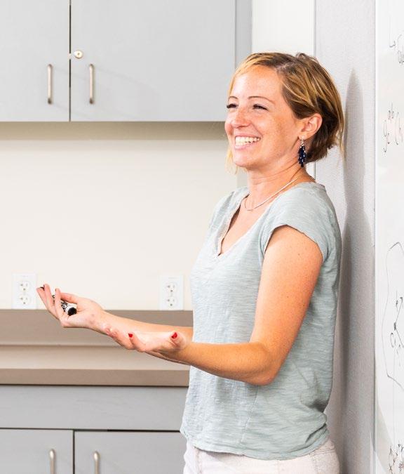
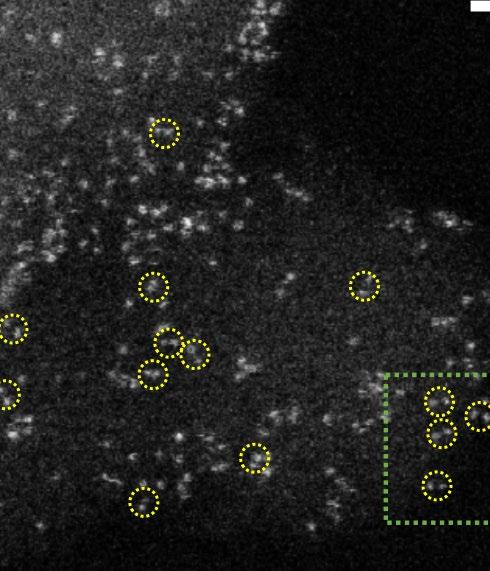
Elsewhere in this issue, you will find articles about the College of Engineering’s world-class collection of researchenabling electron microscopes (“Tech Edge,” page 15) and about collaborative research (page 28) linking Nina Miolane, an assistant professor in the Electrical and Computer Engineering Department, and distinguished professor of biology Denise Montell, who are working together to develop a new method to describe 3D live images of cellular activity with quantitative precision. You’ll also find a Q&A (page 9) with innovative computer science professor and new College of Creative Studies interim dean, Timothy Sherwood; and a report (page 12) on chemical engineering professor Phil Christopher’s new, more efficient “two-atom” catalysis process.
We hope you enjoy the issue, and we wish you a prosperous and healthful 2022-’23 academic year.
Briefs
A collection of news from UCSB engineering and the sciences.
9 Faculty Q&A: Timothy Sherwood
A conversation with the dynamic, curiosity-driven computer science professor. 12 “Pair-site” Catalysis
A new, more efficient way to speed up chemical reactions.
15 Tech Edge: Electron Microscopes
The indispensable enabling instruments of materials science. 18 FOCUS ON: The Bloodworm… …and other marine magicians of biomineralization. 28 The Math
of Cell Movement
Nina Miolane uses geometric statistics to quantify cell dynamics captured in images. 30 Champions of Engineering
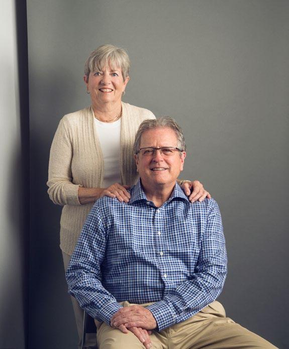
The interdisciplinary giving of Mark and Susan Bertelsen
The Magazine of Engineering and the Sciences at UC Santa Barbara Issue 30, Fall 2022
Director of Marketing: Andrew Masuda Editor: James Badham Art Director: Brian Long Graphic Designer: Lilli McKinney
UCSB Contributors: Sonia Fernandez, Harrison Tasoff
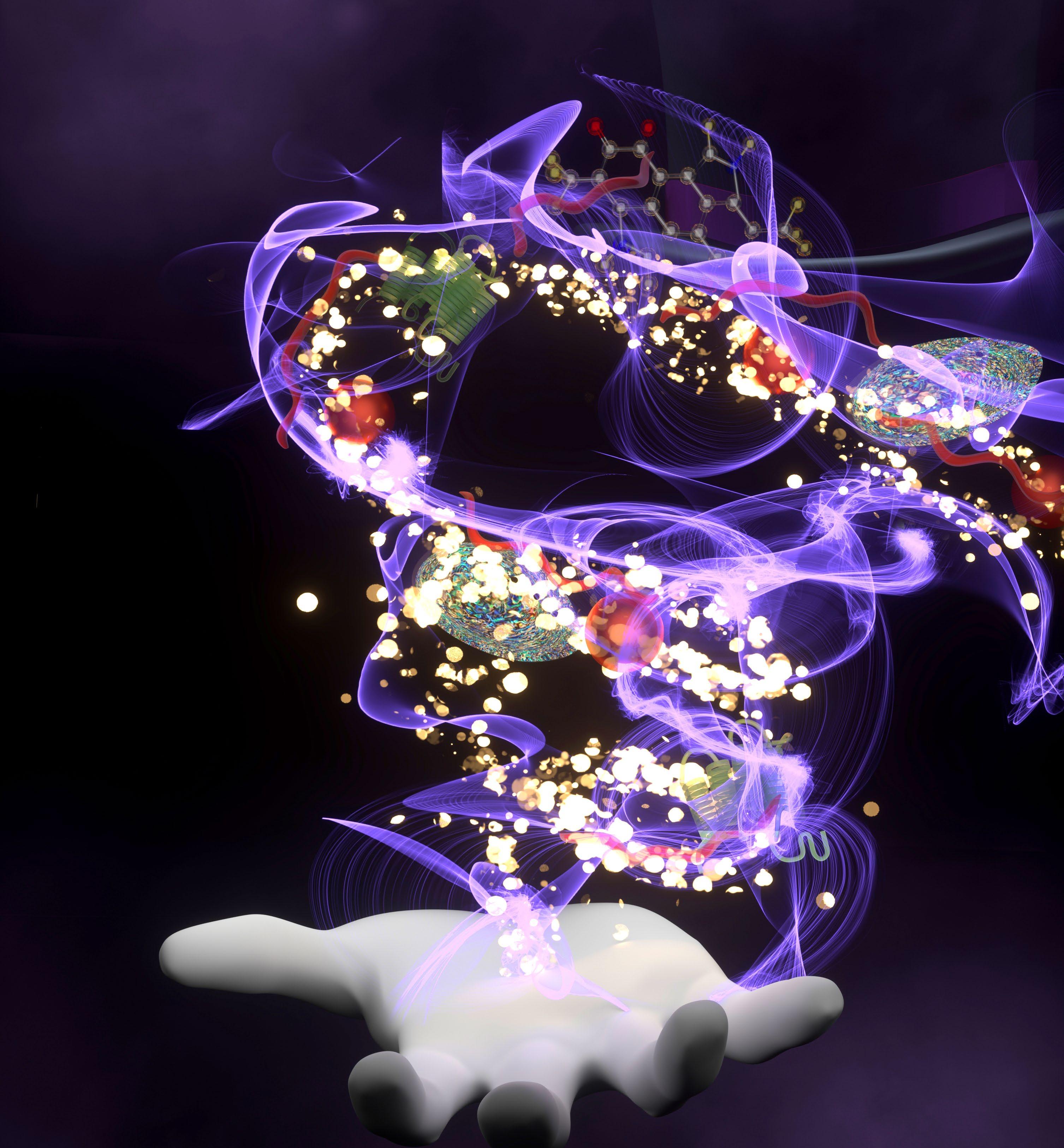
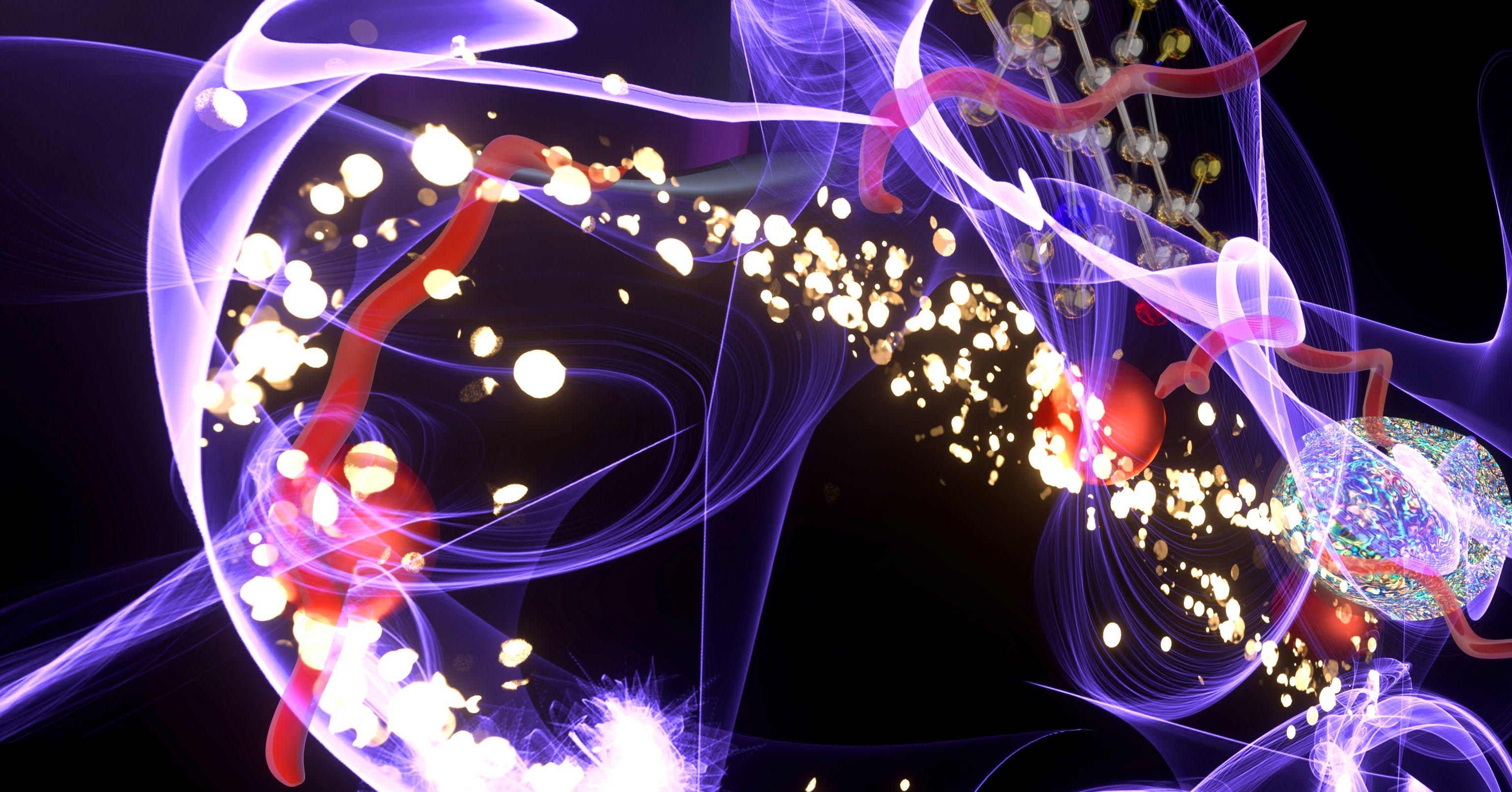
Cover Illustration: Artist’s representation referencing “marine magicians of biomineralization” (page 18), by Brian Long
Photography Contributors: James Badham, Jeff Liang, Lilli McKinney
College of Engineering
NEWS BRIEFS
COE Machine Shop Makeover
Thanks to donor support, the College of Engineering (COE) Machine Shop, a source of valuable support to labs, faculty, and students, is undergoing a major renovation that will transform it into a modern design center.
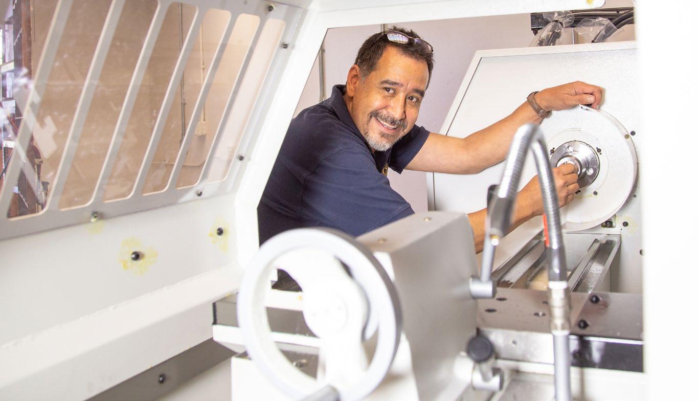
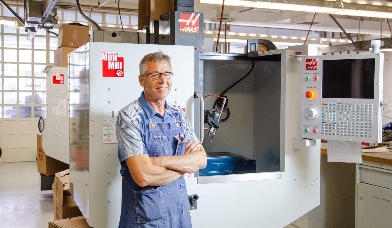
Long the first stop for COE faculty and students needing something built for a lab or an experiment, the shop is also where teams of undergraduate students in mechanical engineering work on their senior capstone projects, making it a critical component of their success both at UCSB and as they prepare to enter the job market.
“The newer technology and the other elements of this important renovation will aid students greatly in their capstone projects, allow them to work better as teams in a safe and productive manner, and enhance the College of Engineering’s competitiveness with other top institutions,” said shop superintendent, Marty Ramirez.
The project, which will increase usable space by 35 percent, is being conducted in phases as funds become available, making it a particularly valuable place for donors to direct their philanthropic efforts. Initial gifts, including one from longtime major UCSB contributor Virgil Elings, are funding the first phase. The goal is to raise enough funds eventually to establish an endowment to maintain the facility far into the future.
Phase I, which got started last spring, includes the removal of old equipment and the installation of eleven new CNC (computer numerical control) machine tools, 3D printers, a laser cutter, electronics fabrication and testing, pneumatics and hydraulics fabrication and testing, and bench and storage space. Phase II will include enclosing the space with glass garage doors and installing keyless entry for students to access the bench workspace and provide security during extended hours, especially at night.
Such a modern design center, says Tyler Susko, the Mechanical Engineering Capstone Instructor, “will introduce students to CNC machining in their freshman year and augment their understanding of those tools in subsequent classes before engaging in the year-long capstone project. We always have safety on our mind, of course, and the new CNC tools are fully enclosed, removing the danger of exposed spinning cutters. I’m looking forward to seeing the growth of our students as they get comfortable with the new tools, which will enable them to create things that were unfeasible in previous years.”
Big Numbers for Computer Science
10,655
1,521
2012 2022
A Boom in CS Undergraduate Applications
It’s no secret that computer science is a sizzling-hot major these days, a fact reflected in the following recent statistics from the College of Engineering’s (COE’s) own Computer Science (CS) Department. With 1,805 applicants for Fall 2022, the CS graduate program received the most applications of any graduate program in the entire university, and an impressive 19 percent of all graduate applications to UCSB.
• The 10,655 undergraduate applications received represent a seven-fold increase over the past decade, from 1,521 applications in 2012.
• The new undergraduate CS class includes 39 Regents Scholars out of a total of 54 in the COE. Regents’ Scholarships are among the UC’s highest honors, being awarded to students who have demonstrated academic excellence and leadership and show exceptional promise.
Bioengineering Welcomes FIRST PhD Students
TecHnology management Has a New Chair and its First PhD Grad Placement
The Technology Management (TM) Department in the UC Santa Barbara College of Engineering has a new chair, Professor Paul Leonardi. He follows Professor Kyle Lewis, who spent five years in the position.
Leonardi, who has been with TM since 2014, designed and led the Master of Technology Management program from its beginnings in 2015 through 2019 and has also served recently as PhD director.
“We are well positioned to build on the foundation laid by Kyle and become the preeminent department in the world focusing on the management of technological innovation in its many forms,” said Leonardi. “I believe we are building something unique, powerful, and important.”
Leonardi also announced that the department had placed its first doctoral graduate, Virginia Leavell, who completed her PhD in June and accepted a position as an assistant professor at the University of Cambridge’s Judge Business School. Leavell found the TM program (it was not yet a department) while studying for her MA in Sociology at UCSB.
This fall marked a big moment for the UC Santa Barbara Biological Engineering program, when it matriculated its first six doctoral students. Five of the six are women. They are: Gianna Gathman, who earned her BS in Bioengineering at Santa Clara University, specializing in biodevice engineering; Shaylee Larson, who received her BS in Chemical Engineering from the University of Utah; Elana Muzzy, who did her undergraduate work in Bioengineering at UC Santa Cruz; Zsofia Szegletes, who earned her BS in Biological/Biological Systems Engineering at Cornell University; and
From London in July, she said, “I am so proud to represent Technology Management as its first PhD graduate! I came back to school in search of a place where I could study how technological change affects work. In the TM Department, I found a community of world-class scholars who trained me in research methods and gave me the theoretical foundation to begin to answer the questions about the changing nature of work that keep me up at night. Technology Management is an exciting, rigorous, intellectual, and collegial place to study, and I hope to carry the culture of the department forward with me in my career.”
In her research, Leavell employs ethnographic methods and socialnetwork analysis to investigate how ideas about the future influence work and organizing during the lead-up to the implementation of digital technologies, as well as how organizations use digital technologies to make predictions about the future.
Lauren Washington, who studied Industrial and Systems Engineering at the University of San Diego. The group also includes UCSB alumnus Samuel Feinstein, who earned his undergraduate degree in Biochemistry and Statistical Analysis.
The students will be able to take advantage of a pair of training programs, both of which count as credits toward the PhD — the NIH T32 in Quantitative Mechanobiology program and the NSF Data Driven Biology Predoctoral Training program. Both provide focused coursework and professional development, while supporting students to “undertake research rotations in multiple labs so that they begin right away to build a network and a community,” said Beth Pruitt, director of UCSB Biological Engineering. “The program is designed around the idea of having the flexibility to explore different labs to help them decide on their PhD paths and projects.”
Enrolling PhD students is an important step in the process required for Bioengineering to become a full-fledged department, the seventh in the UCSB College of Engineering. “We are very excited about it,” Pruitt said. “We’ve been building the curriculum and all the pieces for years, and this fall, we officially start up!”
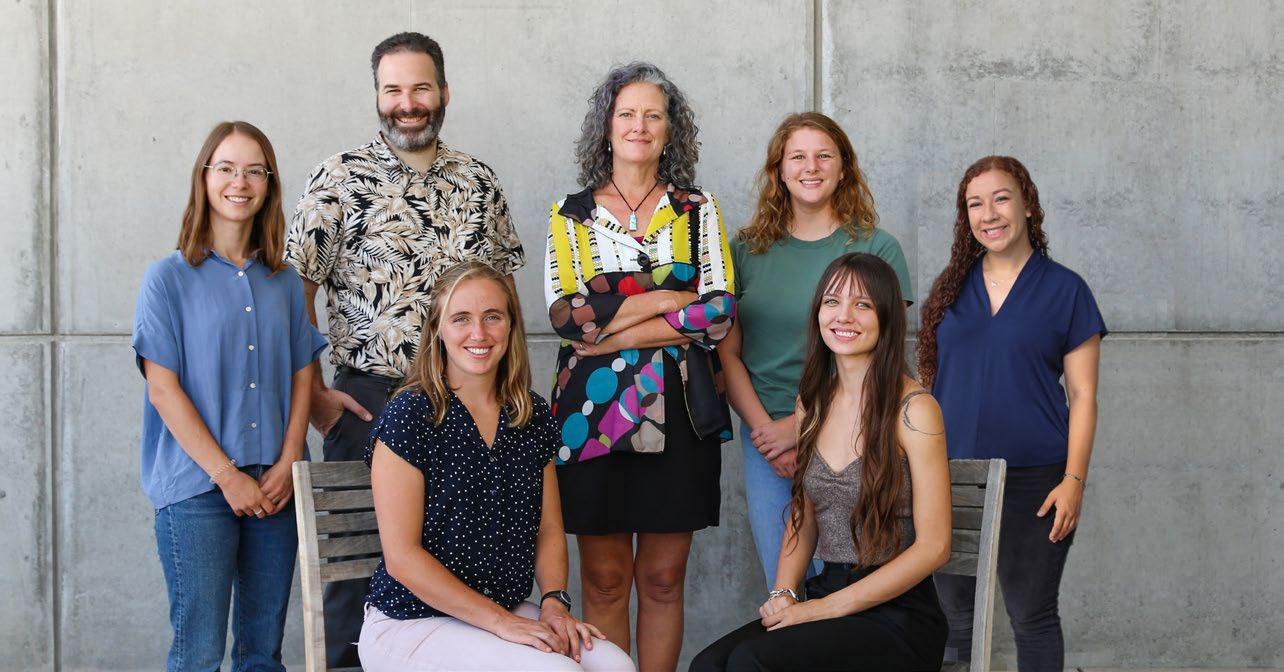
At last summer’s commencement (right), new Technology Management Department chair, Paul Leonardi congratulates Virginia Leavell, the program’s first placed PhD graduate.
“When we launched our PhD program in 2017, our goal was to train students to become leading scholars, researchers, and teachers of technology management at top universities around the world,” said Leonardi. “Virginia embodies everything we hoped for in our students: she is whip smart, her dissertation breaks new theoretical ground while also being immensely practical, and she earned a tenure-track job at one of the top business schools in the world. Her placement demonstrates that, even though we are a young program, the strength of our faculty gives even the most storied universities confidence that our students will excel as professors. Virginia is the first in what will undoubtedly be a long string of such students.”
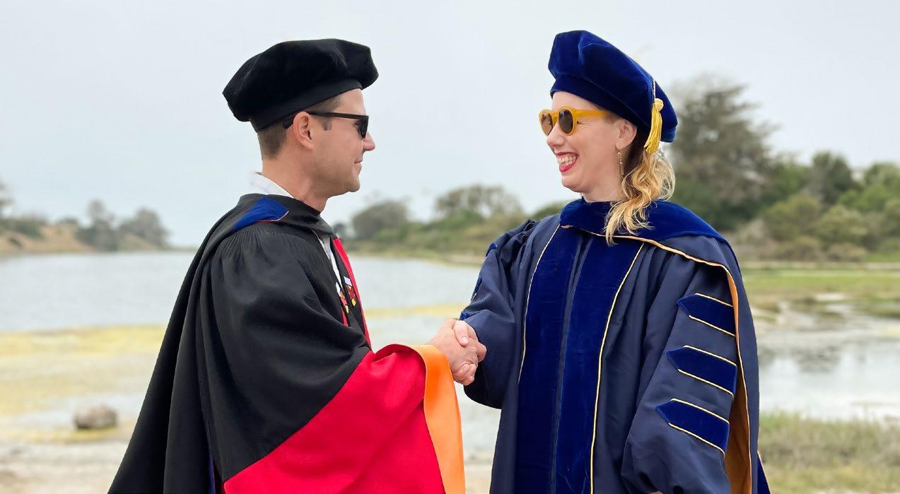
“We’ve been building the curriculum and all the pieces for years, and this fall, we officially start up!”Professor Beth Pruitt (back row, center) with the first PhD students in biological engineering (clockwise from top left): Zsofia Szegletes, Samuel Feinstein, Gianna Gathman, Lauren Washington, Shaylee Larson, and Elana Muzzy.
NEWS BRIEFS
Cell Cleaner extraordinaire?
extraordinaire?
A giant leap
Researchers in UC Santa Barbara neuroscientist Kenneth S. Kosik’s lab have discovered a novel organelle that helps to clean up faulty proteins so that cells can function in times of stress. The findings, reported in the June 2 issue of Nature Communications, have implications for treating Alzheimer’s disease, Parkinson’s disease, and other neurodegenerative conditions that result from misfolded proteins.
“People have known for quite a while that there are a few objects floating around in cells that don’t have membranes,” Kosik said. “Until relatively recently, it has not been clear how they’re held together, what they are, or what they’re doing.”
UC Barbara S. lab faulty can function times in June Communications, have for treating disease, from proteins. a while are Kosik “Until relatively it how they’re held doing.”
A mechanical jumper developed in the lab of UC Santa Barbara engineering professor Elliot Hawkes has jumped higher — roughly 100 feet (30 meters) — than any jumper to date, engineered or biological.
“The motivation came from a scientific question,” said Hawkes, who as a roboticist seeks to understand the many possible methods for a machine to navigate its environment. “We wanted to understand the limits on engineered jumpers.”
Using advanced imaging techniques, the researchers were able to observe biomolecular condensates, which don’t have a recognizable cell membrane enclosure but are, instead, separated from the surrounding cytoplasm as the result of a difference in density, similar to how the different densities of oil and water keep them separated. That separation creates a specialized environment for certain functions and reactions, such as those provided by what is called a stress granule, a membraneless organelle that appears when the cell is under stress. Its job is to sweep up RNA in the cytoplasm, storing those genetic instructions and pausing their translation into proteins.
“A cell under stress wants to shut down from making proteins to conserve energy and get past the stress,” Kosik explained.
But, what about the proteins that are already in a stressed cell? “If they’re under stress, some of them could get damaged and misfold,” Kosik says. Misfolds of the tau protein, for example, can become pathological and turn into the neurofibrillary tangles that characterize Alzheimer’s disease.
This is where the researchers’ newly discovered “BAG2” condensate comes in. Named for the BAG2 protein it contains, the organelle can sweep up these faulty proteins in the cytoplasm and stuff them into a proteasome — the cell’s version of a trash can — located in the organelle.
“A few proteins form a little barrel, and as the protein is threaded through that little cylinder, it gets degraded,” Kosik said. This inactivates and breaks down the protein. Many proteasomes are present in cells at any given time, he added, but what makes this particular one, labeled 20S, special is that it can accept proteins that are already somewhat misfolded and would not fit in the other cellular trash cans. Kosik suspects that the BAG2 protein may have a role in helping to organize the messy protein before it goes into the 20S proteasome.
“BAG2 is considered a co-chaperone in that it works with molecular chaperones to help proteins fold,” he said. In a previous study, researchers in the Kosik Lab demonstrated BAG2’s ability to target and clear tangled tau proteins in cell cultures. “The BAG2 condensates seem to actually travel to the damaged tau and gobble it up. It would be nice to figure out how we can shuttle tau into this condensate at the early stages of its damage for the cell to get rid of it before it gets worse.”
researchers able to of difference different water separated. a specialized functions reactions, as those what under stress. to sweep up proteins and the explained. are under stress, some Kosik the protein, pathological neurofibrillary tangles disease. where researchers’ Named organelle can and stuff proteins the is said. cells he makes 20S, that are already in other cans. the in to messy before it into the considered works molecular fold,” study, target clear tau in cultures. condensates actually condensate at early stages damage rid it before
In the biological world, the maximum achievable jump is limited by the amount of energy the system can give to pushing the body off the ground. In engineered jumpers, however, motors that ratchet or rotate can be used to multiply the amount of energy a jumper can store in its spring, an ability known as work multiplication.
“This difference between energy production in biological versus engineered jumpers means that the two should have very different designs to maximize height,” said Charles Xaio, a PhD candidate in the Hawkes lab.
Animals should have a small spring — only enough to store the relatively small amount of energy produced by their single muscle stroke — and a large muscle mass. “In contrast, engineered jumpers should have a spring that is as large as possible and a tiny motor,” Xaio said.
The team designed a 30-centimeter-tall jumper in which the spring, relative to its motor, is nearly one hundred times greater than what is found in animals. In their spring, carbon-fiber compression bows (the four black curved elements shown below) are squashed while rubber bands (the white elements) are stretched by a line wrapped around a motor-driven spindle.
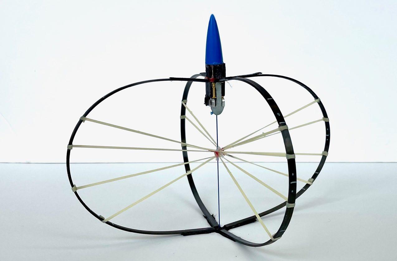
The mechanical jumper can accelerate from 0 to 60 mph in 9 milliseconds — an acceleration force of 315 g. (NASA astronauts experience up to about 6 g.) The 100-foot height it reached is, the study reads, “near the feasible limit of jump height with currently available materials.”
The jumping ability of this design sets the stage for reimagining jumping as an efficient form of machine locomotion, with jumping robots able to go where, currently, only flying robots can. The team calculated that, in a low-gravity environment, such as the Moon’s, the jumper should be able to reach 125 meters in height while achieving a kilometer of forward motion. That, Hawkes could not resist suggesting, à la first man on the moon, Neil Armstrong, “would be one giant leap for engineered jumpers.”
Ready for launch (above): The jumper built in Elliot Hawkes’s lab set a record for a jumping robot; (left): differences in density prevent water and oil from mixing; molecular condensates behave in a similar way, providing a favorable environment for stress granules.NEWS BRIEFS
UCSB Hosts System-wide Bioengineering Event
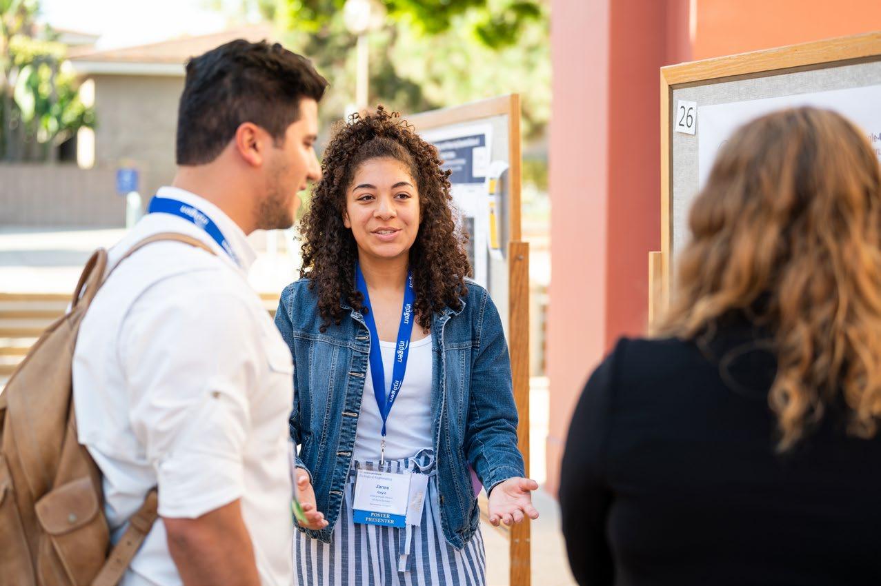
More than three hundred people attended the 22nd annual University of California Systemwide Symposium on Bioengineering and Biotechnology Showcase, known as BIC, held August 8-10 at UC Santa Barbara. The three-day event, which had been on hold during two years of the COVID pandemic, featured diverse opportunities for graduate students to interact closely with industry representatives and like-minded graduate students from all ten UC campuses, and to learn about diverse paths to success in the industry.
Major Awards for Chem-E Chair
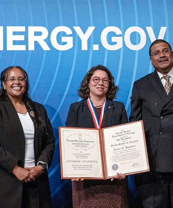
Over the course of a few days last spring, UC Santa Barbara chemical engineering professor and department chair, Rachel Segalman, won two of chemical engineering’s most prestigious awards. The first was the 2021 Ernest Orlando Lawrence Award in Condensed Matter and Materials Science, the U.S. Department of Energy’s highest scientific honor. The second was the American Institute of Chemical Engineers’ (AIChE) Andreas Acrivos Award for Professional Progress in Chemical Engineering. Then, in September, a third honor followed when Segalman was elected a fellow of the AIChE. And finally, in October, she was elected a fellow of the Royal Society of Chemistry, the oldest chemical society in the world.
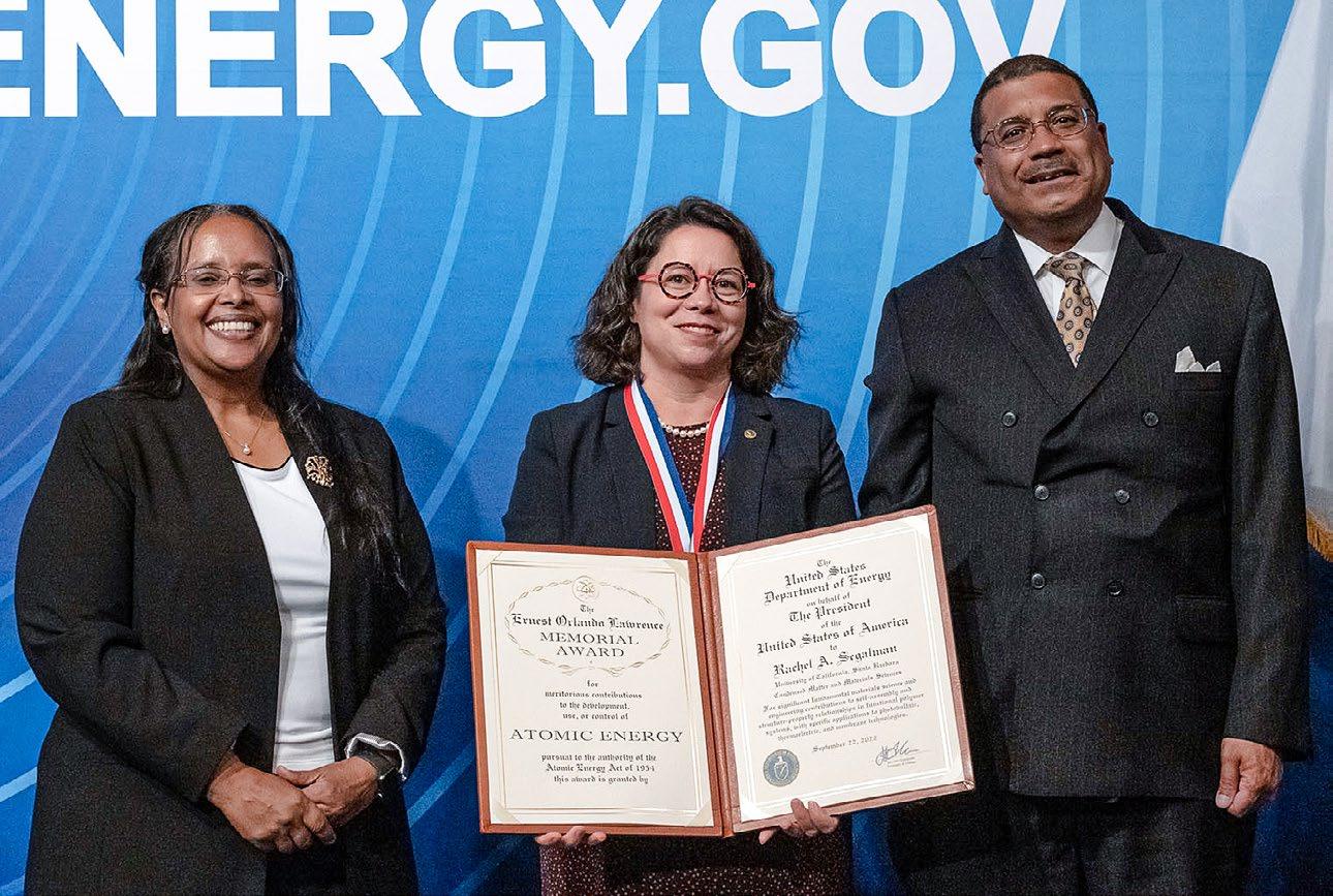
Segalman received news of the first award by way of a phone call from the Secretary of Energy herself, Jennifer Granholm. “All I could think about during the call was how this was the award that many of the all-around scientists and leaders that I looked up to as a young scientist had won,” said Segalman. “The attribute common to all of the prior recipients is that they are great scientists who had a significant impact in identifying and solving basic, fundamental scientific problems of critical energy consequence. They are also generous, insightful mentors and educators. For me, receiving this honor comes with a huge sense of accomplishment and responsibility.
“Much like the Lawrence Award, the Acrivos Professional Progress Award is special because some of my personal heroes have won it,” Segalman said of the second award, referencing Frances Arnold (2004) and former UCSB College of Engineering dean Matthew Tirrell (1998). “While the Lawrence Award is special because of its stature in the U.S. government, the Acrivos Professional Progress is a recognition from my peers!”
“The industry component made this event much more than an academic conference,” said Ryan Stowers, assistant professor of biological engineering and mechanical engineering and an organizer of the event. “This is a huge draw for the students who are more on the industrial track, giving them the opportunity to meet people and see what’s new in the field. And beyond the science, the speakers offered remarks about how they transitioned from grad school and how they got to their positions now. The students saw that there’s not just one route to a position and gathered insight into how some successful people navigated the professional terrain.”
The event included many invited speakers presenting on a wide range of topics, a poster session, a panel of professionals who took questions about pathways to biotech careers, and a kind of “speed dating” analog in which students paired with industry experts for ten minutes of rapid-fire exchange before switching to a different expert.
One junior faculty member from each of the ten UC campuses competed for the prestigious Shu Chien Early Career Lecturer Award, which went to UC San Diego assistant professor Daniela Valdez-Jasso, with Jury Awards going to UCSB assistant professor Siddarth Day and UC Davis assistant professor Randy Carney.
“Rachel Segalman has been at the forefront in materials and chemical engineering, making major contributions to our general knowledge of block polymers and hybrid thermoelectric materials,” said Tresa Pollock, the UCSB College of Engineering’s interim dean and Alcoa Distinguished Professor of Materials. “We are extremely proud that the Department of Energy has recognized the impact and significance of her research, and we are thrilled to extend to her our deepest congratulations.”
Segalman’s research is focused on controlling self-assembly, structure, and properties in functional polymers, work that paves the way for the development of sophisticated materials for such energy applications as photovoltaics, fuel cells, and thermoelectrics.
Students from all ten UC campuses participated in a poster session at BICNEWS BRIEFS
the right approach, not the popular one
New strategic development leader
The UC Santa Barbara College of Engineering (COE) has hired Meredith Murr as the new Director of Strategic Programs and Corporate Development, a position crucial to securing important industry partnerships that support faculty research at the leading edge of discovery.
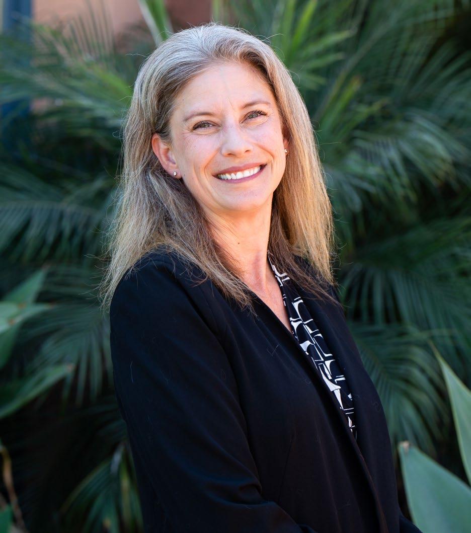
“I’m really excited,” said Murr, who started her new position in August. “I have spent a large portion of my life at UCSB. I started my PhD here in 2001, and I was on campus straight through 2018, either with graduate school or various jobs. It definitely feels like coming home.”
Murr earned her PhD in UCSB’s Department of Molecular, Cellular, and Developmental Biology in 2006. She spent the next twelve years working at the university, including nine years with the Office of Research, where she rose to become the assistant vice chancellor of research development and strategic planning. In that position, she worked with the vice chancellor and academic deans to match strategic research goals with funding opportunities. She left UCSB in 2018 to launch a consulting business, where she advised academic institutions and national laboratories on strategic planning and proposal development.
UCSB materials professor and Nobel laureate Shuji Nakamura tenaciously pursued his vision of a GaN LED, despite prevailing opinion suggesting he was wrong.
At the Japan Society of Applied Physics conference in 1992, approximately five hundred individuals attended the zinc selenide sessions [related to using the compound to create a blue LED), but only about five people attended the gallium nitride (GaN) sessions.
“Not only was zinc selenide more popular at the time [as a likely candidate for creating the then-elusive blue LED], but gallium nitride was actively discouraged, with researchers stating, ‘Gallium nitride has no future,’ and ‘Gallium nitride people have to move to zinc selenide.’”
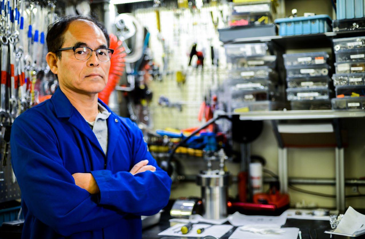
One year later, Shuji Nakamura, a dedicated “gallium nitride person,” would use his GaN platform to invent the blue LED, which would revolutionaize lighting and earn him the 2014 Nobel Prize in physics.
From a September 8 article on forbes.com about the importance of challenging conventional wisdom to achieve great things.
“We are thrilled to welcome Meredith to the college,” said Tresa Pollock, interim dean of the College of Engineering and Alcoa Distinguished Professor of Materials. “She is familiar with the college and our faculty, and she knows the importance of engaging industry partners in pioneering research. I am confident that her abundance of relevant experience and her clear strategic vision for advancing interdisciplinary research and enhancing external support will allow her to make a significant impact.”
Murr’s responsibilities in the position include two major components: working to interest industry partners in funding research, and meeting directly with faculty to brainstorm about the next grand challenges in their fields and how to solve them in ways that play to UCSB’s strengths. Once faculty-driven ideas are established, Murr’s team will present them to members of industry and collaborate with the Office of Development and the Office of Research to pursue private or federal funding to establish research centers on campus. The overarching theme of her director position, Murr says, “is to do the groundwork to help raise funds that enable college faculty to expand the frontiers of their research.”
“My scientific background and my work experience will allow me to speak with potential funders in a way that combines a compelling scientific story informed by a firm understanding of the scientific details,” said Murr, who did quite a bit of strategic planning as a consultant. “I also know how to bring people together and build consensus by ensuring that people feel heard and excited about moving forward.”
Murr will also oversee the Corporate Affiliates Program, which is designed to facilitate the interactions of corporate members with faculty, students, and other campus stakeholders. When it comes to industry partnerships, she says, “The college has a huge potential for growth.”
“In the departments that are really growing — computer science, biological engineering, and technology management, for instance — there are tremendous opportunities to develop new partnerships,” said Murr. “There’s also a lot to build on with the already-excellent industry relations that are in place in the Chemical Engineering, Materials, Mechanical Engineering, and Electrical and Computer Engineering Departments.”
Murr encourages industry representatives to contact her to learn more about the college’s many areas of expertise, the diverse investigations being undertaken with them, and the state-of-the-art facilities that enable UCSB’s world-class research enterprise.
“Our office serves as a gateway for industry partners,” she says. “Once we understand what they are looking for, we can identify the key researchers and resources available within the college to explore prospective research collaborations and accelerate innovation.”
QA & Faculty
TIM SHERWOOD
A conversation with dynamic, curiosity-driven computer science professor Timothy
 Sherwood
.
Turn the page to read some of his thoughts on temporal logic, the importance of communication and representation, curiosity-driven research, and more.
Timothy Sherwood is a busy man on campus.
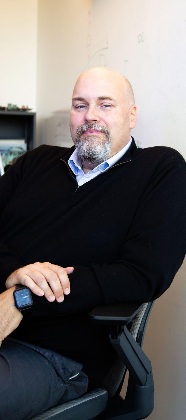
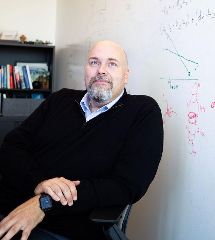
The engaging UC Santa Barbara computer science professor leads a full research agenda focused on cybersecurity and energy-efficient computing, mentors about six graduate students at a time, and runs a program with Associate Vice Chancellor for Diversity, Equity and Inclusion Sharon Tettegah aimed at creating a new interdisciplinary, student-driven foundation for understanding issues of personal significance through a diverse lens. He recently spent five years as Associate Vice Chancellor for Research and, in September, began his tenure as interim dean of the College of Creative Studies, while still teaching an introductory class on computer architecture to about 130 students. We spoke with him in August.
TIM SHERWOODConvergence: Can you talk a bit about your recent research on what is referred to as temporal logic?
TS: Any time you do arithmetic, whether as a machine or as a human, you need a way to represent a number. As humans, we use the written digits zero to nine and then string them together to represent a number. A computer represents a number in a similar way but uses only one or zero. Here, instead, we’re representing the number as an amount of time that has passed. So, you might have two signals that are like a pulse. There’s an event, and you encode the value as the time elapsed between some reference time and when that event happens. One event might be encoded as three and another event that occurred closer to the reference time as a two. That’s the basic idea.
Now, you need to do arithmetic. So, you define arithmetic partially based on your system of representation. For instance, as a human, we have rules for how to carry values in addition, etc. A computer does arithmetic in an analogous way. But when you’re dealing with these time-encoded values, you do arithmetic in a very different way. One of the interesting things is that, in this encoding system, some operations become easy and some become hard, and some of the things that become easy are bioinfomatics and certain signal-processing applications, which lots of people care about.
C: Have there been further advances in this area? If so, are they related to improving the energy efficiency of computing?
TS: We’ve been doing some work related to how you can do more general machine-learning operations incredibly efficiently this way, which may be closer to how your brain works. Your brain doesn’t calculate by storing ones and zeroes; it does it some other way that we don’t fully understand, but it seems clear that spikes interacting over time is part of the story. Clearly, the brain is doing something right, as it is hard to think of a more efficient “computing system.” So, here we have a spiking network that does arithmetic, and we know the rules of this new way of doing arithmetic, which is closer to how the brain works than the traditional way of doing computation with zeroes and ones. We now have preliminary data showing that this approach is even more computationally powerful than we thought it was. For example, we can use this logic to approximate arbitrary functions, which, in turn, implies new ways to perform machine-learning operations and points toward new and even-more-energy-efficient ways of performing computation. Given how much energy is used each day by computer hardware, this type of logic has a great deal of potential — plus, I just think it is so interesting to find this completely new way of looking at things.
C: Mention cybersecurity, and many people probably think first of software, but you have a reputation for your work on defending against attacks that occur via hardware. Can you explain?
TS: Fifteen years ago, my PhD students had a simple curiosity-driven question, which was, how exactly does information flow through hardware? It’s a good question, and we didn’t know the answer. After a lot of work, it turned out that you can define it mathematically at the hardware level far more cleanly than you could ever hope to at the software level. In that work, we discovered all kinds of security vulnerabilities, including some that hadn’t been invented yet. Several years later, these two huge vulnerabilities came along, Spectre and Meltdown, which were a very big deal and caused hardware companies to lose billions of dollars. Digging into our curiosity-driven question had made us the only people in town who knew how to reason through something like that, so suddenly it went from, ‘This is an interesting little thing’ to ‘Oh my gosh, can you help us?’”
C: That kind of “just wondering” about a question reminds me of when UCSB materials professor Carlos Levi described our interim COE dean, Tresa Pollock, as an especially “creative” scientist. Does that word resonate for you?
TS: It does, and creativity is something people have mentioned in awards I’ve gotten. I think that curiosity-driven research in engineering is not given enough consideration, and yes, we’re very problem-driven people, and at the end of the day, every one of my students wants to solve real problems to make the world better. But at the same time, you have to be open to these curiosity-driven questions. Sometimes they don’t go anywhere, but they can also go someplace really interesting. There are projects that result from just wanting to look behind this door to know what’s back there. And it turns out that behind that door are a whole bunch of other interesting doors, and you just keep pushing and they keep giving, and you push some more, and then one day you find that you’re in this whole undiscovered area where no one has been before. Gosh, what an amazing feeling that is! You get to help define some whole new space for people to explore.
C: You have been involved in addressing issues of diversity, equity, and inclusion (DEI) on campus, including starting the Sustaining Engagement & Enrichment in Data Science (SEEDS) program with UCSB’s Vice Chancellor for DEI, Sharon Tettegah. Can you talk about the aims and importance of those efforts?
TS: Representation is one of the most important questions we should be addressing as engineers. My field, computer science, is a powerful lens for viewing the world. It reveals hidden truths in data, giving you the ability to learn things no one else can. So, a critical question is, who gets to look through that lens, and what problems do we use it to address? As engineers at such an important research-intensive university, we have a responsibility not only to provide access to what we already know how to do on the problems we already look at, but also to help others turn this lens on the problems they care about and the issues that are impacting their communities — and we have a particular responsibility to those who have been systematically marginalized and disenfranchised.
Doing that takes humility, first and foremost. I have to realize that my life experience does
not equip me even to have all the questions, let alone the answers to everyone’s problems. One of the things I see that we need to do is empower more people to be engaged in defining what are “important” questions and identifying what we need to do to understand them better and ultimately address them.
C: Can you talk a little about the importance of graduate students to the success of your lab, which has produced a considerable amount of awardwinning research?
TS: Probably the most important thing is that what we do in my lab is possible only because of our outstanding PhD students and our faculty collaborators, such as computer science professors Jonathan Balkind and Ben Hardekopf, and electrical and computer engineering professor Dmitri Strukov. My group consists of about six PhD students and a professional researcher, which allows me to spend a lot of one-on-one time with my students. I think that’s central to how you come up with really interesting “out-there” stuff. And it allows them to take their ideas and really develop and drive them. I just help to facilitate their vision.
C: The writing on your UCSB web page is friendly and light in style, conversational and clear. Is that by design?
TS: Yes, absolutely, and it is a result, first, of my desire to share. If you come up with something cool in your lab and it just stays there, it never does anyone any good, so communication is really important. Part of what we’re here to do is to share our understanding with the world, and with a broader audience — not just with people who already have PhDs in my subdiscipline.
The writing in my lab is something I take seriously. Although I’m constantly wondering if I’m wasting everyone’s time by emphasizing communication, when I talk to my former PhD students about the skills they learned in my lab that have served them well, they are very clear that one of those things is how to communicate their work. They tell me that it has been incredibly powerful in their careers: to be able to communicate with diverse audiences what it is they’re doing, why they’re doing it, and why it’s important.
...then one day you nd that you’re in this whole undiscovered area where no one has been before. Gosh, what an amazing feeling that is! You get to help de ne some whole new space for people to explore.
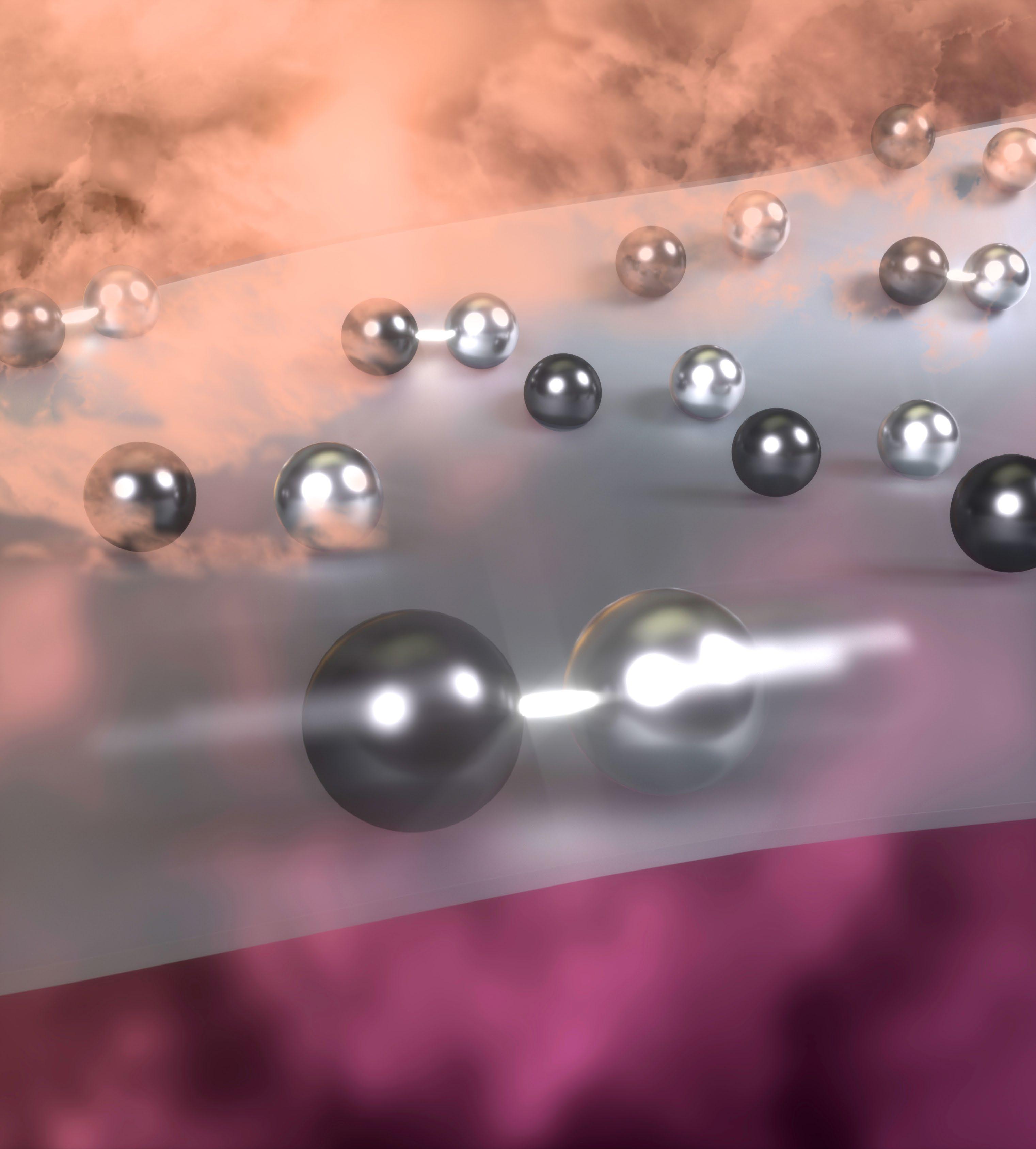
PAIR-SITE ” CATALYSIS
More Efficient Way to Speed Up Chemical Reactions
Catalysis is an enormous field comprising thousands of complex materials — catalysts — that speed up and direct chemical reactions and make it possible to produce everything from yogurt and laundry detergent, to paper and plastic, to gasoline and fertilizer. “The configurations and arrangements at the atomistic scale of the different components you put in catalysts dictate how reactive they’ll be for the different kinds of chemistries they are required to perform,” says Phil Christopher, professor of chemical engineering at UC Santa Barbara, an expert in catalysis, and a UCSB Mellichamp Chair in Sustainable Manufacturing. “The interactions between the catalyst and the chemicals being converted are an interesting and complex dance that can be challenging to understand.”
Many solid catalysts are porous materials that contain metal structures at nanometer length scales in their pores, where the reaction occurs as the material to be catalyzed encounters them. Often, the chemical reaction being catalyzed occurs as multiple chemical species interact on the catalyst. Accordingly, researchers spend a great deal of time pondering how those chemical species can find each other on the surfaces of nanometer-sized metal domains located inside porous materials and then react to form the products of interest.
One longtime goal in the field has been to make the metal domains increasingly smaller, thus increasing the efficiency of metal use in the catalytic process. “If you have a big chunk of expensive metal, most of that metal is in the middle, not at the surface, so, you use only a small fraction of the material to do catalysis,” Christopher explains. “If you shrink the metal domains down to one atom, then every atom has the chance to do chemistry.
“Furthermore,” he continues, “researchers often combine multiple metals in nanometer-sized domains to optimize catalyst performance. The ultimate limit of that would be to place two different metals next to each other in pairs of atoms that exist across a porous carrier, called a support. The hope would be that the pairs of atoms could work in cooperation to efficiently catalyze the reaction of interest.”
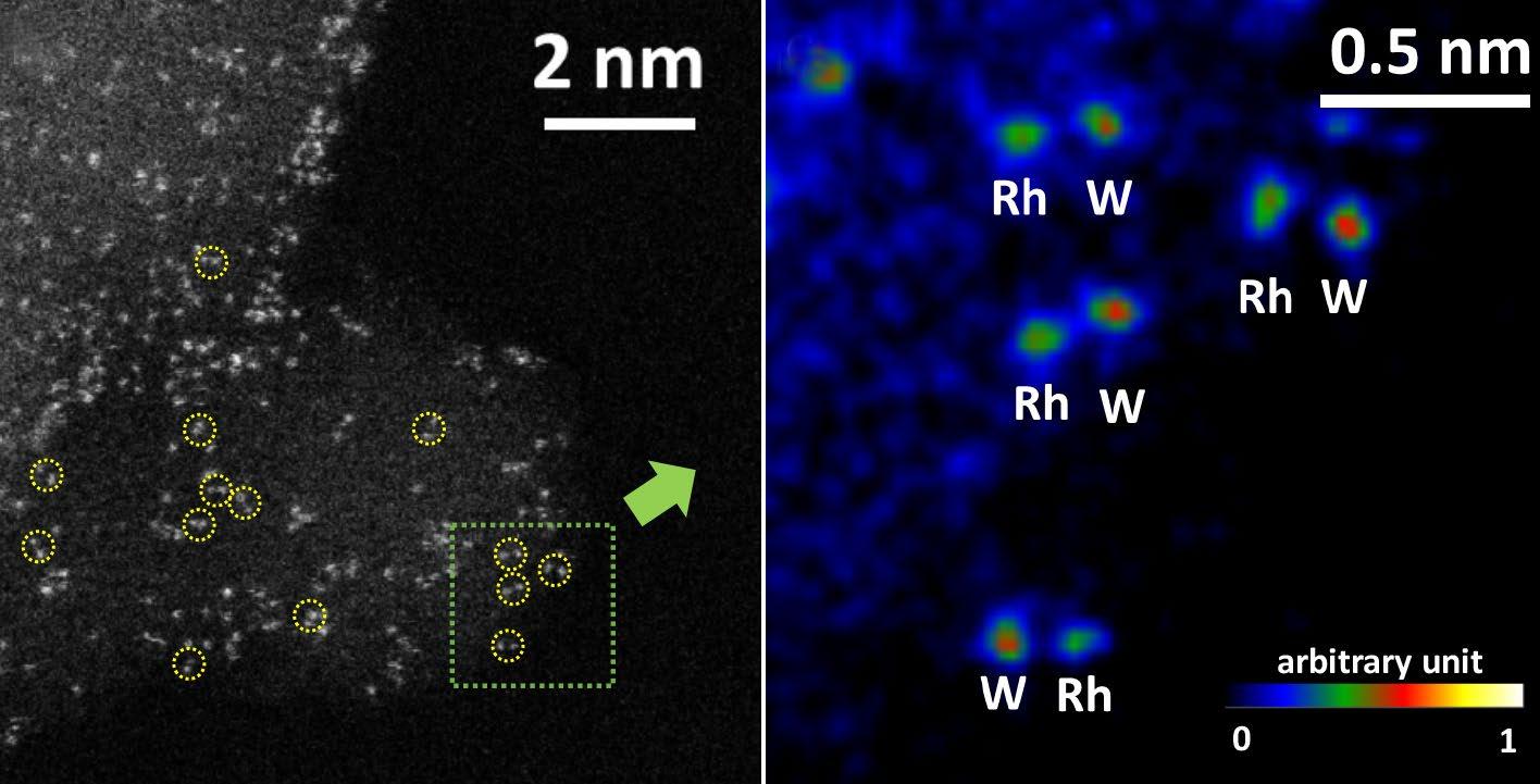
About four years, ago, Christopher began working to realize the goal of creating a catalyst of that kind. Now, he and his colleagues have synthesized such structures and identified rhodium and tungsten as elements that could work together to catalyze specific reactions. The research was funded through one of the U.S. Department of Energy’s Energy Frontier Research Centers, named the Catalysis Center for Energy Innovation.
A paper about the work, co-authored by Christopher, a former
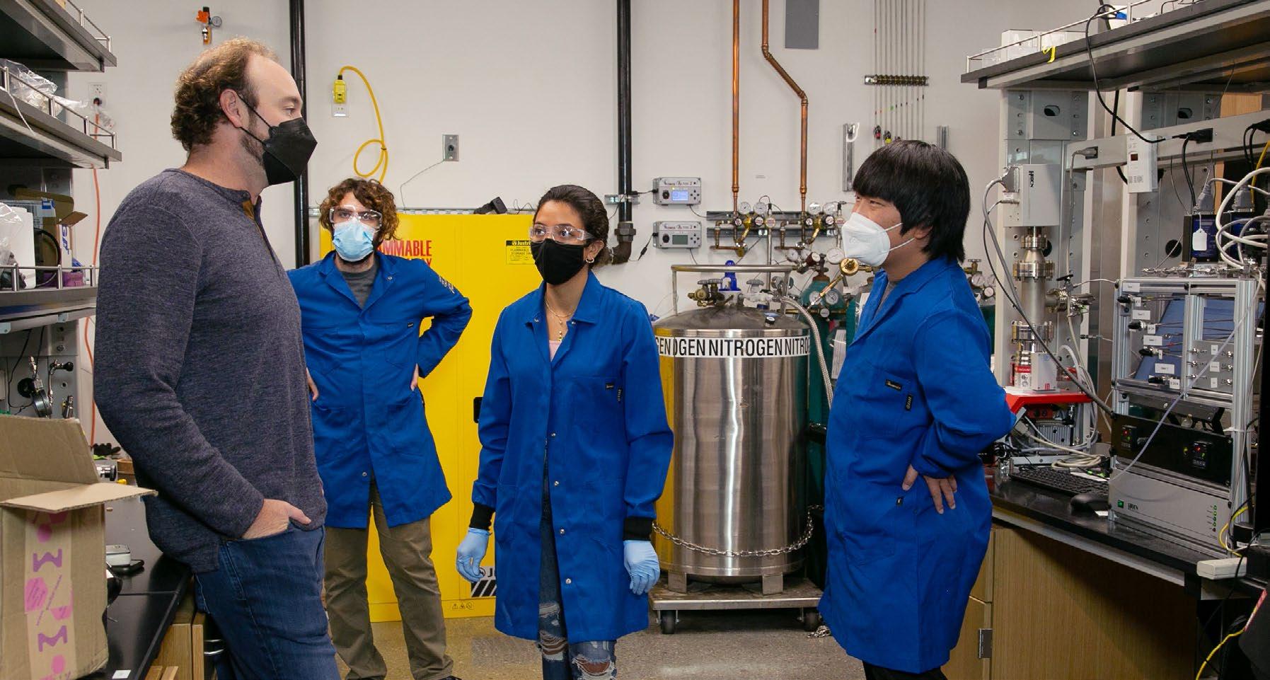
postdoctoral researcher in his lab Insoo Ro, now-graduated PhD student Ji Qi, current PhD student Gregory Zakem, and undergraduate student Austin Morales, along with colleagues from the University of Delaware, Columbia University, and UC Irvine, was published in the September 7 issue of NATURE. Christopher says that it describes “an observation of pairs of atoms that catalyze the conversion of three different chemical species in a cooperative process to form a single, desired product.
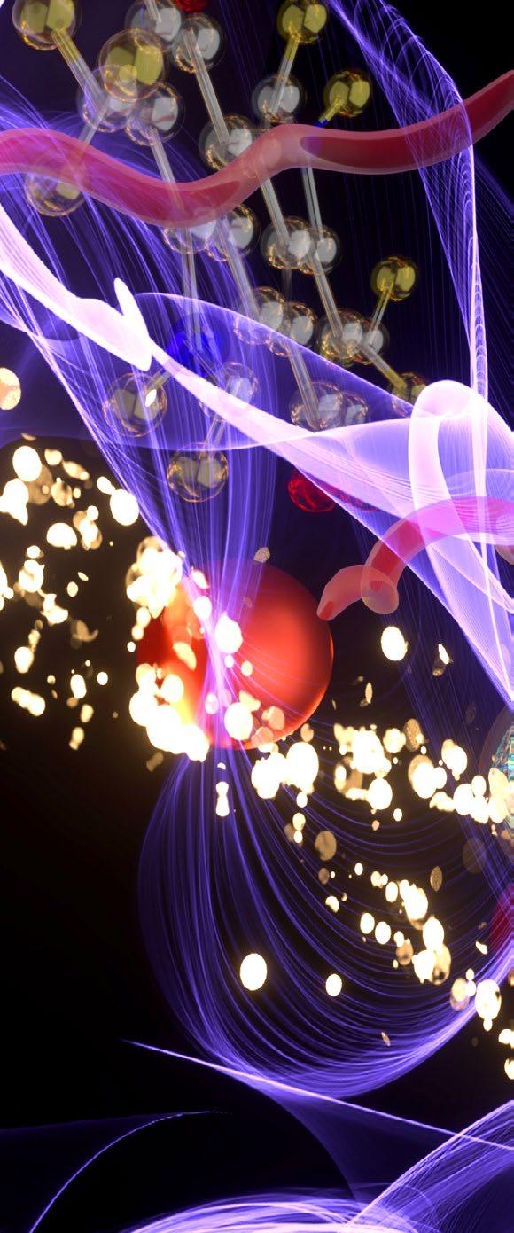
“Through our analyses,” he adds, “we hypothesized that one reactant prefers to bind to rhodium, while another binds to tungsten, and that the third binds at the interface between rhodium and tungsten. The atoms then transfer reactants among each other, while dynamically moving closer to
and farther
A Cleaner,
NOT ONLY DID WE PUT THE ATOMS NEXT TO EACH OTHER, BUT WE DID IT IN A WAY THAT TAKES THEM BEYOND BEING JUST A STATIC PAIR SITTING THERE. THEY ARE WORKING COOPERATIVELY TO ENABLE EFFICIENT REACTIVITY.
—PHIL CHRISTOPHER
from each other, ultimately enabling formation of the desired product. In catalysts that don’t contain pairs of rhodium and tungstem atoms, it was observed that only two of the reactants can combine, forming undesired products rather than the desired product.”
So far, the researchers have used their “pair-site” catalysts for only one specific but important and widely performed industrial reaction: hydroformylation, in which hydrogen, carbon monoxide, and an alkene (in this case ethylene) react to form the aldehyde propanal, aldehydes being building blocks for a wide range of commodity chemicals. “Oil or natural gas that you pull out of the ground contains hydrogens and carbons — hydrocarbons — and to make many consumer goods from them, you have to start by adding oxygen onto hydrocarbons,” Christopher says. “Hydroformylation is one of the first processes used in industry to do that.”
Making catalytic surfaces on which this chemistry can occur efficiently is extremely difficult, so current process chemistry instead employs liquid-phase catalysis. It requires liquid-soluable catalysts, which must be separated from products after catalysis.
Doing catalysis on surfaces could simplify the process. “The gas-phase reactants come into a reactor, convert to the product at the solidcatalyst surface, and come out the other side of the reactor,” Christopher explains. “Unlike in liquid-phase catalysis, the catalyst is stationary, eliminating the need for post-catlysis separation.”
Further, the on-surface method would, by definition, almost certainly be more environmentally friendly, because, Christopher notes, “removing sometimes-challenging separation steps means that less energy needs to be spent to produce products, so the process may cause fewer emissions.”
However, he notes, “Even though our catalysts work more efficiently than most other catalytic surfaces, they are currently much less effective than liquid-phase catalysts, so there is still a long way to go before industry would potentially adopt these new materials.”
Christopher believes that the paper was published in NATURE for a couple of reasons First, he says, “It’s hard to make a material where pairs of different metal atoms are stabilized next to each other across a support. We figured out how to do that and demonstrated that the material exhibited interesting behavior for a challenging rection.”
Second, he adds, “The rhodium and the tungsten work together in a very interesting way. They form a bond for a little while, they break apart, and they pass one of the reactants between them. So, not only did we put the atoms next to each other, but we did it in a way that takes them beyond being just a static pair sitting there; they are working cooperatively to enable efficient reactivity.”
Christopher explains how his group makes the material: “A highly porous supporting material called alumina is suspended in water containing a source of tungsten, which is then dried and treated at high temperature. The alumina, containing dispersed tungsten, is then suspended in water that contains a source of rhodium at a basic pH of 10. This pH was found to enable the deposition of rhodium next to tungsten. We’re still a little murky on how exactly rhodium and tungsten pairs form, but we have some ideas.”
Preparing their experimental catalytic setup involved “loading about one gram of the catalyst into a tubular quartz reactor, elevating the temperature to approximately one hundred degrees Celsius [212 degrees Fahrenheit], and flowing the reactant mixture over the catalyst. This
amount of catalyst contains more than a quintillion [1018] pairs of rhodium and tungsten atoms that were doing chemistry.”
Colleagues at the University of Delaware performed simulations based on quantum chemistry that provided details about the mechanism of the reaction, and electron microscopy done at UC Irvine allowed the team “to see the pairs of atoms across the porous alumina support with adequate resolution to be able to say, ‘That’s tungsten, and that’s rhodium in a pair.’
“This combination of analyses provided detailed insights,” Christopher continues. “Not only could we make a material where we thought we had these pairs, but we could also observe them and then understand how they’re working together to do this chemistry much more efficiently than could, say, either one of them separately or other versions of this that we tried before.”
Of course, new findings raise new questions, and, as Christopher says, “One question I get often is whether we have seen experimentally the dynamic behavior of the atoms moving to drive this chemistry. The answer is: not currently. The reactions happen on ultra-fast time scales, too quickly to be seen with a microscope. The best we can say is that the theory made numerous predictions that are consistent with experimental observations, so, if you believe that the coincidence is convincing, here is the mechanism that we think is at work.”
This research has also led Christopher’s group to consider whether there might be other recipes, pairs of other atoms, that might work as well or better than the current combination, or whether these materials could drive other important reactions, and also whether it would be “so challenging each time we try to make a new pair that it’s prohibitive. We’re thinking about it.”
Imaging the Ultra-Small
UCSB’S ELECTRON MICROSCOPES ENABLE ELITE MATERIALS SCIENCE
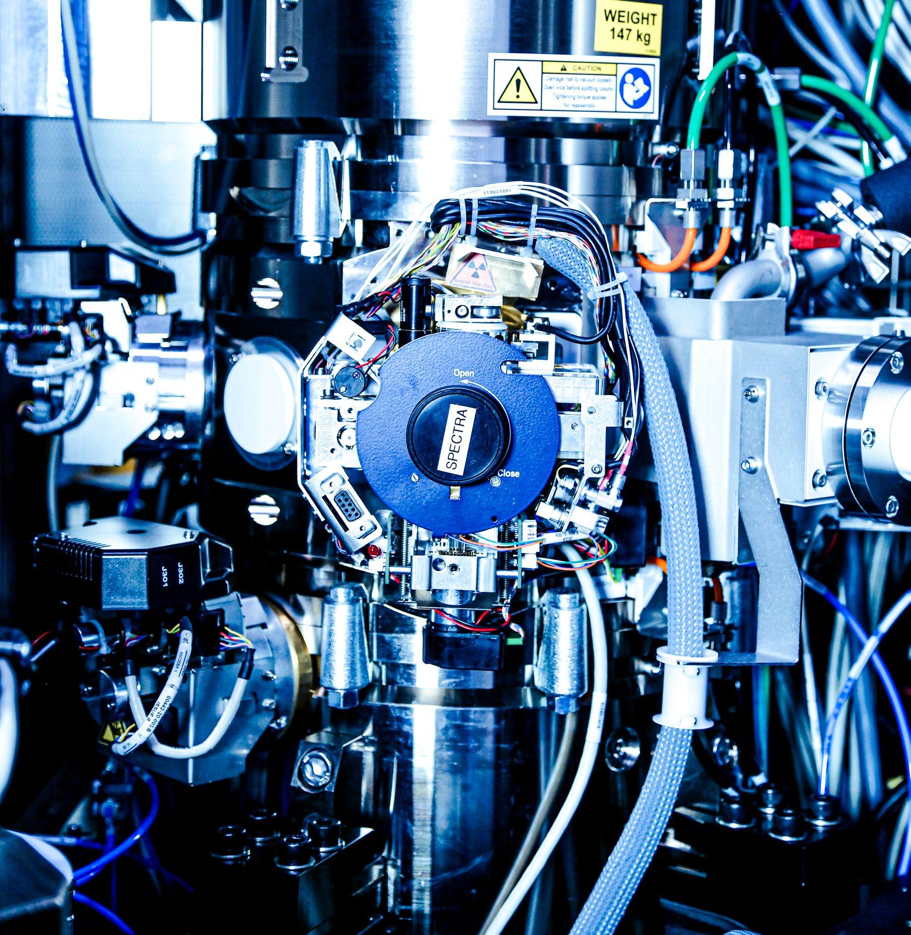
Electron microscopy (EM) makes modern materials science possible. The field took a giant step forward after the invention of the electron microscope, which dates to 1931 and earned a 1986 Nobel Prize for Ernst Ruska, although it wasn’t until the 1950s that the transmission electron microscope (TEM) had advanced enough to enable the indispensable imaging of defects in materials. The field expanded further with the invention of the scanning electron microscope (SEM) in 1965, which enabled close examination of surfaces.
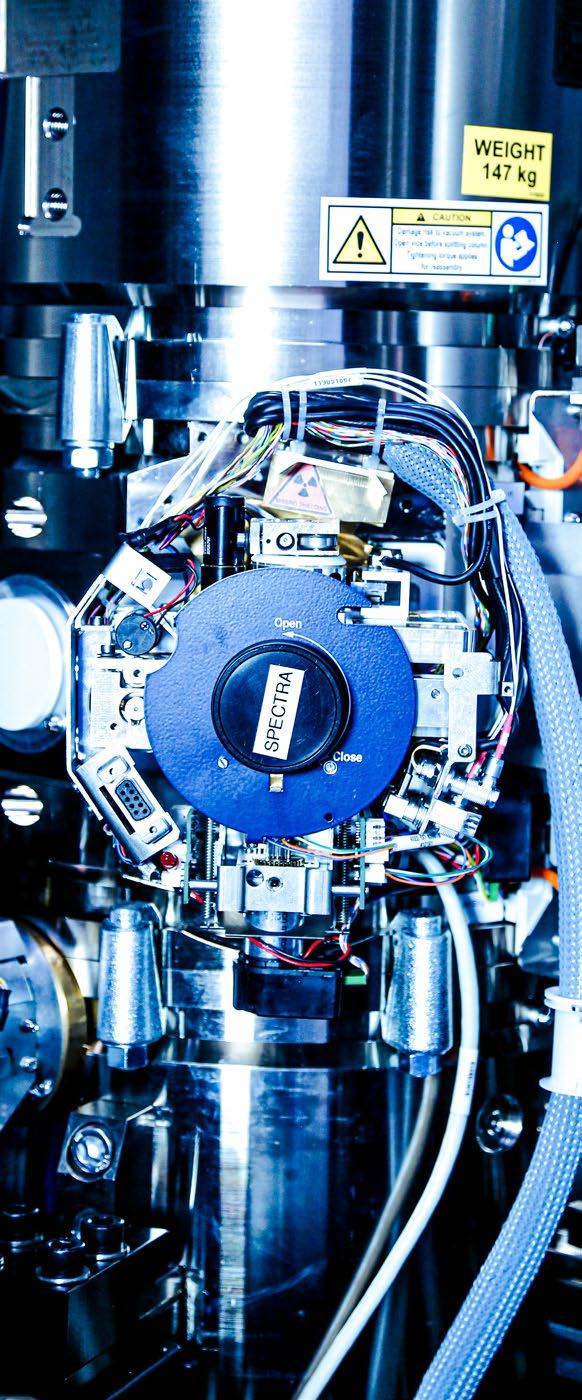
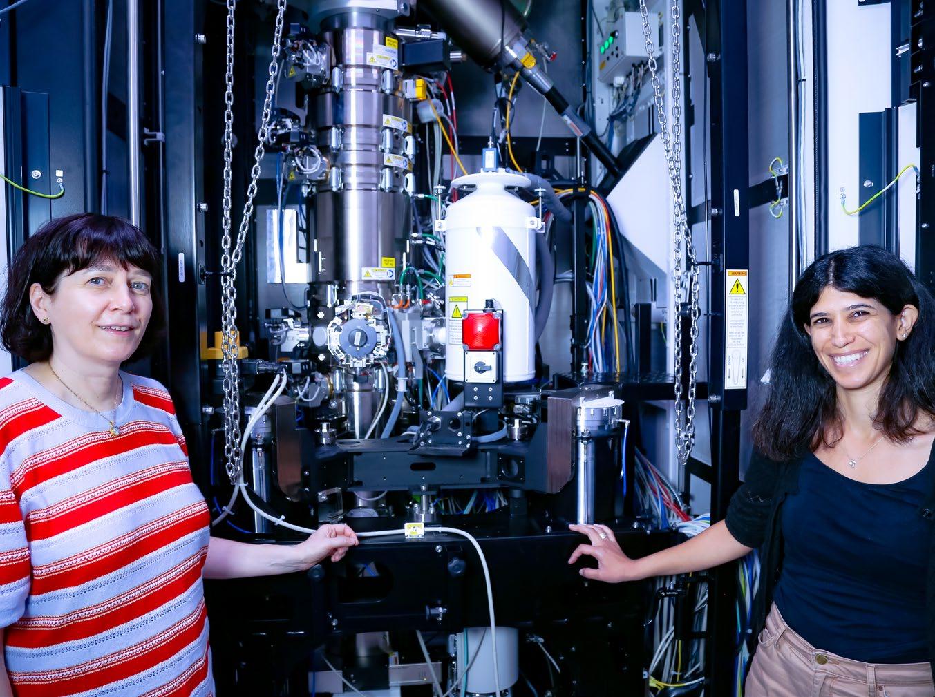
The Materials Department in the UC Santa Barbara College of Engineering (COE), which consistently ranks among the top five such programs in the world, houses an impressive array of electron microscopes, including a new stateof-the-art instrument called Spectra. The seven EM instruments in Elings Hall — three SEMS, two TEMS, and two dual-beam microscopes — are used each year by approximately 230 internal and external researchers who represent 91 research groups, work on more than 100 projects, and spend some 11,000 hours on the various instruments. EMs are mission-critical technology.
Mechanical engineering assistant professor Bolin Liao has another new instrument in his lab. The scanning ultrafast electron microscope (SUEM) is the only one of its kind in operation at a U.S. university. It couples a femtosecond pulsed laser with an SEM, enabling time-resolved imaging of microscopic energy transport processes with high spatial and temporal resolutions. The instrument will become available for shared use once development is complete.
EM: The Essential Tool
“At the core of materials science is the extent to which the internal structure controls the properties of materials,” says UCSB materials professor Susanne Stemmer, an EM expert who led the effort to obtain the Spectra instrument, a Thermo Fisher scanning transmission electron microscope (S/TEM). “It spans semiconductors, structural materials — a whole range of materials that require you to control imperfections all the time.
“Materials exhibit various types of imperfections; a simple one is a missing atom,” Stemmer continues. “For example, to get electrical carriers into a semiconductor, you have to introduce
With its increased resolution, says Ram Seshadri, the Spectra, newest addition to the Elings Hall suite of electron microscopes, has already “revealed previously obscured details of the structure of useful materials.”
those imperfections by doping; you might put in one type of impurity atom [called a dopant] to replace an atom of silicon or something else. The same is true for strengthening metals. There’s a certain defect, called a dislocation, that enables metals to plastically deform. The fact that we have steel strong enough to construct buildings, bridges, and ships is a result of our ability to strengthen the material by controlling imperfections.”
Electron microscopes make it possible to see
those imperfections, also called defects. “Basically, you look at the internal structure of a material, at length scales ranging from the atomic — how the atoms are organized — to larger-scale aspects, such as compositional inhomogeneities, phases, and interphases,” Stemmer says. “Using the electron microscope, we can determine where the individual dopant atoms are. It’s the technology that gives you the most information about your material. We can’t do our work without it.”
“Which technique you choose, whether SEM or TEM, depends on what problem you’re trying to address,” says materials professor and COE interim dean, Tresa Pollock. “There are different kinds of defects, and they exist at different length scales. For instance, if you bend a metal bar, you cause millions of dislocations, called line defects. There are different microscopy techniques for looking at those kinds of defects versus, say, point defects, where you just have an atom in the wrong place.”
Complexity at Work
“As complexity of instrumentation goes, electron microscopes are way up there,” Stemmer says.
In classic optical microscopy, visible light is used to illuminate a sample, and a magnified image is obtained through a glass lens. In EM, however, a sample is imaged using a powerful electron beam. Each material scatters electrons in a way that amounts to a unique signature of the structure of that material. While the most powerful light microscope can achieve nearly two-thousandtimes magnification, electron microscopes achieve magnification of ten million times.
The lens in an EM is composed not of glass but, rather, of finely tuned electromagnetic fields. By way of explanation, Stemmer picks up an old lens that serves as a door stop in her office.
“We run current through the copper wiring of this piece to generate a magnetic field at the position of the sample, which controls the beam
and acts as a lens, with focusing action and magnifying action,” she says. “In many ways, it’s equivalent to a glass lens, in that it’s supposed to magnify your object.”
But it’s not as simple as that, because, she says, “The lenses can have aberrations that distort the image in a nontrivial way. If you have a crystalline sample with periodically arranged atoms, that aberration affects certain spacings more than others, which can lead to an image that is not only blurry but also misrepresents the sample.”
Like lenses in optical microscopes, EM lenses also control resolution. Microscopes are good at magnification, making something small appear much larger. But if an instrument’s resolution — its ability to distinguish one object from another — is poor, powerful magnification is of little use.
NASA found that out with the Hubble Space Telescope. Its lenses contained imperfections, resulting in images that had superior magnification but were useless as a result of poor resolution. For electron microscopes, fixing the problem took years, and, Stemmer says, “For some time, it was not clear whether they would solve it. It was complex, because it was done by adding
additional lenses, and each additional lens also has imperfections, so the more lenses you add, the more complex the problem becomes.” A solution arrived in the late 1990s, in the form of a technique called aberration correction, which greatly improved resolution. “We do not have that in our older TEMs, but the new Spectra has it,” Stemmer says, “increasing resolution from 1.4 angstroms to 0.6 angstrom, a factor of more than two.”
TEM vs SEM
SEMs and TEMs use very different accelerating voltages — the power that propels the electrons — reflecting their different processes. An SEM’s highest voltage is 40 kilovolts (kV), whereas a TEM can run at 200 kV, an order of magnitude higher.
“That is because the electrons in TEM have to penetrate through the material, which requires more power, whereas in SEM, some electrons are scattered, and some penetrate,” says Ravit Silverstein, a materials scientist and the staff development engineer for UCSB’s EM facility.
“In general, the higher the voltage, the better the spatial resolution, but sometimes a lower accelerating voltage is needed, even in TEMs, to reduce beam damage to sensitive materials.”
— Susanne StemmerBecause SEM is a scanning process, Pollock explains, “There is much more space around the sample. That makes it possible to set up all kinds of different stages, where we can pull on the material or do other dynamic testing, things you can’t do in
The electrons in TEM have to penetrate through the material, whereas in SEM, some electrons are scattered, and some penetrate.
the very confined space of the sample while you’re imaging on a TEM. Looking at things that are static, you don’t always learn the same things as you do when they’re in action.
“In general, electrons can transmit through only 100 to 300 nanometers of material, and that is what happens in the TEM,” Pollock says. “To do that, you need to carefully fabricate very, very thin foils. This allows you, for example, to look at fine-scale chemistry at material interfaces. However, some important features exist only at higher length scales, in the micrometer range. To study these, the SEM is needed.”
“An especially useful aspect of most SEMs is that you have different types of detectors, essentially different types of cameras that form an image,” says Silverstein. “They can be set to form the same image with different contrast, and the contrast has meaning. The strength of the lenses can also be changed. So, you can collect the scatter and generate an image or change the strength of the magnetic field and collect the scatter pattern from a different region.”
As for TEM, she says, “One of the most amazing things about it is the ability to collect an image of your microstructure and simultaneously collect a diffraction pattern, which provides information about crystallinity and the type of symmetry it exhibits. You can also collect information about the chemistry of the material. When dealing with a new material, TEM can provide answers to most of the unknown questions in one session.”
In both SEM and TEM, the signatures of the electron interaction with the sample are magnified by the objective lens system of the microscope, creating an image that can be recorded or viewed remotely on a screen.
To facilitate her own research on highperformance materials, and especially metal alloys used in, for example, high-stress aerospace applications, Pollock incorporated an EM into an instrument she invented, named the TriBeam. It has a femtosecond laser to shave off layers of materials, an ion beam to “polish” the newly exposed layer, and an electron beam for SEM imaging.
The three elements make it possible to compile images that provide a much more detailed view below the surface of a bulk material, allowing reconstruction of 3D datasets that are cubic millimeters in scale, with nanometer resolution. Such datasets permit the study of rare features of materials that can, for example, cause them to fail prematurely. The TriBeam is now being manu-
factured commercially by Thermo Fisher Scientific. The original instrument resides in Elings Hall with all of the other EMs.
S/TEM: The Newest Instrument
Earlier this year, the advanced Spectra instrument, which combines scanning transmission electron microscopy and TEM capabilities with high resolution, was installed in Elings Hall, enhancing the EM capacity on campus. “S/TEM is a TEM, but it uses a finely focused electron beam for imaging what is scanned,” Stemmer explains. “This allows for different types of atomic-scale imaging modes and also for analyzing the chemical composition with extreme spatial resolution. The Spectra can form an electron beam that is less than one angstrom in diameter.”
“The acquisition of an aberrationcorrected transmission electron microscope for a university with so much research activity into the structure of matter has been highly overdue,” says Ram Seshadri, UCSB professor of materials and chemistry and the director of the Materials Research Science and Engineering Center (MRSEC). “Already, the new microscope is revealing previously obscure details of the structure of useful materials. The efforts expended by Susanne Stemmer to bring this new facility to us for the benefit of the campus and the larger user community are particularly commendable.”
“The Spectra instrument represents a landmark achievement in modernizing the shared, centralized Microscopy & Microanalysis Facility by adding a state-of-theart electron microscope capable of resolving and quantifying the finest-scale features — at the atomic scale — that form the building blocks of matter,” says materials professor Dan Gianola, who was involved in many of the discussions that paved the way for the new instrument to arrive at UCSB.
“Much as adaptive optics transformed the field of space astronomy and turned otherwise blurry images into crisp images, the Spectra is able to unveil the details that underpin the properties of some of the most interesting materials relevant to quantum technologies, advanced structural materials slated for extreme environments, and energy materials,” he adds. “Open to university, community, and regional users, it marks a new era for microscopy on campus and carries forward a long UCSB legacy of providing the most modern characterization facilities.”
“You get higher resolution on the new
system in either mode — TEM or S/TEM — than on any of the other instruments we have,” says Pollock. “It’s very good for the sorts of materials that the quantum-computing people and the semiconductor heterostructure folks are interested in: controlling, doping in very small amounts, and looking very specifically at an interface and trying to control the electrons and holes that they find there. For them, the details of what sits at any interface matter tremendously, so they have to really control their materials.”
The Spectra is also equipped with what is called a super x-ray detector, which makes it possible to collect x-rays while simultaneously doing electron scanning, to produce a chemical analysis without having to stop the scanning process.
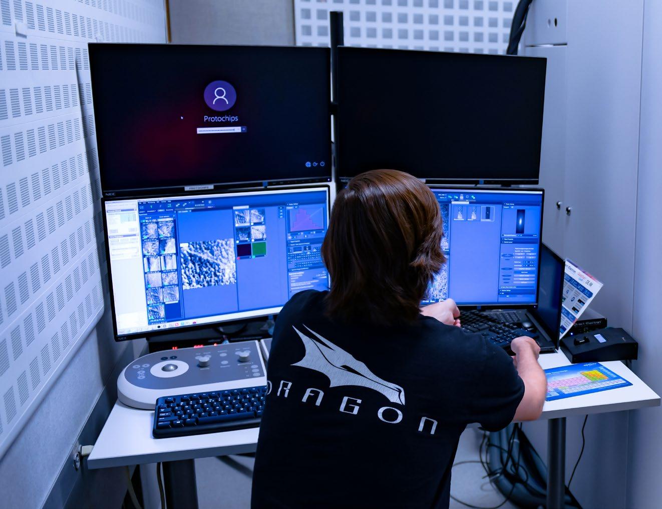
Electron microscopes are extremely sensitive to environmental factors, such as temperature, humidity, and motion. The Spectra is completely enclosed within a large “box” about ten feet tall that protects it from sound, magnetic fields, temperature changes, and so on. The case itself is located inside a room fitted with special panels to absorb sound and minimize airflow. It also has a remote console, so that the operator no longer has to sit in the dark room with the instrument, thus enhancing comfort and further reducing environmental impacts.
Prior to receiving the Spectra, Gianola says, “There were also lots of critical discussions about what renovations would be needed to create the exceptional space needed, in terms of vibrations, temperature control, and electromagnetic interference, to achieve the correct specifications and accommodate this very high-end instrument.”
The Spectra reflects what Stemmer says she finds so enticing about electron microscopy, aside from its indispensability to her research, which is “the rate of technology improvement. It keeps improving so fast. There are new inventions that improve what we can see and therefore learn about materials. There are other instruments we use that are not improving nearly as rapidly.”
Speaking to the significant effort it took to obtain the Spectra, Pollock says, “These instruments that enable scientific breakthroughs are essential for faculty research. They are also very expensive, and staying at the forefront requires a lot of fundraising. It is a team effort among faculty, campus, federal agencies, and external donors. We are grateful for all of their important investments.”
THE BLOODWORM ... AND OTHER MARINE MAGICIANS OF BIOMINERALIZATION
Last April, The New York Times ran an article about a marine creature known as the bloodworm, or Glycera dibranchiata. Upping the Eek! factor of the creature, which is not especially handsome or friendly, was a horror-film-fodder headline proclaiming that the roughly seven-inch-long worms, which burrow into intertidalzone mud and are a favorite bait for New England fishermen, “grow copper fangs and have bad attitudes.”
The article did eventually mention that copper makes up only one to ten percent of the worm’s fanglike jaw, depending on location, and it quoted a pair of researchers from UC Santa Barbara, where the vast bulk of bloodworm research has occurred: Herb Waite, a renowned marine biochemist and a professor in the department of Molecular, Cellular and Developmental Biology (MCDB); and William “Billy” Wonderly, one of Waite’s PhD students, who graduated in December 2021 after writing his dissertation on the composition of the worm’s jaw. A chapter of that work, published last June in the journal Matter, inspired the newspaper article.
As a daily news outlet, the Times necessarily focused on only a tiny segment of what is actually a very long arc of interconnected research that began in the 1970s with investigations into materials produced by other marine creatures.
That longer story says a lot about the often-unpredictable evolution of scientific inquiry. It speaks to the brilliance of nature as creator and to the value of both the undiluted curiosity of people like Waite in bringing oddities such as the bloodworm into the scope of scientific investigation, and to the tenacious application orientation of engineers who build on that knowledge to develop new highperforming materials. And it sheds light on the kind of highly collaborative, long-term interdisciplinary scientific investigation, so prevalent at UCSB, that has allowed the College of Engineering (COE) and STEM disciplines across campus to develop top-tier international standing without enormous research endowments.
Finally, the full, ongoing story of the bloodworm research, as well as related long-term investigations of other high-performing biomaterials, speaks to the value of the National Science Foundation’s (NSF) Materials Research Science and Engineering Center (MRSEC) model and the Interdisciplinary Research Groups (IRGs) it mandates. (See sidebar on page 27.) As a PhD candidate, Wonderly presented regularly in IRG 3, “Resilient Multiphase Soft Materials,” co-led by mechanical engineering professor and co-director of the California NanoSystems Institute (CNSI) at UCSB Megan Valentine and professor of chemical engineering Matthew Helgeson, who, with Waite, is listed as a co-author on Wonderly’s paper in Matter.
UCSB has leveraged collaboration not only to generate numerous high-impact scientific breakthroughs, but also, increasingly, to attract multiple large awards from the NSF and other funding organizations to support a range of important centers and institutes built upon collaborative interdisciplinary models.

A UCSB tale of curiosity-driven faculty, tenaciously applied expertise, and the pursuit of new materials via long-term collaborations that blur the lines of disciplinary divides
Collaboration and Diverse Systems
Like the Times, Waite, Wonderly, and their colleagues are interested in the copper in bloodworm jaws, but as part of an intricate system in which it interacts with other natural components — especially a melanin-like substance and a protein that has an unusually simple structure — to create an extremely hard and shatter-resistant functional biomaterial. UCSB materials scientists are interested in using that knowledge to develop new synthetic materials.
“Hardness is something you want in a blade or a tip that you use to penetrate something, but high hardness usually comes with high stiffness, which means brittleness,” Waite explains. “The properties we saw in the worm jaw were really counterintuitive, a combination of high hardness but relatively low stiffness. A penetrating or cutting tool made from such material is going to be tougher and less brittle.”
“A common denominator that interests us in all these materials is their amazing strength given their light weight,” says Paul Hansma, UCSB emeritus professor of physics, whose innovations in atomic force microscopy (AFM) enabled it to become an indispensable tool for atomic-level characterization of biologically produced minerals.
Hansma developed an AFM technique to measure the step growth of a crystal — the deposition of atoms as it grows — and suggested using it to view the crystal structure of abalone shell.
Through the years, the bloodworm research has spilled over to, inspired and been inspired by, and occurred alongside of or in conjunction with related long-term investigations into an array of biomaterials, including human bone, spider-web silk, sticky mussel filaments, abalone shell nacre, the pigment cells that enable cephalopod bioluminescence, the silica-based glass spines of certain sponges, and the silica cell walls of diatoms (single-celled algae).
“What we’re trying to do in IRG 3 is understand how the processing of the material occurs — not just what chemicals are there but how they have been put together and the order of operations and how they are mixed and handled,” says Valentine. “We want to know how that piece of it leads to the mechanics. What’s the recipe?”
Regarding the bloodworm, which was highlighted in the UCSB proposal for a previous six-year round of additional MRSEC funding, she says, “We knew that the extraordinary jaw material existed and that the organism could make it. Some of the components and some of the composition were known, but the details of exactly how this was done and, more importantly, how we might be able to think about leveraging them to work in an engineered environment — those were things we didn’t know.”
“If you can extract out and identify the critical components that an organism uses that are different, then you can understand how the natural system operates and how you can design synthetic systems to take advantage
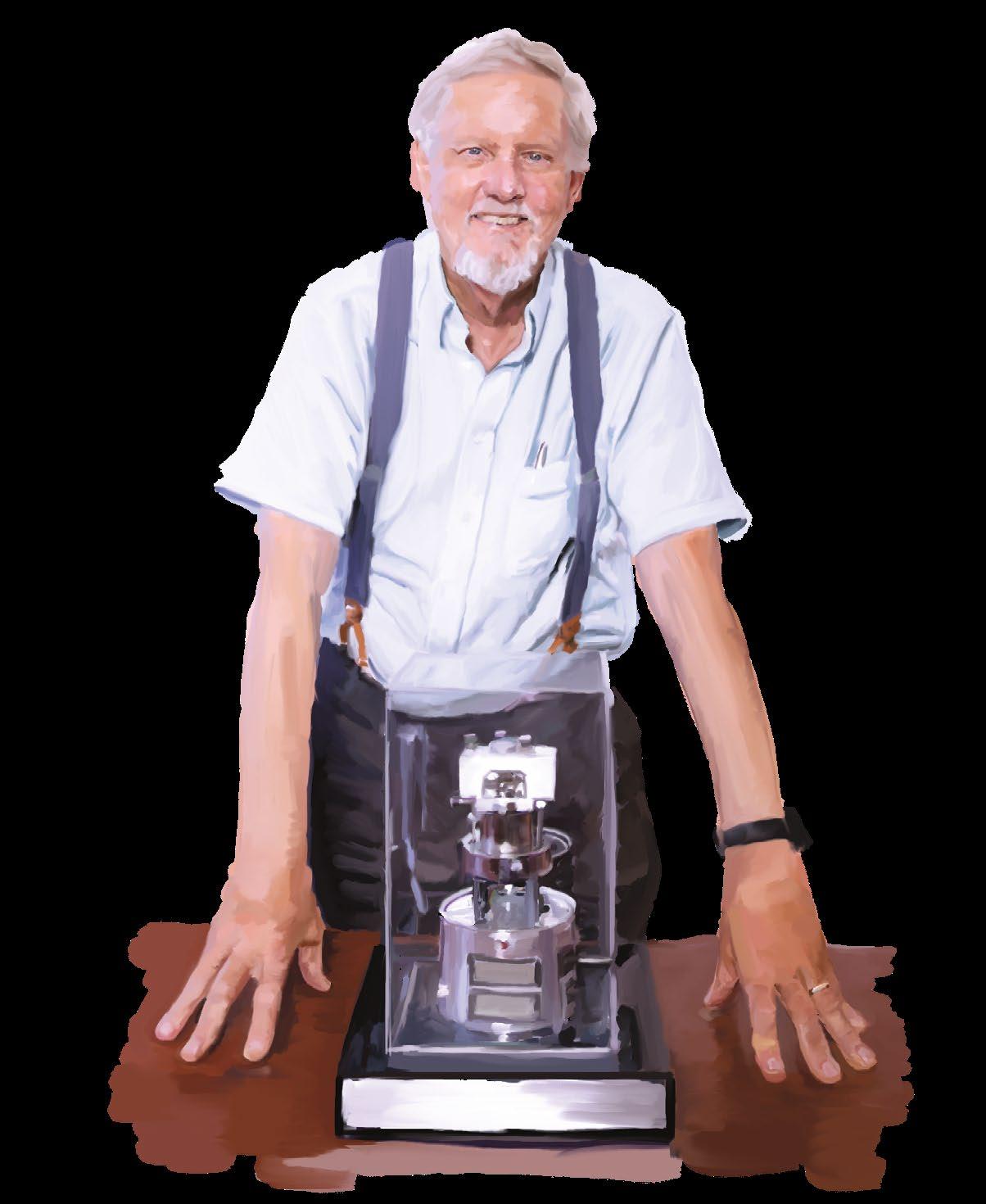
Physicist Paul Hansma with an atomic force microscope he built. During years of biomineralization research, Hansma enabled multiple breakthroughs and developed innovative techniques that led to the instrument’s widespread use among biologists.
of that,” says Craig Hawker, co-director of the CNSI at UCSB, the Alan & Ruth Heeger Chair in Interdisciplinary Science, and a Distinguished Professor in Materials.
Professor of materials and chemistry
Galen Stucky brings yet another perspective to the topic. “The creation of life on Earth is based on the dynamical integration and cooperative assembly of inorganic and organic species into composite living biosystems,” he says. “The interest for me is understanding the chemistry of that assembly, the functional interfacial relationship between the organic and inorganic components of the resulting biological systems, and how biosystems interact with their surrounding ecosystem. We’re always asking, How does a dynamic, functional system evolve from atomic and molecular parts?”
“There’s something really fascinating in an interdisciplinary way about what these organisms have evolved to do, but these biological systems are constrained to specific conditions or applications that can benefit from some evolutionselected advantage,” says UCSB chemical engineering professor Brad Chmelka. ”We want to understand the physicochemical origins of the biological processes that form such fascinating, often multifunctional, materials and their corresponding atomic-level compositions and structures. From there, we seek to exploit such insights to develop new synthesis strategies for preparing non-biological materials having novel properties. That’s the biomimetic angle, which can liberate us from the requirements that are otherwise set by the physiology of an organism and the conditions it might need for survival. Such biomimetic approaches open new opportunities for expanding the conditions by which new multifunctional materials are formed,”
“It’s research that brings together two different types of people,” says Waite, who began working on the unique materials produced by marine organisms as a PhD student in the 1970s, conducting research on the byssal threads that mussels use to cling to rocks in turbulent tidal zones, a major focus of MRSEC-funded research at UCSB for more than a decade. “On the one hand, you have the creature people, and then you have the materials people. It’s a meeting and a collaboration.
“There are very few engineers who are interested in the general idea of creatures making adaptive structures to survive in their daily challenges,” Waite adds. “However, if you report a bizarre materials property, that’s where they usually get hooked. UCSB is special in that there are a lot of engineers here who are at the cutting-edge of materials analysis and are fascinated by working on bio-inspired materials.”
The Worm’s Turn
When Wonderly began his doctoral work at UCSB, he entered something of a research desert. Even figuring out where to begin the search for the bloodworm-jaw “recipe” that Valentine spoke of above proved difficult, because Wonderly had to restart long-dormant research and find a direction for his own work on a subject about which little foundational knowledge existed. “It’s such a complicated system; it took me years to really understand what was going on,” he says.
The first paper relevant to the bloodworm jaw was published in 1980 by a pair of British researchers, who reported copper concentrations of up to thirteen percent in the jaws of a related bloodworm, Glycerasp.
Waite, a collector of such biomaterial-related oddities, kept the article. Then, in 2001, an Austrian postdoctoral researcher named Helga Lichtenegger, who for her PhD research had used high-resolution synchrotron X-ray scattering to study bio-nanocomposite materials, arrived at UCSB to study in the lab of Galen Stucky. He introduced her to Waite, with whom he had previously collaborated on a variety of projects related to biomineralization in marine organisms. Waite shared the jaw-copper paper with Lichtenegger and Stucky. “I kind of reached into my grab bag of zoological model systems and thought of the bloodworm, which is the only organic system apart from lichens to concentrate copper,” Waite recalls. “We knew that, but no one had taken that knowledge further.”
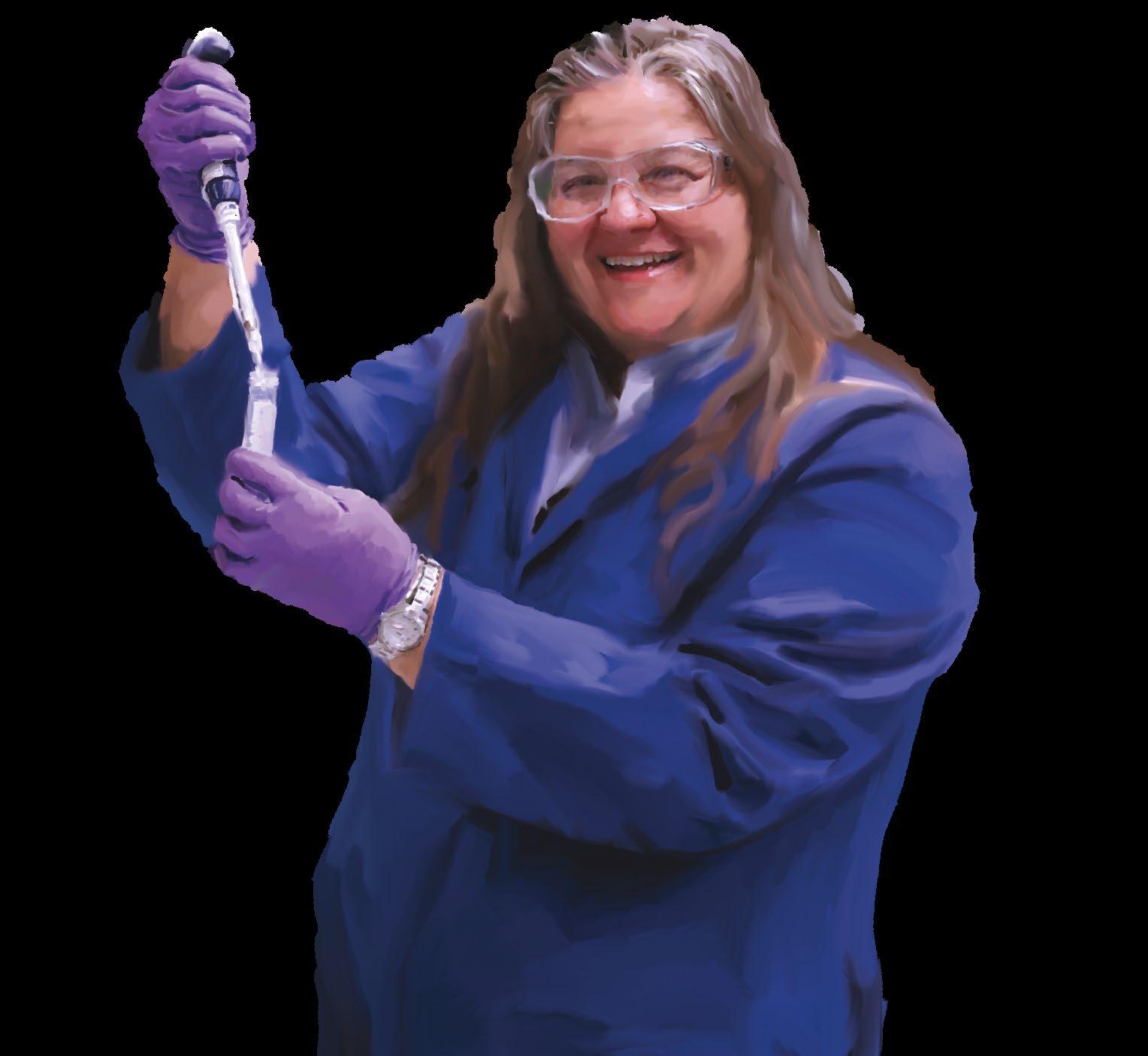
Lichtenegger got to work, and in 2002 she published a paper in Science
that, Waite recalls, “received a huge amount of attention in the press.”
“Helga did the first mechanical studies of the jaw, identifying the protein composition, showing a regional distribution of the copper, and determining that it is present primarily in mineral form, as atacamite,” Wonderly says, adding that Lichtenegger, an expert in X-ray diffraction, did a follow-up study that was “more of a technical X-ray analysis of the jaw.”
Waite notes, “There were informational tidbits that came out of that research period: that parts of the jaw contain a lot of copper in mineral form, that other parts, the best-performing parts, contain just a little bit of ionic copper, and that the other two components are protein and melanin.”
After Lichtenegger returned to Austria, a PhD student in Waite’s lab, Dana Moses, continued the bloodworm research, beginning in fall 2004. She identified the presence of the melanin-like material in the system and determined the composition of the jaw, as well as the role of a surprisingly simple protein — so simple that Waite initially hesitated to call it a protein — that makes up about forty percent of the jaw and that, unlike most proteins, which are far more complex, is composed almost entirely of just two amino acids: glycine and histidine.
When Moses graduated, there was no one to continue the bloodworm work, which then lay idle for about eight years, until 2016, when Wonderly arrived. He became part of a large interdisciplinary research family that had been growing and collaborating for several decades.
Beginnings
Back in the late 1970s, a group of UCSB professors began an unfunded ad hoc collaboration that had its beginnings in a field trip to the Channel Islands.
“A faculty colleague in another department took my wife and me to Santa Cruz Island for a weekend,” recalls Daniel Morse (MCDB), a self-described naturalist who has studied biomineralization processes in marine creatures extensively, from the perspective of the genes and proteins that direct them. While on the island, he recalls, “I looked down into a tidepool and saw hundreds of abalone just bumper to bumper to bumper. It was a natural phenomenon, and I wondered, what causes that? I wanted to see if I could understand it.”
Morse and Waite had been collaborating since the early 1970s, when Waite was still at the University of Delaware and would come to UCSB to work with Morse on research related to another marine protein. As a spinoff from Morse’s abalone research, he discovered the trigger molecule that makes worm larvae settle and metamorphose. The worms use that same molecule as glue to build the tubes where they live. Waite elucidated the structure of the glue protein, discovering it to be very similar to the chemical in mussel glue.
Back in his UCSB lab, Morse started trying to
generate baby abalone to see what controls were involved. He first discovered the trigger that causes them to spawn, releasing eggs and sperm, an investigation that became the cover article of the April 15, 1977 issue of Science.
“We soon had millions of larvae swimming in the plankton, cute little things,” he recalls, “but they wouldn’t settle out of the plankton or metamorphose. They would just die. They required a molecular signal to trigger activation of their genes to metamorphose, develop, and grow.”
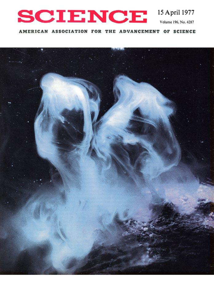
With that clue, he and his wife, Aileen Morse, discovered the natural trigger for their metamorphosis, a molecule produced by algae that cover the tidepool rocks, which the larvae recognize with chemo-sensors. “We isolated the molecule and found a simple substitute for it, so, now we could trigger one hundred percent of the larvae to settle, metamorphose, and start to grow as juveniles,” he explains. (While Morse began the work as a way to study how genes control early development, the discoveries it yielded would prove critical in enabling abalone aquaculture, which saved the California abalone after the wild population was nearly eliminated by disease, pollution, and climate change.)
Some years later, in the early 1990s, Morse recalls, he heard a knock on his door at UCSB. “It was this tall guy who said, ‘I’m Galen Stucky. I’m in chemistry. I understand you’re studying abalone shell,’” Morse recalls. After explaining to Stucky that he was indeed “triggering the development of abalone to study tissue-specific gene expression and protein synthesis leading to development,” Stucky asked, “Have you ever thought of the shell as a material?”
Stucky explained that he had been working with Hansma, making use of his advancements in atomic force microscopy to study how small organic molecules influence and control crystal growth, and that they were interested in studying more biological systems. Hansma’s atomic force microscopy work in that realm would lead to multiple breakthroughs, including the first one in the abalone-shell research.
Soon, Stucky, Morse, and Hansma began meeting every week in the conference room in the Marine Biotechnology Lab. “At first, there were a few students sitting on the sidelines away from the table and three professors at the table,” Morse recalls. “As we discussed how the shell might be built, we’d ask each other questions, and one of the professors would jump to the board and sketch out a possible answer. Soon, the graduate
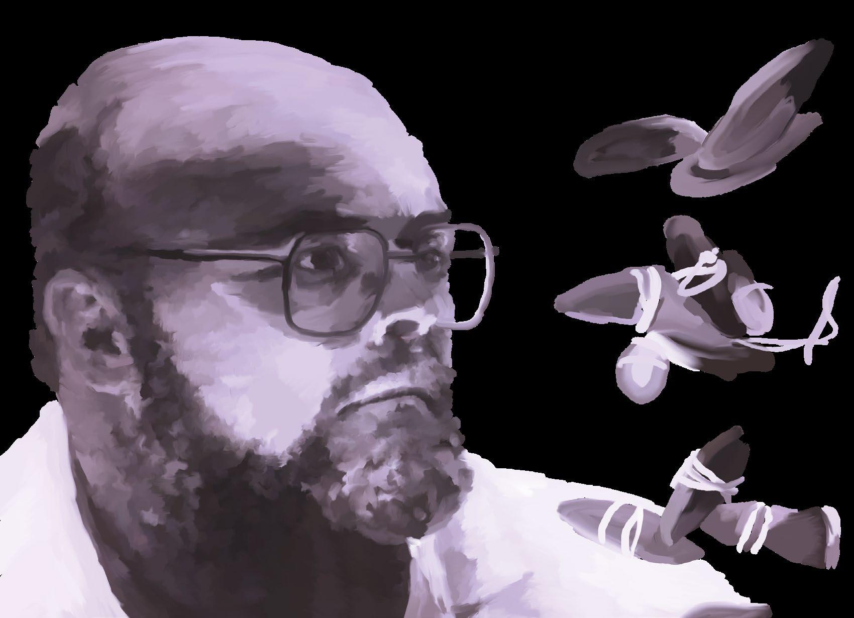
Herb Waite, shown here as a young professor with mussels in an aquarium, brings a sense of wonder to his work. His files are filled with curiosities from the sea, like the bloodworm, the jaws of which may inspire new materials.
students were also going to the board. We were trying to get at the fundamental bio-physical mechanisms behind these materials. Some of us thought of the genes and proteins that must control the process, while others thought about how the minerals and crystal growth completed the picture. This always led to hypotheses, which grad students would test in the lab.”
“It was a great learning experience for everyone,” Stucky adds. “The discipline languages of physics, chemistry, and marine biology, and the different ways faculty thought became translated into a common universal scientific language that opened up new perspectives for all.”
The process resulted in significant findings. “We started getting cover articles in Nature and Science, because we were making breakthroughs and overturning dogma that had stood for more than a hundred years in the field of biomineralization,” Morse says.
Brad Chmelka soon joined the group, bringing expertise in nuclear magnetic resonance (NMR) spectroscopy and chemical interactions at the atomic level, and then came EEMB ecologist and marine oceanographer Mark Brzezinski, whose work on the biological synthesis of silica complemented the lab work of Stucky and Chmelka, and Tim Deming, a UCSB materials professor (now at UCLA) and an expert on functional polypeptide materials who was making synthetic proteins.
Eventually, the group secured a Multidisciplinary University Research Initiative (MURI) grant, with all six members co-equal PIs and funding from the Navy Research Office, the Army Research Office, and NASA, all of which focused on biological and bio-inspired materials.
“All of the projects were based on the remarkable properties of biomaterials, like the abalone shell, which is made so strong by nanocomposites, which are also intimately integrated in the bloodworm jaws,” Morse says. “It’s a similar underlying concept, with different materials. The organism has this kind of nanoscale machinery that temporally and spatially governs the differential secretion of the proteins that direct the final composition of the composite material.”
During that period, Hansma developed an AFM technique to measure the step growth of a crystal — the deposition of atoms as it grows — and suggested using it to view the crystal structure of abalone shell. One of Stucky’s graduate students, Srin Manne, did the microscopy and quickly discovered a conundrum: multiple layers but a single crystal. The mystery, Morse says, was “There’s a very thin layer of protein between the crystal layers, so how could it be that each thin layer is capped by a protein but the crystal continues to grow?”
From there, Morse recalls, “We discovered the presence of nanopores that are located randomly in each protein sheet. Their random placement causes the penetrance from the previous layer to be offset laterally, creating the interlocking brickwork of crystal platelets that contributes to the shell’s incredible toughness, which is three-thousand-times more fracture resistant than chalk, which makes up ninety-five percent of the shell.”
“The idea for a century or more had been that the protein
In a paper published in Nature in 1999, Morse, Hansma, and their co-authors posited another enormously important hypothesis, describing “sacri cial bonds” and “hidden length” in proteins, which o ered a new explanation for the toughness of natural composites like abalone nacre and bone.
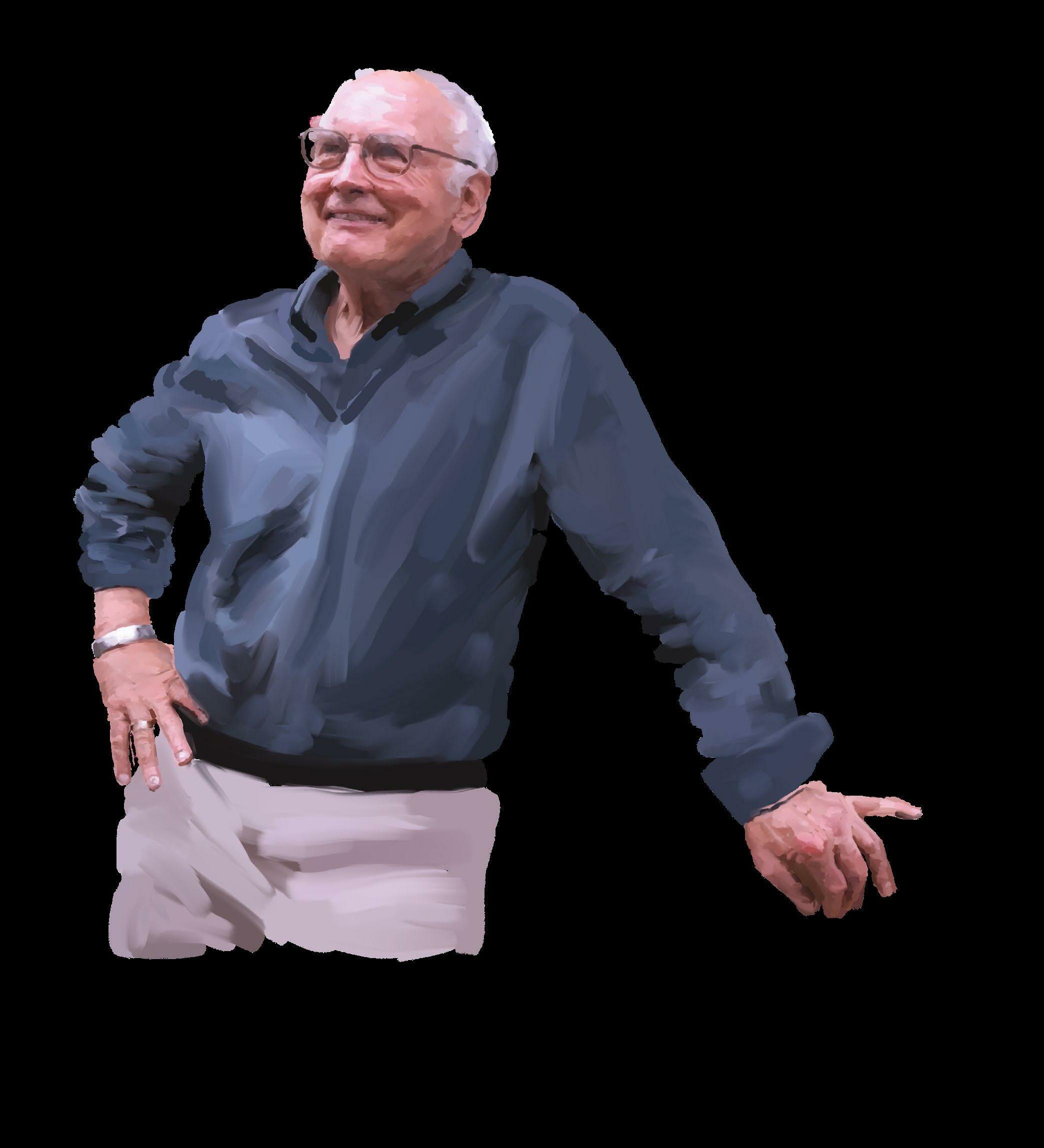 The day that Galen Stucky walked into Dan Morse’s lab and asked, “Have you considered abalone shell as a material?” marked the beginning of a highly successful long-term collaboration.
sheet that covers the first layer of mineral acts as a kind of template to initiate the de novo growth of the next layer, in a repeating process,” Morse explains. “But following Srin’s microscopy work, we said, ‘The new paradigm is this.’”
In a paper in Nature in 1999, Morse, Hansma, and their coauthors posited another enormously important hypothesis, describing “sacrificial bonds” and “hidden length” in proteins, which offered a new explanation for the toughness of natural composites like abalone nacre and bone. Their evidence, gathered by making use of Hansma’s innovations in AFM, showed that some of the proteins are arranged in a tangled state, like a garden hose looped many times over itself, with short, fairly weak bonds connecting the various loops. When the polymer is stretched, such as under a potentially shattering force, those weak bonds break one at a time. When that happens, the energy that was stretching the composite material is dissipated as heat, and the shattering force is reduced back to zero, before it can approach a level that would break the backbone of the polymer, shattering the shell or bone. In this way, such short, “sacrificial” bonds provide resilience to the material. Further, they can reform once the stress on the backbone is relaxed, thus continually healing the material and regenerating that resilience. The ”hidden length” is defined as the part of the molecule — the loop — that was constrained from stretching by the sacrificial bond but lengthens when it breaks.
“It’s been like that ever since Galen knocked on my door,” Morse continues, with collaborations leveraging disparate expertise and leading to important discoveries. “Nature has evolved such complex utilization of such simple ideas in ways you’d never think of, like this offset nanopore stenciling of continuous crystal growth generating all of these properties. There’s the bloodworm, with copper, and there is silicatein, the name I gave to the enzyme that enables some marine sponges to produce a glass skeleton, and make it at low temperature without the need for a furnace or acid or some terribly caustic solvents, so that it’s benign and compatible with life.”
The work on sponges, incidentally, included the discovery of the first identified enzyme that catalyzes the synthesis of any mineral. It also led to major breakthroughs, such as the one resulting from a collaboration involving then-chemistry graduate student Angela Belcher, now the head of the Bioengineering Department at the Massachusetts Institute of Technology, who worked with Morse and Evelyn Hu, then a UCSB materials professor. Together, they developed a high-throughput commercial process for synthesizing designed inorganic nanoparticles using phage displays, a laboratory technique employed in the study of protein–protein, protein–peptide, and protein–DNA interactions.
In terms of engineered materials, further abalone studies informed the development of an external inorganic treated gauze that could control the blood-clotting cascade system of the human body. As of 2021, the resulting commercialized anti-coagulation product remained the most recommended first-responder hemostasis treatment of major arterial bleeding for all the uniformed services in the United States.
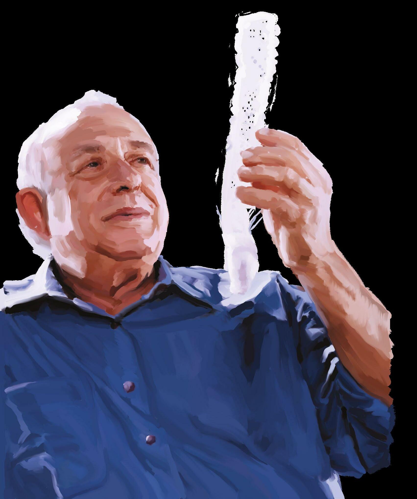
Teasing Out the Protein-Copper-Melanin Trio
Wonderly’s doctoral research uncovered several previously unknown functions of what he called a “multi-tasking protein” (MTP) in the formation and performance of the bloodworm jaw, which is composed of fifty percent protein, up to ten percent copper — most in mineral form, with smaller amounts in solution as copper ions — and up to forty percent melanin.
Melanin has many desirable properties, but because experimental formation of it typically produces very small granules less than 100 nanometers in diameter, applications of melanin in synthetic materials remain limited. For instance, melanin has semiconductive properties, so it could be useful in, say, a solar panel, but it would have to be present at a much larger scale, as a continuous bulk material in crystal, not powder, form. “The worm jaws are amazing because they have contiguous melanin at the millimeter length scale, which is orders and orders and orders of magnitude larger than even most melanin found in a biological context,” Wonderly says.
“And,” Waite notes, “because the melanin and copper in Glycera jaws are correlated with impressive wear resistance, a deeper understanding of the mechanisms of their formation and function could lead to significantly expanded use of melanin in highperformance materials.”
Wonderly achieved a key advance — Waite calls it his “first breakthrough” — when he figured out how to get protein, so that he would have enough for his experiments without having to harvest thousands of bloodworms. “Making recombinant jaw protein was a big deal,” Waite recalls, adding, “Then, in playing around with it in solution, he found how tightly it bound with copper.”
That led to the hypothesis — which was then confirmed through observation and based on the fact that the copper-bound protein shares many characteristics with enzymes known to be involved in melanin synthesis — that the copper-bound protein is what catalyzes the chain reaction that William Wonderly’s PhD research on the bloodworm jaw has generated great interest among materials scientists.
An Argument for Long-term Research
Engineering and science require patience, and functional materials, like those that might one day be inspired by the jaws of the bloodworm, or others having no biological precursor, don’t always arrive on schedule. While a new material may result directly from specific funded research, life-changing advancements, the kind that occasionally earn a Nobel Prize, typically come much later.
Last spring, the journal Nature Synthesis published an article by UC Santa Barbara professors emeritus Anthony K. Cheetham and Fred Wudl, together with Ram Seshadri, professor of chemistry, biochemistry, and materials and the director of the NSF-funded Materials Research Science and Engineering Center (MRSEC), which has funded diverse research into materials synthesized in marine organisms, like the bloodwarm jaw. The article tracks how and why the materials were initially synthesized, as well as how, and when, their utility was eventually recognized.
The authors identify several pathways to materials breakthroughs, with the most common appearing not to involve inventing something new at all. Rather, they tend to occur more often when an existing compound, often one that was synthesized out of simple curiosity, is repurposed in the application for which it becomes known.
“Our thesis is basically to encourage discovery synthesis of materials without worrying too much about the application space,” Cheetham and Seshadri noted. “Funding agencies would like to think that if they give you one dollar, then they will get one dollar — or ten dollars — of utility out of it. Actually, they might get back a million dollars on the dollar, but twenty years from now and for something they never initially envisioned.”
For example, polyacetylene was originally identified in 1866 and was first synthesized by a known polymer synthetic method in the 1950s. But it was only in the mid-1970s that UCSB professor (now emeritus) Alan Heeger, working with Alan MacDiarmid and Hideki Shirakawa, produced a revolutionary conductive thin film that earned each of them a share of the 2000 Nobel Prize in chemistry. That breakthrough would enable the entire field of conjugated polymers, with applications in such areas as thermo-electrics and wearable electronics.
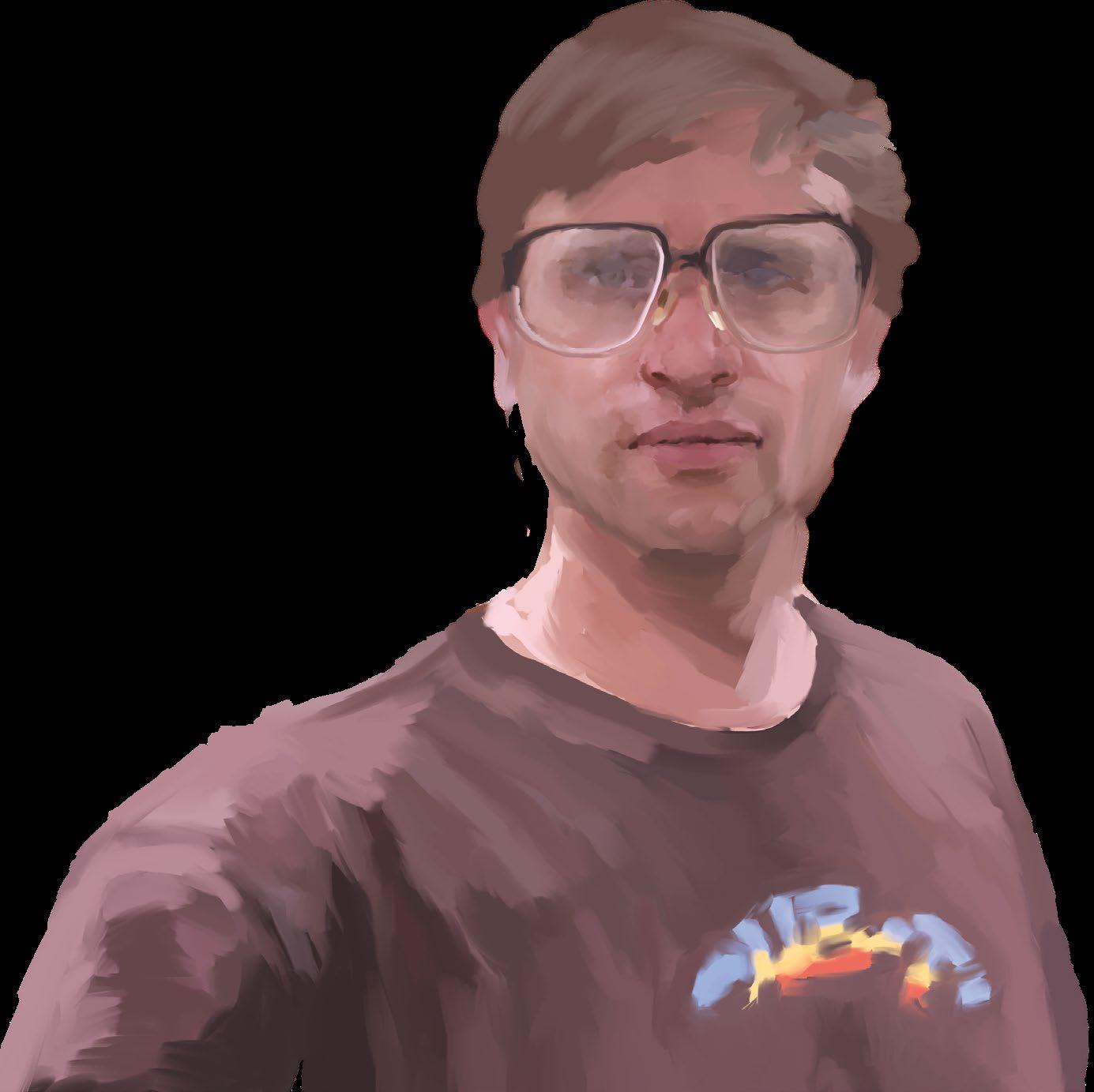
Hybrid 3D perovskites are another example. Initially synthesized out of curiosity in Germany in 1978, it was not until 2009 that the work was done that led to their application as visible-light sensitizers in photovoltaics.
Another example, lithium-ion batteries, are the multigenerational result of research on lithium cobalt oxide. First synthesized in the 1950s, only much later did it find its way into service as the key compound in batteries used globally to power our electronic devices and vehicles.
As the unsurpassed creator of high-performance materials, Seshadri says, “Nature has had the luxury of evolving its processes over a very long period of time, and it has never had a funding agency breathing down its neck, requiring rapid results.”
leads to formation of melanin and the bloodworm’s tough jaw.
Wonderly then determined that once the protein binds copper, a phase separation occurs. That is, he explains, ”When we have soluble protein, and we add copper to that solution, the protein binds the copper, but rather than remain soluble, it forms dense liquid protein droplets separate from the water. The copper-bound protein and water won’t mix.”
Wanting to figure out how exactly the protein droplets turn into a solid, Wonderly went to Matt Helgeson, whose group does lot of rheological measurements, that is, measuring mechanical properties of complex fluids. “What we discovered,” he says, “is that the copper not only causes the protein to condense into droplets, but the droplets then go to the interface and sort of spread to form a layer, but only in the presence of copper.”
More lab work led to the discovery that “If you just have the protein sitting in a jar and add some copper and come back a little later, a film will have formed on top of the fluid where it contacts air,” Helgeson says. “That means that somehow the protein is being recruited to that air-water interface and somehow solidifying. “
“It was super exciting,” says Wonderly. “We went from this thing that we didn’t think was really a protein to finding that, despite its low complexity, it has this high degree of functionality. The protein finds metal, it accelerates melanin formation, it phase-separates, and then that phase separation leads to this melanin formation concentrated at the air-water interface to form this solid melanin-reinforced solid: the jaw.”
Wonderly says that a key challenge in the project was navigating the tension between “the desire to try to come up with something truly unique and the utility of that, while not wanting to miss something that should be included in the project and not wanting either to dilute my own insights or lean entirely on what came before.”
In the end, he says, “Getting an idea of what was going on in this new system required a synthesis of paradigms from a lot of different systems.”
Studying the polymerization process further, Wonderly found that it resembles the manner in which nylon polymer threads form at an air-water interface, suggesting possible material applications of the bloodworm’s own polymerizing mechanism. And recently, Wonderly worked with Chmelka’s lab on sophisticated NMR analyses of the jaws to understand the individual chemical interactions occurring at the atomic level. A paper on that project is in preparation.

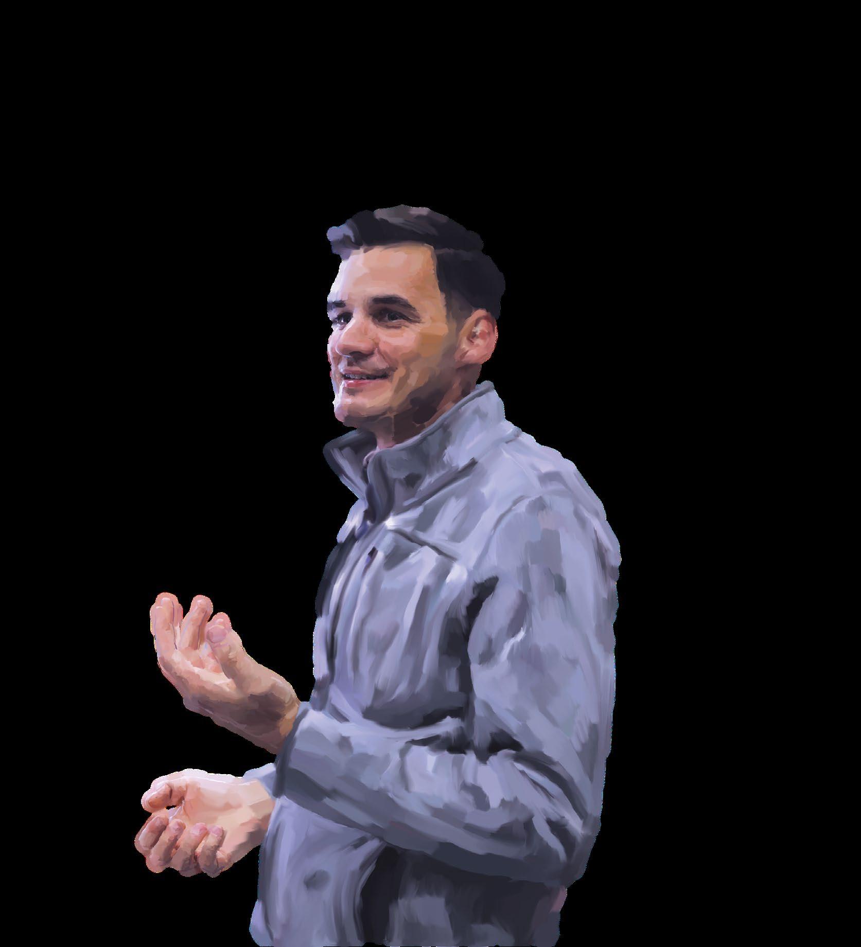
“If I see a material that’s interesting, I want to know why is has the properties it does,” Chmelka says. “What is the atomic-level structure that underlies and accounts for those properties? If we can explain the origins of the properties, then, in principle, we can mimic, improve, or adapt them for diverse engineering applications. In this case, we got the jaws, did the NMR experiments and compared their analyses with those of related materials. From there, we could establish the atomiclevel foundations that appear to account for their very different mechanical properties.”
Incrementally, each of these related research projects contributes to unraveling the recipe, about which much is now known but which, to date, only a bloodworm can prepare.
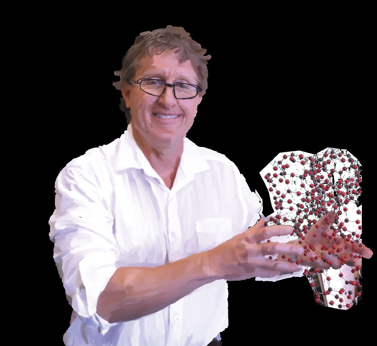
The Collaborative Advantage: Why Working Together Works So Well

Without prompting, every faculty member interviewed for this section spoke to the importance of collaborative research in addressing large challenges. Collaboration is a cornerstone not only of UC Santa Barbara, but also of the National Science Foundation (NSF) MRSEC model and the interdisciplinary research groups (IRGs) that it mandates. The current (sixth) round of funding for the UCSB MRSEC supports three IRGs. IRG 3 — “Resilient Multiphase Soft Materials,” led by mechanical engineering professor Megan Valentine and professor of chemical engineering Matthew Helgeson, is where William Wonderly (PhD ’21) presented regularly on his bloodworm research and received, in return, feedback, ideas, and suggestions reflecting diverse perspectives.
Megan Valentine (Mechanical Engineering)
When UCSB submitted its proposal for renewed MRSEC funding, we wanted to broaden out beyond the mussel to look at a host of other types of organisms. The bloodworm was one of our showcase examples in our pitch to the NSF, because we thought it would allow us to answer a lot of questions about how organisms make materials that have these extraordinary properties.
Matthew Helgeson (Chemical Engineering)
For me, one of the most rewarding things about leading this big collaborative effort is that once you create a space for people to really work together to be creative and think outside the box of the typical training students receive, it’s very autocatalytic in terms of their being able to generate their own research ideas and start pursuing them.
Wonderly (PhD ‘21)
I can’t say enough about how important it is to create these situations where people can interact. It gives them a voice. From my perspective, this project would have been impossible without the IRG.
Morse (emeritus, MCDB)You know what you know, but you also know what you need. A molecular biologist knows that she needs a hard-materials expert and someone to do electron microscopy. Projects require a lot of people who have varied expertise, so that one fact triggers a path of thinking that you never could have imagined. Many PhD students who participate in collaborative research that leads to groundbreaking discoveries get fantastic jobs at other universities and then nucleate the collaborative model there. The path of discovering the unknown is a delightful adventure, so, of course, this is how they work.
Galen Stucky (Materials, Chemistry)
Understanding the assembly, structure, and multicomponent networking of biosystems requires experiment and theory in the spatial and temporal continuum from atoms to macroscale and from milliseconds through the biosystem regenerative life cycle. It also greatly helps to have immediate access to real-life marine biology in the ocean and in the Marine Science Institute. Working in the interdisciplinary project environment has proved to be a great experience for researchers and faculty.
Herb Waite (MCDB)
We had dynamic meetings to talk about mussel adhesion, with equal participation from biology, chemistry, physics, and engineering. Although we knew a lot about chemical structures, the missing link was why the molecules were sticky underwater. You can make mimics easily enough, but the other disciplines and their tools — such as the surface-forces apparatus, atomic-force microscopy, NMR [nuclear magnetic resonance] spectroscopy, and modeling simulation — help you understand the structure-properties relationships and, in the hands of trainees, drive the evolution of the concept.
Ram Seshadri (Materials, Chemistry, MRSEC director)
Right from the beginning, our MRSEC has had an emphasis on looking at this interface between materials and biology, and trying to understand how nature makes materials. Initially, a lot of emphasis was just on finding out what the material was. Increasingly, it has been on how to reproduce the synthetic processes that nature uses to make the materials.
Craig Hawker (Materials, Chemistry)
It’s subtle but important that we tell our students, “You need to attend every weekly IRG seminar. Even if you don’t think the work is important to you, just absorb the ideas; expose yourself to them.” Our junior professors and students are brought up in this culture, so they think it’s normal, but it’s not. There are places that try to mimic it and may get some way toward fulfilling the promise, but I don’t think anyone does it as well as we do. And it’s because people buy into the system. You need the initial buy-in to get the generational buy-in. I give credit to Ram [Seshadri], as leader of the MRL, and to all the others who lead and participate in multiPI programs, because they practice what they preach.
Brad Chmelka (Chemical Engineering)
We’re not all experts on all of these things, so we collaborate; it’s the interdisciplinary culture at UCSB, and it allows each of us to reach beyond our specific expertise and expand the scope of our research to solve high-impact problems. The students become comfortable working beyond the boundaries between disciplines and receive training in the use and utility of different materials-synthesis techniques and instrumentation. This makes it possible to combine multiple methods or approaches, broadening and deepening the results of research and elevating its impact. And it presents fantastic educational opportunities.
William DanThe Math of Cell Movement
MRIs and CT scans have long been valued by the medical community for their ability to convert raw data generated by a scanner into tomography images that reveal the body’s internal terrain. These instruments can capture the detail of tissue and bones and the shape of organs; however, other tools are able to generate much more detailed images of cellular activity.
In the past couple of decades, a new technology, 3D live imaging, has expanded the diagnostic toolkit by making it possible to create videos of cell activity. Because it depends on generating and compiling enormous numbers of digital time-lapse images captured from light microscopy, the technology has, like others that rely on very large data sets, benefited from faster and more powerful computers.
But that is not the end of the story. If a picture is worth a thousand words, one might ask, what are the right words to describe a picture, especially one that reveals nuanced changes in such characteristics as the shape, size, and orientation of a cell? While new live 3D imaging technologies generate amazing videos of cellular processes, describing what is shown in those videos remains a challenge.
Nina Miolane, a UC Santa Barbara applied mathematician and an assistant professor in the Electrical and Computer Engineering Department, has developed a new approach, pioneering the field of computational geometric statistics to precisely quantify every nuance of cell activity captured in live 3D images. She has a grant proposal pending to test her methodology further. So far, she has used the software to quantify only static 2D images of cells, and funding would enable her lab to apply the same approach to 3D
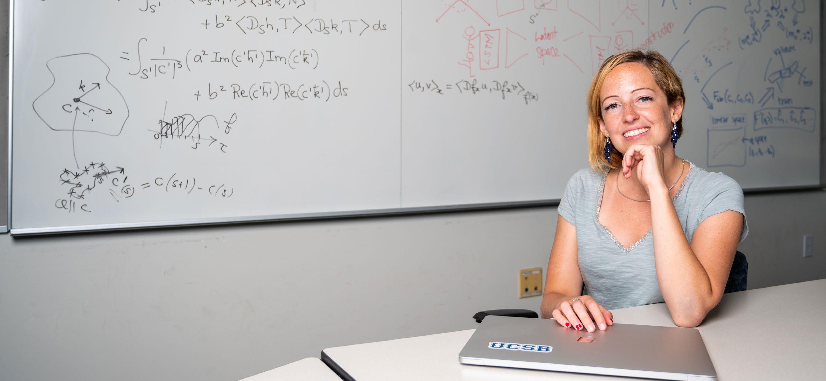
videos, which Miolane describes as “a much more challenging and realistic setting.”
Miolane’s work may prove to be of enormous benefit to biologists and medical professionals who require a common, precise language to describe what they see. Geometrical statistics provide that language, the value of which is evident to Allison Gabbert, a sixth-year PhD student in the lab of Denise Montell, a distinguished professor in the Department of Molecular, Cellular and Developmental Biology at UCSB who is working with Miolane. “You can see things under a microscope and describe them subjectively,” Gabbert says. “In a gene-expression study, I would knock down or over-express a gene and see that the cells were changing, but I couldn’t describe the change precisely. Nina’s lab did a different analysis on the images that provided us with a way to quantify what we saw.”
Developing efficient clinical strategies requires filling this knowledge gap, which, in turn, requires next-generation computational tools that can accurately — that is, quantitatively — describe live images of moving cells.
—Nina MiolaneCell migration
Biologists know that cells move by using their cytoskeleton to stretch and retract their membranes in response to mechanical forces both inside and outside the cell, but the exact mechanics of those processes are still not understood.
In one important type of cell migration linked to the metastasis of cancer, a cell creates a protrusion that causes the cell membrane to extend. Sometimes, a cell can produce several protrusions. It will then migrate by extending and retracting the protrusion(s) until it reaches its destination and returns to a non-migratory state, although, Gabbert says, “The specific mechanisms vary depending on the cell type and on whether it is a single cell or group of cells.”
Researchers in the Montell lab are studying migratory cells in the ovaries of fruit flies as a model for the migration of cancer cells, which can migrate as single cells, strands of cells, or cell clusters. Some migrate more effectively than others, speeding metastasis. Therefore, Gabbert says, “If cells could be bio-engineered to become less motile [prone to migration], cancer progression could be slowed.”
She and Montell discovered that one way to prevent migration of the cells they are studying is to over-express the protein septin, which prevents protrusions from forming. Researchers would like to better understand this cellular process, correlating changes in gene expression with their impacts on protrusion size and shape and the cell’s migratory ability. New knowledge in this area could have significant translational application.
“Developing efficient clinical strategies
“
Nina Miolane uses geometric statistics to quantify cell dynamics captured in images
requires filling this knowledge gap, which, in turn, requires next-generation computational tools that can accurately — that is, quantitatively — describe live images of moving cells,” says Miolane.
Currently, she and Montell are collaborating to use 2D images, generated in Montell’s lab, to observe the behavior of migrating cells after septin has been overexpressed or knocked down.
Origins and the Process
Research in the field of geometric statistics began in the 1950s, and several decades ago, code was written for it, but, “It was seldom tested and could not be easily re-used,” Miolane notes. Her lab’s important advance, she says, has been “to implement the theory and create opensource code,” thereby “democratizing geometric statistics as a class of methods that can be used to automatically quantify shape motions and deformations from images.” As of August 2022, the open-source software she wrote for her method had been downloaded more than ninety thousand times since being released in December 2020.
Complicating the step up to video analysis is the fact that generating a live 3D video of a cell requires that hundreds of thousands or even millions of digital light-microscopy images be converted to an outline of the cell comprising every one of the points on the outline of it — generally, about two hundred for a 2D image of a cell and many, many more than that for a 3D image — each
of which is represented by its coordinates in 3D space. Miolane’s software then provides a precise mathematically quantified representation of the image, making it possible to objectively describe any shape or change.
“Shape analyses performed in biology still require a lot of user input,” Miolane explains. ”Biologists often look at images individually, processing them using partially automated, partially manual techniques. Such a time-consuming, laborintensive process is not practical with the enormous number of images required for 3D imaging. If you have only, say, ten images, you can look at them and maybe trace the contour of the cell and then say that in one video, the cell is moving and deforming rapidly and in another video, it is not deforming much. But if you have perhaps a million images, you won’t be able to look at them individually and sort them like this. You need a method that can tell you, for example, that all of these half-million cells are deforming faster when they move faster, and all of these other half-million are going slowly and do not deform much. If instead, however, we had a smarter algorithm that could detect geometric changes, then you wouldn’t need to manually label anything.”
Miolane uses the word geometry to define not dimensions of a feature of cell biology, but rather, in the framework of statistical theory, to indicate what she calls “the geometry of the data space.”
“Suppose you wanted to use traditional statistics to compute the mean blood pressure of ten patients,” she begins by way of explanation.
“You would do that by adding the individual blood pressure of all ten patients and then dividing by ten. Now, suppose that you were to apply that same formula to do a mean average of your patients’ heart shapes. You take each patient and use 3D imaging to extract the shape of each of their hearts. Now, you add all the coordinates of the heart surfaces together and divide by ten. But you don’t get a nice, neat mean; you get something that doesn’t look like a heart at all. The traditional equations don’t work here. It’s hard even to know what it means to compute the differences between two hearts, or the ‘average’ heart shape. It requires a brand-new area of statistics.”
People realized quickly that using traditional equations for something like averaging heart shapes gives results that are, Miolane says, “so bad that they cannot be used. But then you find tricks around it. So, maybe you can align all of the hearts and then find that this one was too big, so you make it slightly smaller, and you ‘account’ for the differences. But then you get something that looks like a heart, but what does it really mean? If you do so many computational tricks to arrive at something that looks like a heart, is that really a meaningful ‘average’ heart? And if the goal is human health, what are you going to tell your patient? ‘Your heart looks like the average healthy heart, because after we did a bunch of tricks, it does appear normal?’”
The point, Miolane says, is, “We were lacking the theory to be able to say, ‘This is what an average heart actually looks like.’ Our work provides that theory, that framework to accurately account for all of the differences that might exist. Applied to cell biology, we will be able to claim: this is how the cancer dynamics of an average cell actually look.”
For her methodology, Miolane relied on the same mathematics, developed by Bernard Riemann, that Einstein used to reach his theory of relativity. “Einstein used it to explain gravity. We’re using it to explain biology,” Miolane says.
An illustration depicting cells that have developed multiple protrusions associated with cell migration, which can occur in metasticizing cancer cells.
The grant she has applied for is intended to fund research that carries some level of risk. The risk in Miolane’s work, she says, is, “We don’t know for sure whether a finer description of shape dynamics is going to provide more biological knowledge than just saying ‘There is a protrusion.’ I really think it will, but I can’t be one hundred percent certain. We will have to see how medically valuable the 3D images we generate actually are. However, even if we cannot find mechanistic insights on these specific images, the funding would allow us to develop our methods and make them available to the biomedical community as a whole. With datasets of live imaging growing in size, I expect our methods to reveal fundamentally new biology, and I cannot wait to see what the mathematics give us.”
CHAMPIONS OF ENGINEERING
Mark and Susan Bertelsen
For many years, Mark (‘66) and Susan Bertelsen (’67) have supported a wide variety of programs, institutes, centers, chairs, and activities at UC Santa Barbara, both in and well beyond the College of Engineering (COE). Mark, a member of the COE Dean’s Cabinet, spent fifty years as an attorney at Wilson Sonsini Goodrich & Rosati, where at various times he served as chairman of the management committee, managing partner, and a member of the firm’s executive committee, retiring as a senior partner in January 2022. The Bertelsens’ diverse philanthropy has benefited some twenty entities at UCSB, ranging from an endowed chair and the Institute for Energy Efficiency to the Natural Reserve System and even UCSB baseball. We spoke with Mark in August about their gifts to the university and the vision that continues to motivate them.
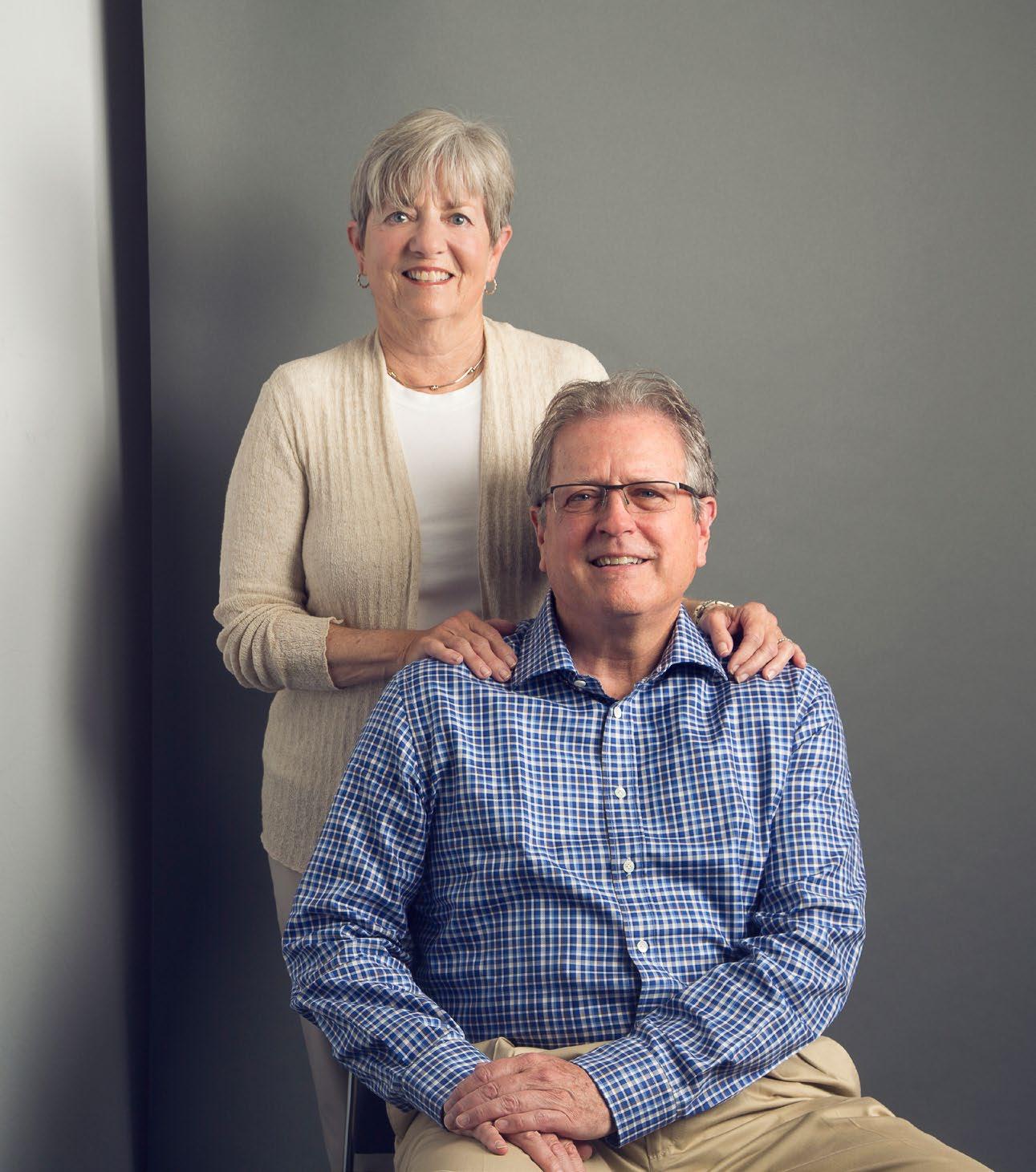
Convergence: You represented Silicon Valley technology clients for your entire career. You must have seen a lot of change.
MB: I spent my career at a law firm located in Stanford Research Park in Palo Alto, ground zero for the financial aspects of Silicon Valley. Our clients are predominantly technology companies. We did the Apple IPO in 1980, and were involved in all that went on before that following the commercialization of the integrated circuit and the microprocessor in the seventies, and the development of ever more powerful microprocessors and the software to run on them. Then, networks came along. We did the Cisco IPO in 1990. The 1990s and forward brought the advent of client server technology, the internet, and e-commerce with Netscape and browser technology; and Amazon, Pixar, and Netflix in entertainment; Google and the democratization of data; Tesla; social networks and the cloud and associated security and privacy issues — all the trends that have changed society over the past several decades.
C: Can you talk a bit about the roots of your engagement with UCSB?
MB: Susan and I made some small donations in the 1980s, but our first real gift went to Computer Science. They had received an $1 million anonymous gift, and the idea was to create four chairs at about $500,000 each for younger faculty, people who would not normally get chairs. So, our gift was combined with funds from the anonymous gift to create the chair in the name of Eugene Aas, Susan’s father, who was a World War II veteran and an electrical engineer in computer science.
My deep involvement with the COE began when Matt Tirrell was dean and they were building the Engineering Sciences Building, to which we contributed. We weren’t expecting to give at the level that Matt requested, but he came to my office, sat down, looked me in the eye, and told me that the College of Engineering was highly respected and was doing good things. He said, “You’ll feel good about your investment.” It was a powerful thing, so we made the commitment.
C: You and Susan have also provided generous support to the Institute for Energy Efficiency (IEE). What motivated that?
MB: When Jeff Henley, who was a classmate at UCSB and is also on the Dean’s Cabinet, gave his large gift for Henley Hall, it really accelerated the project. With respect to the IEE, the idea was to address the issues associated with the effects of climate change and energy use by supporting scientific research and technological innovation that could enable more efficient use of energy and less waste. I thought, here’s a very practical idea that could yield significant returns and make people’s lives and society better.
C: What do you see as the biggest challenges to higher education today?
MB: Obviously, for UCSB, state support is a huge issue. There may be budget surpluses in California occasionally, but in the longer term, to be competitive you’re going to have to rely more
on philanthropy and other sources of revenue, including how you monetize the intellectual property, if you will, of the institution. One of the things I’ve suggested for UCSB — and I understand that you can’t do everything — is to start some of what I call venture capital funds. There are various iterations of this, but the university gets a portion of the proceeds paid out from the fund’s investment.
communication skills and education? We found out some impacts during COVID. Yes, we can do some of this remotely, and some of it works well, and some doesn’t. There are things going on there that we should be more informed about, and that’s what CITS is all about.
C: What is the unifying vision or goal behind your diverse contributions?
MB: In my view — and this goes back to the ancient Greeks — universities are not just about pure academics. It’s also science, the arts, and also the body, mind, and spirit. Baseball, for example, is a program that is not rich at UCSB, yet it competes with the top programs in the country and has been to regional tournaments and the College World Series. Our players are actually students and athletes. They don’t come here to do their freshman year and then go to the pros. I played freshman baseball, so I understand a little about the game. At UCSB, we have an inadequate field and therefore can’t host regional tournaments. For a relatively modest investment, we could have a facility, not a Taj Mahal but one that could host regional tournaments, and we could build a softball field for the women. We could have community outreach, clinics for boys and girls, especially in the underserved communities. What’s the unifying thought? It is that this is what a university is supposed to do: serve multiple needs of the immediate campus population and the greater community to which it’s connected.
C: Can you talk a bit about your support for the Center for Information Technology & Society (CITS), which was established at least partially to investigate and understand “ethics and technology.” What does that mean to you?
MB: Globalization and technology, which to a certain extent have gone hand in hand, have resulted in a great benefit to a lot of people, including myself. It hasn’t necessarily had that effect universally. Millions of middle-class manufacturing jobs have become service jobs. That has affected the economics of society. Given that and the velocity of change, and a political discourse that, in my view, has been weaponized and cheapened, you ask, how do we move forward and maintain the social compact? Technology is important to that. At the CITS, the mission is to “apply knowledge from diverse perspectives to understand and guide the development and use and effects of technology in contemporary society.” That means, what are we doing here and how does it affect future generations, and how do they deal with things like
It’s the same with supporting the UC Natural Reserve System. Again, the reserves can host important environmental research and also be assets for underserved communities. That gets us to the UC Disaster Resilience Network (DRN), which tries to harness depth and breadth of knowledge at all of the UC campuses to prepare for and recover from disasters. What we liked about that, again, was that it seems a better way to use the university resources. UC doesn’t usually work as a whole; campuses do their own thing. You want to support expertise that has an objective of improving people’s lives, and if it can have a public-service component to it, that’s important, too.
C: Why do you think that it is important for people, like you and Susan, who have the ability support the university, to do so?
MB: I was seven years at the University of California: four at UCSB and three at Berkeley for law school. And even today, when you think people would understand, you hear them say, “Why should I give to UC; my tax dollars go there.” And I tell people that state support makes up only about twenty percent of UCSBs budget. Even private institutions like Stanford, Harvard, and Yale are crunched from time to time, but they have these $30 billion and $40 billion endowments. I spent seven years at a place that wasn’t free, but with scholarships and other things, I could pretty much fund my education. Great public institutions deserve support. If I look around and ask, what am I going to invest in, I’d say that investing in UC provides a return that benefits our communities and society in general.
The unifying thought is that this is what a university is supposed to do: serve multiple needs of the immediate campus population and the greater community to which it’s connected.
— Mark Bertelsen
College of Engineering
University of California Santa Barbara Santa Barbara, CA 93106-5130
You Make the Difference
The philanthropy of individuals like you helps to ensure that breakthroughs in science and engineering remain the order of the day across our beautiful UC Santa Barbara campus. Large gifts, small gifts, estate gifts — they all matter, because each advances academic excellence and leading-edge research that, together, help pave the way to a better tomorrow. Please consider making a difference by making your gift today!
To learn more about opportunities for the College of Engineering and the Division of Math, Life and Physical Sciences, please contact Lynn Hawks, Senior Assistant Dean of Development | 805-893-5132 | lynn.hawks@ucsb.edu
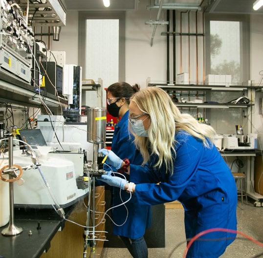
The University of California, in accordance with applicable Federal and State law and University policy, does not discriminate on the basis of race, color, national origin, religion, sex, gender, expression, gender identity, pregnancy, physical or mental disability, medical condition (cancer related or genetic characteristics), ancestry, marital status, age, sexual orientation, citizenship, or service in the uniformed service. The University prohibits sexual harassment. This nondiscrimination policy covers admission, access, and treatment in University programs and activities. Inquiries regarding the University's student-related nondiscrimination policies may be directed to the Office of Equal Opportunity & Sexual Harrassment/ Title IX Compliance, Telephone: (805) 893-2701.
