SWT/KWS MT KENYA MOBILE VETERINARY UNIT
MARCH 2024


11 Cases in March


5 Poaching Cases 6 Elephant Cases
March 2024 Report by Dr. Dr Jeremiah Poghon
During the month of March 2024, there was an increase in the number of cases in the Mt. Kenya and Southern Laikipia area. The veterinary unit was involved in the treatment of an elephant bull with lameness of the left forelimb, an elephant at with a wound on the left hindlimb, another elephant with a wound on the left hindlimb and an adult male white rhino for injuries sustained during a territorial fight. Several necropsies were carried out on both rhinos and elephants. The Unit also collared 3 male lions and 1 lioness at Loisaba Conservancy.
Acknowledgement
The Mt. Kenya Mobile Wildlife Veterinary Unit thanks the Senior Assistant Director, Mountain Conservation Area and the Head of Veterinary Services, Kenya Wildlife Service for providing leadership and technical expertise. The veterinary team also appreciates The Sheldrick Wildlife Trust (SWT) for providing the financial and logistical support that enables the Unit to fulfil its mandate.
Case Details
1-Mar-24
4-Mar-24
Causes Sustained injuries after being involved in a territorial fight with another bull Successfully Treated
5-Mar-24
5-Mar-24 Antelope Laikipia Snared De-snaring an oryx
6-Mar-24
Chololo Ranch Spear Postmortem revealed injuries caused by a sharp object (most likely a spear) Poaching Death
6-Mar-24 Lion Loisaba Ranch Collared Lions have been causing problems to the newly translocated Black rhinos Task Successful 19-Mar-24 Rhino White Ol Pejeta Conservancy Postmortem The carcass was found on left lateral recumbency and had a poor body condition Died
20-Mar-24 Rhino White Solio Ranch Postmortem Numerous superficial wounds on face, front limbs, chest area and torso. Died
Date Species Area Found Reason for Intervention Outcome
Elephant Ol Jogi Conservancy Natural Causes Lameness of the left forelimb Prognosis Poor
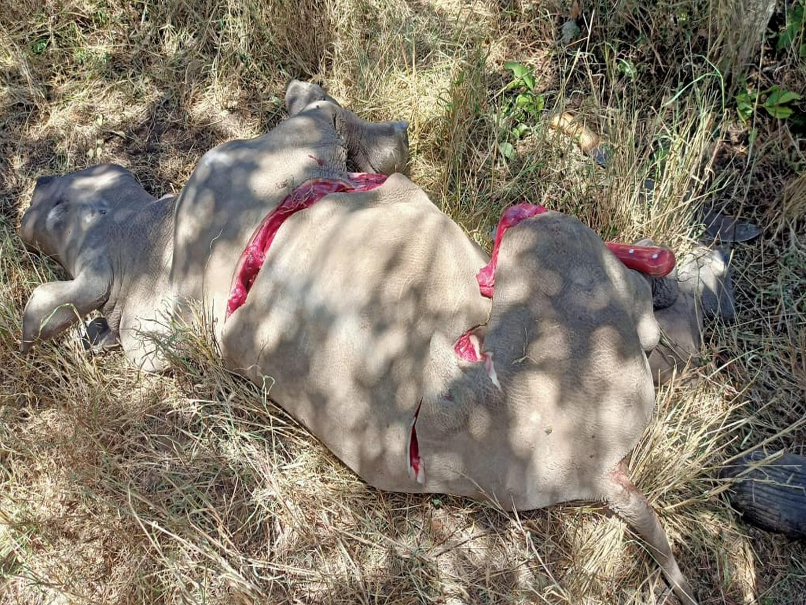
Elephant Imenti Forest Spear Postmortem revealed a deep cut might have been caused by a spear Poaching Death
Elephant
Poaching Postmortem Inconclusive but
poaching
Poaching
2-Mar-24
4-Mar-24
Naivasha
might be due to
because the tusks were missing
Death
Elephant
Poaching
Inconclusive
Poaching
Natural
Naivasha
Postmortem
but might be due to poaching because the tusks were missing
Death 5-Mar-24 Rhino White Borana Wildlife Conservancy
Elephant Lewa Conservancy HWC the elephant had an infected degloving injury Successfully Treated
Successfully
Treated
Elephant
Introduction
SWT/KWS Mt Kenya Mobile Vet Unit Treatment Locations
March 2024


Case 1 – 1st March 2024
Elephant Natural Causes
Ol Jogi Ranch
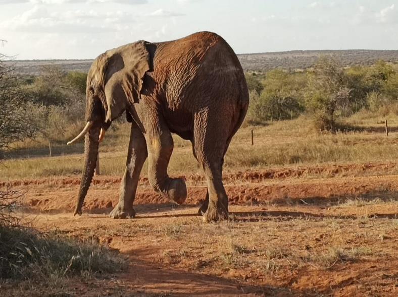
An elephant bull at Ol Jogi Conservancy was reported to be having lameness on the left forelimb. The carpal joint was flexed and the animal was totally unable to use the limb.


Immobilisation, examination and treatment
The elephant was darted with 18mg Etorphine hydrochloride delivered using a Dan-Inject CO2 rifle, fired from a vehicle. The dart was delivered on the left rump but it did not fully discharge, only about 10 mg was delivered. The elephant went down after 3 minutes onto left lateral recumbency. Ropes were used to turn the elephant and position it into right latreal recumbency to allow assessment of the left forelimb.
On examination, there were no open wounds on the forelimb but there was flexion of the carpal joint and on palpation a hollowness on the dorsal aspect of the joint indicating a torn extensor tendon. Systemic cover was done using; 15,000mg Amoxicillin (I.M) and 2,000mg Flunixin Meglumine (I.M).
Revival and prognosis
Reversal was done at using 200mg of Naltrexone intravenously and the elephant was up in 4 minutes. Prognosis is guarded.

.
Case 2 – 2nd March 2024
Elephant Spear
Imenti Forest
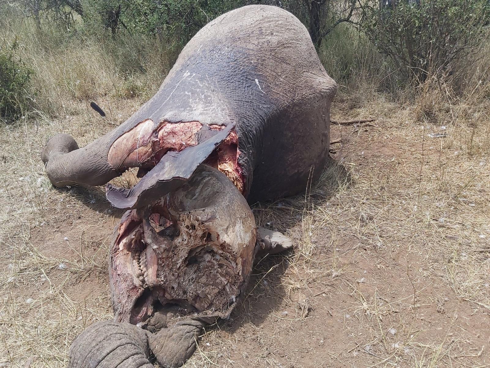
The animal submitted for necropsy was an adult male elephant. The postmortem interval was estimated to be approximately 2 days therefore rigor mortis was absent.
Post-mortem Findings
The body condition scoring was impossible due to bloating of the carcass. The carcass was lying in right lateral recumbency.
There was a penetrating wound about 30cm long with straight margins on the dorsal thoracic wall. There was putrefaction and maggot infestation of the wound, indicating that the wound had been there for some time. The rib just below the wound was completely fractured and had been pushed into the thoracic cavity, puncturing the right lung. There was a focal region of fibrosis on the right lung at the point of puncture by the fractured rib which was also infested with maggots.
Cause of Death
The penetrating wound and the completely fractured rib indicate that the wound was inflicted from above by a sharp object with force great enough to fracture a rib. The fractured rib punctured the right lung leading to contamination and bacterial infection leading to septic shock and death of the animal.

.
Case 3 & 4 – 3rd March 2024
Elephant Poaching Post-mortem Kimende, Naivasha
On the 3rd of March 2024 Aberdare investigation attended and processed the scene of two elephant carcasses reported by WPD team based at Kimende. The vet team then conducted an autopsy on the two carcases
Post-mortem Findings
The first carcass was lying on the right side and at advanced stage of decomposition. The head had sharp object cut marks with both tusks removed. The neck was cut halfway The skin was opened and subcutis examined on the left flank, but no penetrating wound was seen. Gastrointestinal system was not perforated, and the peritoneal cavity was normal. The liver was autolysed. Spleen was also decomposed. The kidney was decomposed but with good consistency. Stomach was opened and the ingester was normal Liver, kidney and stomach contents were sampled for toxicological analysis at government chemist.
The second carcass was lying on the right flank and the head was decapitated. The carcass was at an advanced level of decomposition with the carcass body score 4/5. The skin was opened on the left flank and no wound was seen on the subcuties. Abdominal cavity was opened and had some ingester but no rupture of intestines. Peritoneal cavity appeared normal apart from the ingester. Liver, kidney and spleen were decomposed Intestines appeared normal but decomposed and when opened had large amounts of water ingested. Stomach was full of ingester and appeared normal. Colon had feed material which appeared normal. Tissue samples were collected for molecular forensic analysis in case the tusks are recovered and can be linked to the carcass. Liver, kidney and stomach contents taken for toxicological analysis at the government chemist.
NB. A metal detector was used to scan the carcasses for metallic objects but returned negative. The collected samples were submitted on the 4th of March 2024 to the government chemist for toxicological analysis in order to rule out poisoning as the cause of death.
Cause of Death
Cause of death could not be determined by post-mortem but due to the lack of tusks and no external injuries, poisoning with the intent to poach is suspected.


Case
White Rhino Natural Causes
Borana Conservancy
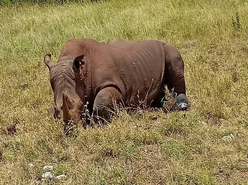
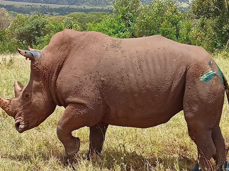
An adult male white rhino was reported to have sustained injuries after being involved in a territorial fight with another bull. The rhino was also reported to be having lameness on the right hindlimb.
Immobilisation, examination and treatment
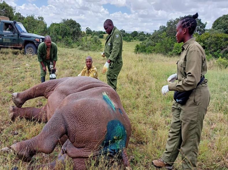
The rhino was darted from a helicopter with 5.5 mg Etorphine hydrochloride in combination with 60mg Azaperone. The rhino went down onto right lateral recumbency and a rope was used to further secure the animal 25mg Butorphanol was administered intravenously and oxygen insufflation started intra-nasally to improve cardio-respiratory function.
On examination, the animal had good body condition (score of 4out of 5). The animal had sustained a deep penetrating wound just next to the tail, and abrasions on both hindlimbs at the level of the digits. Lameness of the right hindlimb was suspected to be caused by a torn extensor tendon. The wounds were quite fresh therefore they were irrigated with Iodine, green clay was packed into the deep penetrating wound close to the tail, then topical Oxytetracycline spray applied. Systemic cover was done using; 15,000mg Amoxicillin (I.M) and 3,000mg Phenylbutazone (I.M) and 30ml Catosal I.M.
Prognosis
Prognosis is good to guarded.

5 – 5th March 2024
Case 6 – 1st March 2024
Elephant Spear
Lewa Conservancy
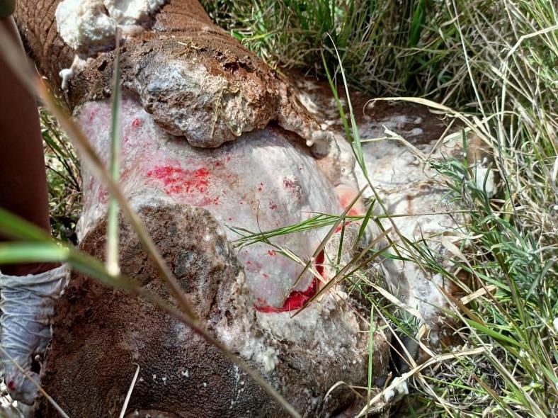
An adult female elephant at Lewa Conservancy was reported to be having lameness on the left hindlimb caused by a wound on the limb.
Immobilisation, examination and treatment
The elephant was darted with 15mg Etorphine hydrochloride delivered using a Dan-Inject CO2 rifle, fired from a vehicle. The dart was delivered on the left rump and the elephant went down after 6 minutes onto right lateral recumbency. Water was poured on the animal to cool it down.
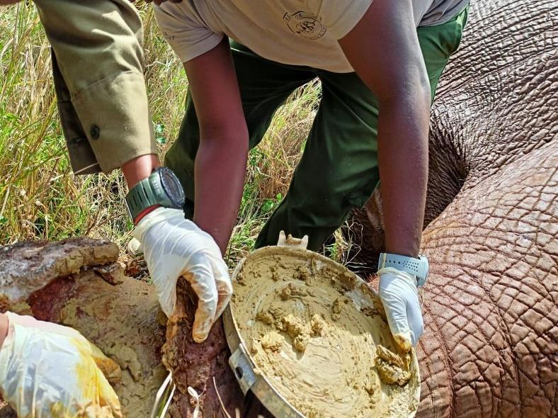
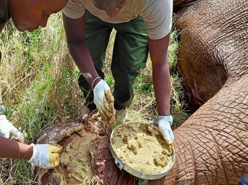
On examination, the elephant had an infected degloving injury extending from the metatarsus to the digits. The wound was covered by a thick layer of pus and had a foul odour. It was scrubbed with dilute Hydrogen peroxide, then rinsed and irrigated with Iodine. After cleaning, green clay was packed in the wound and topical oxytetracycline spray applied. Systemic cover was done using; 15,000mg Amoxicillin (I.M) and 2,000mg Flunixin Meglumine (I.M).
Prognosis
The wound is possibly a spear injury. Prognosis is good. However, it will take time to heal due to the location.

The Unit attended a oryx with a snare.
Immobilisation, examination and treatment
The oryx was found and darted with Etorphine.
The loose snare was removed from the right foreleg and the animal was then reversed from the anaesthesia.
Prognosis
Prognosis is good


Case 7 – 5th March 2024
Oryx Snared Lakipia
.
Case 8 – 6th March 2024
Elephant Spear
Chololo Ranch
The animal submitted for necropsy was an adult male elephant. The postmortem interval was estimated to be approximately 1 day, rigor mortis was absent.
Post-mortem Findings
There was a trail of dried-up blood leading to the site of death. The body condition score was good (4/5). The carcass was lying on left lateral recumbency but it was later turned to right lateral recumbency for complete observation and assessment of lesions.
The left forelimb was missing as a result of scavenger activity. Scavenger activity was also evident on the head, neck and the skin of the abdomen. There was a perforating wound with straight margins on the right pinna. The wound went through the pinna and into the neck. Bilateral diffuse hemorrhage was observed in the supraspinatus muscles.
Cause of Death
The sharp object (most likely a spear) used to inflict the wound went through the pinna and into the neck, piercing either the jugular or internal carotid, or both. The cause of death is hypovolemic shock due to massive bleeding.
Lion Collaring Loisaba Conservancy
Lions have been causing problems to the newly translocated black Rhinos at Loisaba Conservancy.

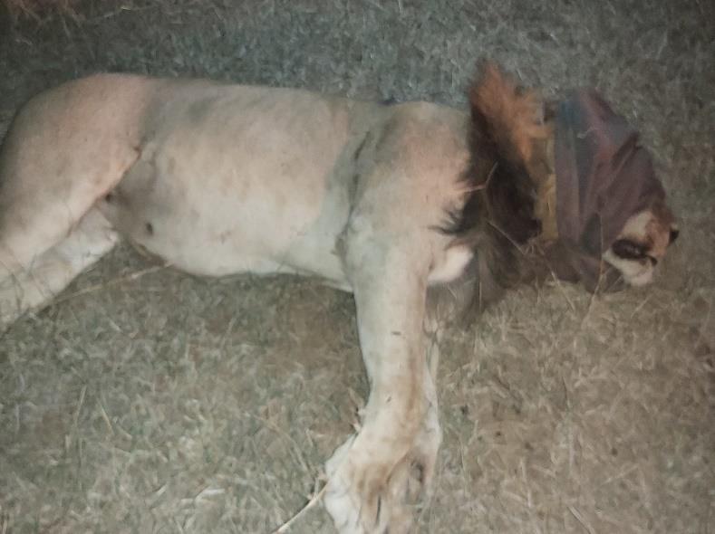
Immobilisation and collaring
Recently 2 male lions were translocated to Kora National Park after attacking 2 newly introduced rhino. A further plan was made to collar 4 lions for monitoring purposes in the conservancy.
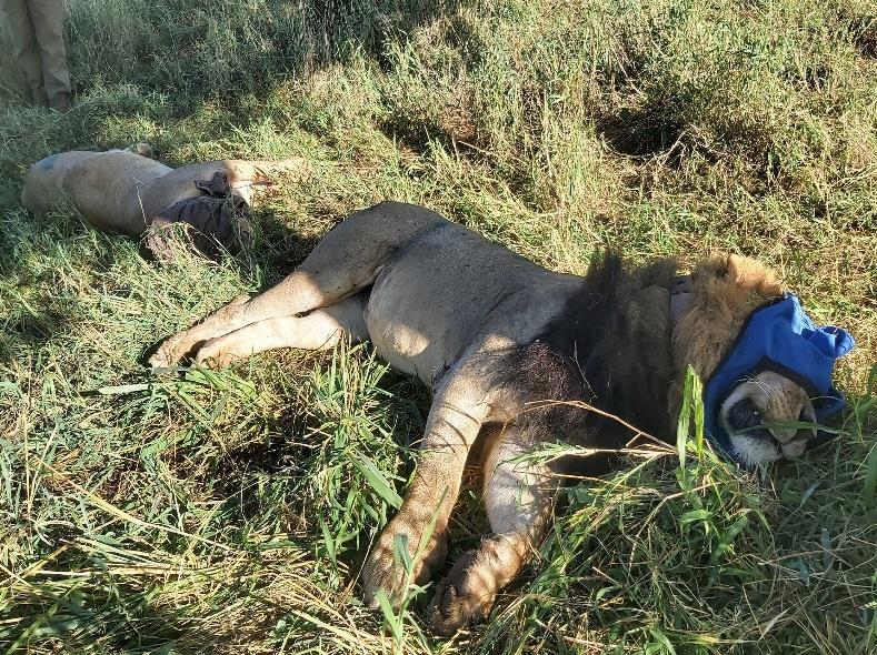
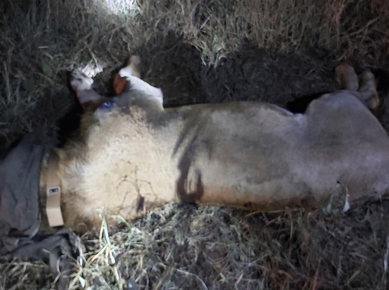
A bait and callback system were set up in the evening and early morning. Sounds of herbivores in distress were played near a carcass tied tightly onto a tree. The lions were darted using a Dan-Inject CO2 rifle fired from a vehicle. One adult male received 600mg Ketamine and 8mg Medetomidine (in two darts) whereas the other two adult males received 350mg Ketamine and 8 mg Medetomine and the lioness 300mg Ketamine and 6mg Medetomidine. The nose-tail length, shoulder height and the paw and canine measurements were taken during the collar fitting exercise. Tissue samples were also collected for DNA analysis. A collar was fitted on the neck. Cloxacillin (Opticlox) was infused into the dart wound and topical OOOxytetracyclline spray applied. 3000mg Amoxicillin was administered intramuscularly.
Prognosis
After the effects of immobilization drugs were allowed to diminish through metabolism the anesthesia was reversed. About 30 to 40 minutes after reversal, though a little drowsy, the lions rose and walked away.
9 – 6th – 8th
Case
March 2024
Case 10 – 20th March 2024
White Rhino
Post-mortem
Solio Ranch
An adult male white rhino was reported to be recumbent by the KWS security. It was struggling to get up, with weakness on its hind quarters. There were signs of struggle for about 200 metres where the animal was pulling itself along the ground. The hindlimbs were suspected to have been fractured. The veterinary unit arrived and found the animal still alive but struggling to get up but the rhino succumbed shortly after. A postmortem was conducted and the trophies were removed and handed over to the Solio KWS team.
Post-mortem Findings
There were numerous superficial wounds on the face, front limbs, chest area and torso. The left hindlimb was held up and could be bent meaning it was fractured. The skin was opened on the rear aspect and massive haemorrhage was observed in the subcutis and broken bones were observed just below the knee joint. The tibia and fibula were fractured. The haemorrhage was observed from the knee joint all the way to the digits. Severe hematoma was observed on the thigh muscles of the left hindlimb. Comminute fracture of the proximal tibia and fibula just below the knee joint. The humerus was intact. There was all diffuse haemorrhage on the serosa of the small intestine and colon. The stomach and small intestine were empty. Colon was filled with desiccated ingesta. Mucosa of the colon was diffusely hemorrhagic.
Cause of Death
Territorial fight with another male that lead to fracture of the hind limb and hemorrhage, shock and death.

.
Case 11 – 20th March 2024
White Rhino Post-mortem
Chololo Ranch
A rhino calf carcass was reported by the patrol team near the Ol Pejeta dam area on left lateral recumbency. The post-mortem was carried out by the KWS regional, Ol Pejeta and Mount Kenya vet team.
Post-mortem Findings
The carcass was found on left lateral recumbency and had a poor body condition of < 2.5 out of 5. There were no signs on external injury. There were numerous ticks around the armpit, pelvic area and around the perineum. There was vomitus(grass) around the mouth area.
The lungs were bilaterally congested and haemorrhagic. The heart also had highly injected superficial vessels and the pericardial tissue highly haemorrhagic (myocarditis) There was scanty to no peritoneal fluid indicating that the animal was possibly dehydrated. There was generalized haemorrhage along the intestines and prominently injected vessels. The mesenteric lymph nodes were also largely reactive. The liver was slightly enlarged with areas of localized haemorrhages. Spleen was slightly pale and had areas of localized haemorrhages Stomach contents were mainly grass with no evidence of any milk contents. The glandular part of the gastric mucosa had localized areas of haemorrhage. The small intestines were empty with areas of haemorrhages and ulcerations along the mucosa The serosa of the bladder had areas of haemorrhage
Cause of Death
The rhino died from an infection leading to septicemia, pneumonia (congested lungs) and the hemorrhages .
