Avian Pathology
Bacterial Diseases Avian Pathology Bacterial Diseases
Prof.Dr. Al-Sayed R. Al-Attar
Prof.Dr. Al-Sayed R. Al-Attar
Professor of Pathology
Professor of Pathology
Avian pathology Bacterial disease
Avian pathology Bacterial disease
Salmonella infections
Salmonella infections
Pullorum disease and fowl
Pullorum disease and fowl
typhoid
typhoid
The Enterobacteriaceae are a large family of Gram-negative bacteria that includes, along with many harmless symbionts, many of the more familiar pathogens, such as Salmonella, Escherichia coli, Yersinia pestis, Klebsiella, and Shigella. Other disease-causing bacteria in this family include Proteus, Enterobacter, Serratia, and Citrobacter. Members of the Enterobacteriaceae are rod-shaped, and are typically 1–5 μm in length. They appear as small grey colonies on blood agar. Like other proteobacteria, enterobacteria have Gram-negative stains,[5] and they are facultative anaerobes, fermenting sugars to produce lactic acid and various other end products. Most also reduce nitrate to nitrite, although exceptions exist (e.g. Photorhabdus). Unlike most similar bacteria, enterobacteria generally lack cytochrome C oxidase, although there are exceptions (e.g. Plesiomonas shigelloides). Most have many flagella used to move about, but a few genera are nonmotile. They are not spore-forming.
Catalase reactions vary among Enterobacteriaceae.
The Enterobacteriaceae are a large family of Gram-negative bacteria that includes, along with many harmless symbionts, many of the more familiar pathogens, such as Salmonella, Escherichia coli, Yersinia pestis, Klebsiella, and Shigella. Other disease-causing bacteria in this family include Proteus, Enterobacter, Serratia, and Citrobacter. Members of the Enterobacteriaceae are rod-shaped, and are typically 1–5 μm in length. They appear as small grey colonies on blood agar. Like other proteobacteria, enterobacteria have Gram-negative stains,[5] and they are facultative anaerobes, fermenting sugars to produce lactic acid and various other end products. Most also reduce nitrate to nitrite, although exceptions exist (e.g. Photorhabdus). Unlike most similar bacteria, enterobacteria generally lack cytochrome C oxidase, although there are exceptions (e.g. Plesiomonas shigelloides). Most have many flagella used to move about, but a few genera are nonmotile. They are not spore-forming. Catalase reactions vary among Enterobacteriaceae.
Many members of this family are a normal part of the gut flora found in the intestines of humans and other animals, while others are found in water or soil, or are parasites on a variety of different animals and plants. Escherichia coli is one of the most important model organisms, and its genetics and biochemistry have been closely studied.
Many members of this family are a normal part of the gut flora found in the intestines of humans and other animals, while others are found in water or soil, or are parasites on a variety of different animals and plants. Escherichia coli is one of the most important model organisms, and its genetics and biochemistry have been closely studied.
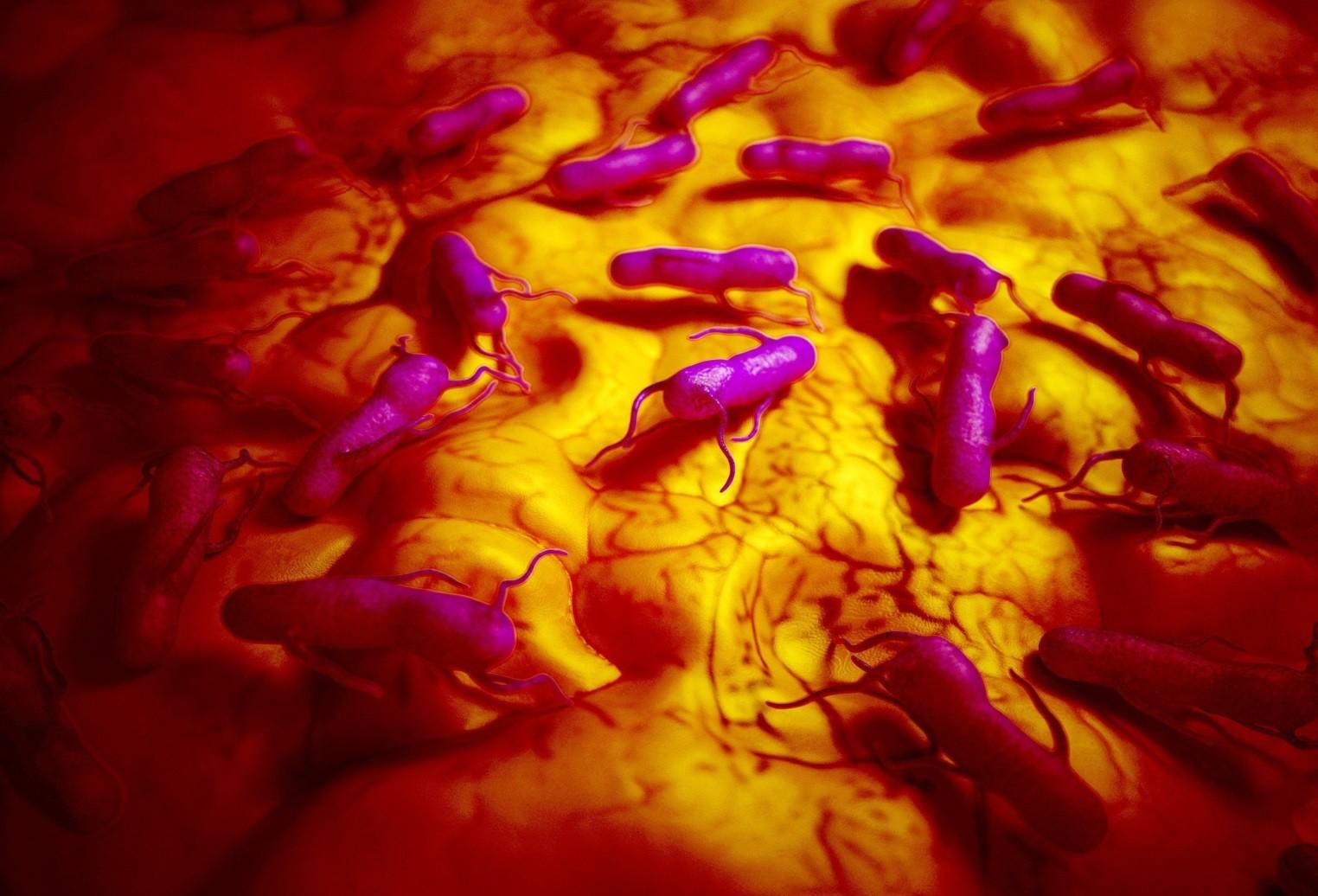
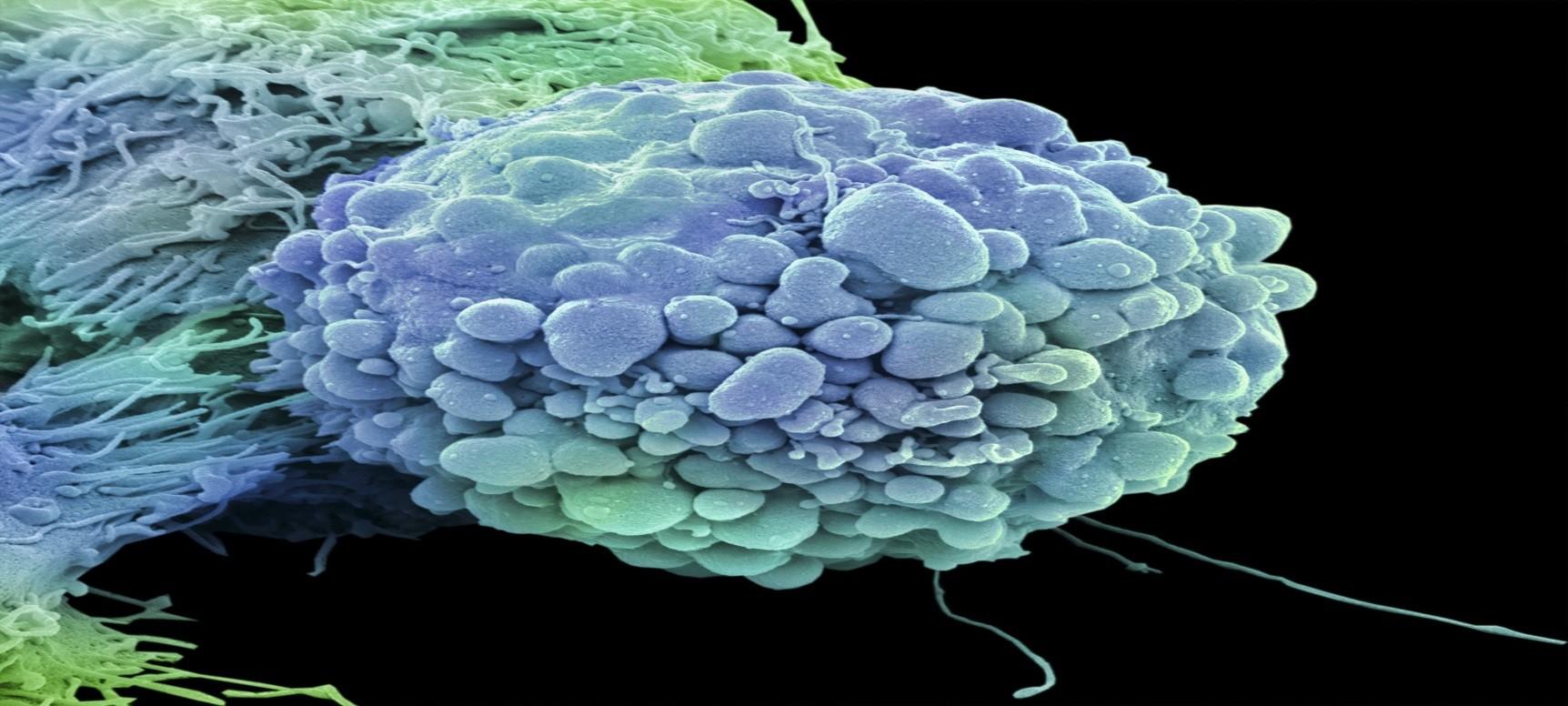
Salmonella bacteria could be used to kill cancer Salmonella bacteria could be used to kill cancer
Bacteria found to cause a common food poisoning bug could be used to kill cancer, medical experts have revealed. Bacteria found to cause a common food poisoning bug could be used to kill cancer, medical experts have revealed.
How? Well, according the scientists, the bacteria can infiltrate tumours and flag the cancer cells up to the body’s immune defences, making them a target for attack.
How? Well, according the scientists, the bacteria can infiltrate tumours and flag the cancer cells up to the body’s immune defences, making them a target for attack.
The break through could massively help fight cancer – which at the moment is avoided by the immune system because it is seen as foreign’.
The break through could massively help fight cancer – which at the moment is avoided by the immune system because it is seen as foreign’.
But food poisoning, you ask? Luckily, the salmonella strain engineered by the South Korean researchers is a million times less potent than the version of the bug that causes sickness – so have no fear. n mice with bowel cancer, more than half the animals were completely cured without any side effects. Previous studies have looked at using bacteria to carry anti-cancer drugs into tumours.
But food poisoning, you ask? Luckily, the salmonella strain engineered by the South Korean researchers is a million times less potent than the version of the bug that causes sickness – so have no fear. n mice with bowel cancer, more than half the animals were completely cured without any side effects. Previous studies have looked at using bacteria to carry anti-cancer drugs into tumours.
The discovery arose when scientists found that bacteria attacking shellfish produced a protein that triggered a strong immune response.
The modified salmonella releases the same protein to spur the immune system into action.
The discovery arose when scientists found that bacteria attacking shellfish produced a protein that triggered a strong immune response. The modified salmonella releases the same protein to spur the immune system into action.
Professor Kevin Harrington, from The Institute of Cancer Research, London, said: ‘It has been known for some time that certain types of bacteria, including strains of salmonella, are able to grow in tumours but not in normal tissues.
Professor Kevin Harrington, from The Institute of Cancer Research, London, said: ‘It has been known for some time that certain types of bacteria, including strains of salmonella, are able to grow in tumours but not in normal tissues.
‘However, until now, attempts to use bacteria as anti-cancer therapies have had limited success, both in the laboratory and in the clinic.’
‘However, until now, attempts to use bacteria as anti-cancer therapies have had limited success, both in the laboratory and in the clinic.’
The clinical signs, epizootology, lesions and control and eradication procedures, pullorum disease and fowl typhoid have many similarityes. The difference in etiology pullorun caused by salmonella pullorum and fowltyphoidcausedbyS.gallinarum.
The clinical signs, epizootology, lesions and control and eradication procedures, pullorum disease and fowl typhoid have many similarityes. The difference in etiology pullorun caused by salmonella pullorum and fowltyphoidcausedbyS.gallinarum.
Bothdiseasesaresepticemicdiseaseaffecting primarily chickens and turkeys but other speciesaresusceptible.
Bothdiseasesaresepticemicdiseaseaffecting primarily chickens and turkeys but other speciesaresusceptible.
Etiology: Pullorum disease and FT are caused by s. pullorum and S. gallinarum respectively. However these 2 bacteria are currently placed in a single serovar. S. enterica subsp.
Etiology: Pullorum disease and FT are caused by s. pullorum and S. gallinarum respectively. However these 2 bacteria are currently placed in a single serovar. S. enterica subsp.
Entrica serovar pullorum- gallinarum of the family
Entrica serovar pullorum- gallinarum of the family
Enterobacteriaceae which is highly host adapted to birds.
Enterobacteriaceae which is highly host adapted to birds.
Natural hosts:
Natural hosts:
Chickens are the natural hosts for both S. pullorum and
Chickens are the natural hosts for both S. pullorum and
S. gullinarum, however, naturally occurring outbreaks of PD and FT have been described in turkeys, gunea fowl, quail, pheasants, sparrows and parrots.
S. gullinarum, however, naturally occurring outbreaks of PD and FT have been described in turkeys, gunea fowl, quail, pheasants, sparrows and parrots.
Transmission:
Transmission:
Horizontal from infected birds (reactor and carrier)
Horizontal from infected birds (reactor and carrier)
Vertical from infected eggs (contaminated ovaries)
Vertical from infected eggs (contaminated ovaries)
Clinical signs:
Clinical signs:
Chickens and poultes:
Chickens and poultes:
PD and FT are primarily disease of chickens and poults. FT is more significant disease in growing and adult chickens and turkeys than PD.
PD and FT are primarily disease of chickens and poults. FT is more significant disease in growing and adult chickens and turkeys than PD.
Chickens and poults hatched from infected eggs characterized by dead embryo in the incubator
Chickens and poults hatched from infected eggs characterized by dead embryo in the incubator
The birds can manifest somnolence, weakness, depressed appetite, poorgrowth and adherence of chalky white material to the vent.
The birds can manifest somnolence, weakness, depressed appetite, poorgrowth and adherence of chalky white material to the vent.
Mortality usually peaks during the second or third week of life. Also, some birds
Mortality usually peaks during the second or third week of life. Also, some birds
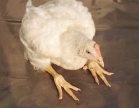
huddle together under heater having droopy wings and distorted body appearance. Labored breathing or gasping may be observed. Blindness and lameness due to swolleng of joints has been described.
huddle together under heater having droopy wings and distorted body appearance. Labored breathing or gasping may be observed. Blindness and lameness due to swolleng of joints has been described.
Survivor birds became carrier at maturity.
Survivor birds became carrier at maturity.
Growing and mature fowl:
Growing and mature fowl:
Acute outbreaks of FT may begin by sudden drop in feed consumption, with birds being droopy and showing ruffled feathers and pale and shrunken combs.
Acute outbreaks of FT may begin by sudden drop in feed consumption, with birds being droopy and showing ruffled feathers and pale and shrunken combs.
Other signs, such as a drop in egg production decreased fertility and diminished
hatchability may be observed in both PD and FT. Death may occure within 4 days
Other signs, such as a drop in egg production decreased fertility and diminished hatchability may be observed in both PD and FT. Death may occure within 4 days
PI. Also anorexia, diarrhea, depression and dehydration could be seen in semimature and mature flocks.
PI. Also anorexia, diarrhea, depression and dehydration could be seen in semimature and mature flocks.
Morbidity and mortality:
Morbidity and mortality:
Both morbidity and mortality are highly variable in chickens. Mortality from PD may vary from 0% to 100% Mortality range from 10% to 93% in chickens due to FT.
Both morbidity and mortality are highly variable in chickens. Mortality from PD may vary from 0% to 100%
Mortality range from 10% to 93% in chickens due to FT.
Morbidity is often much higher than mortality.
Morbidity is often much higher than mortality.
Gross lesions:
Gross lesions:
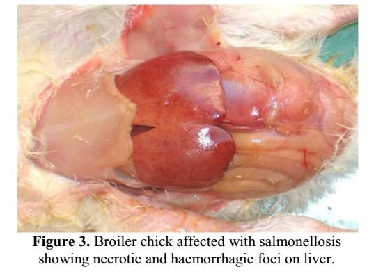
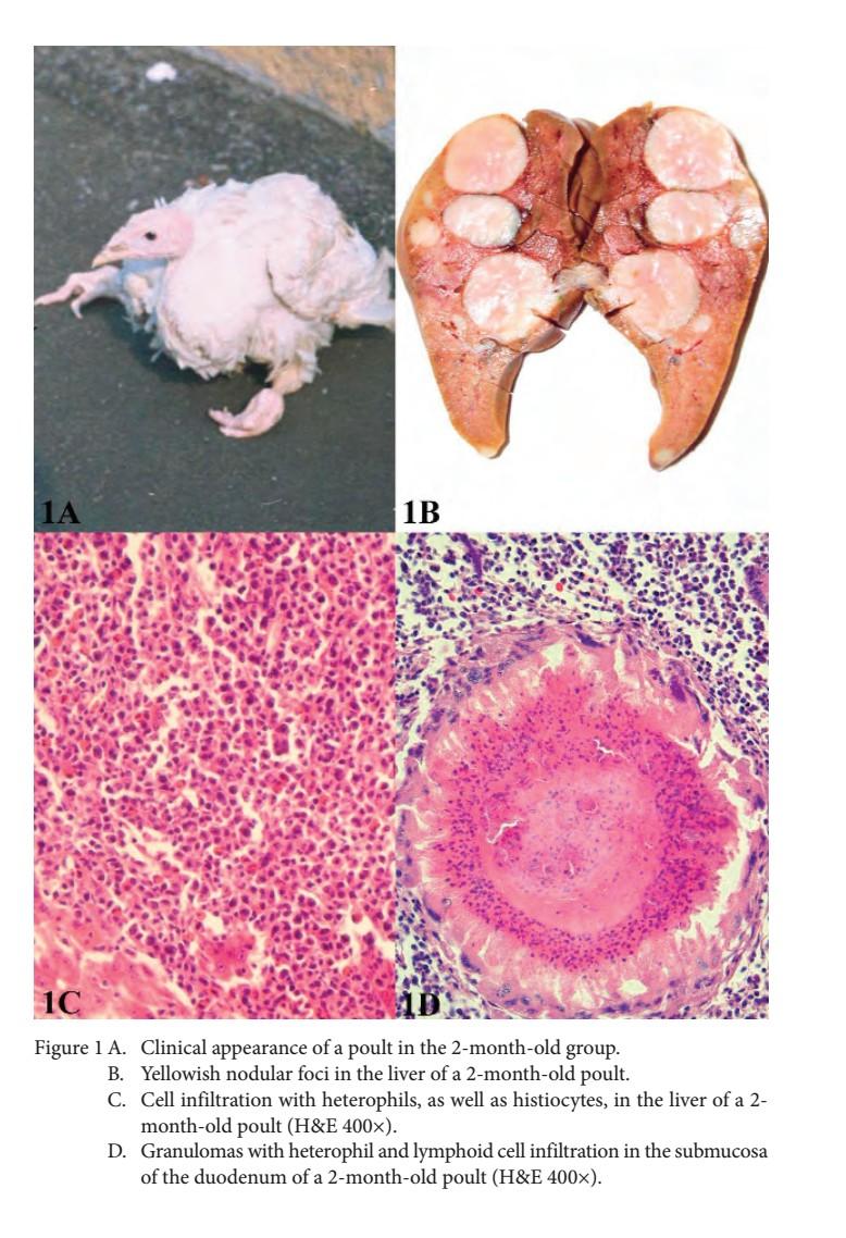
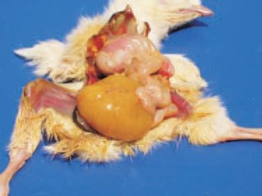
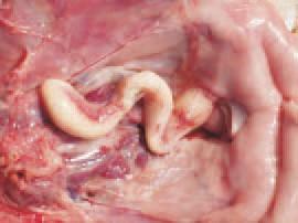
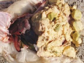
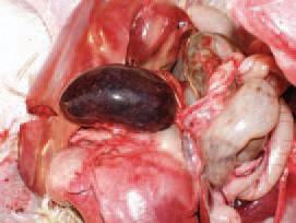
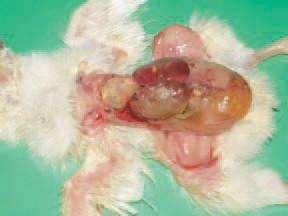
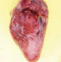
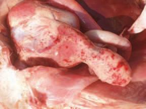
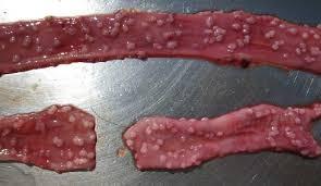
Chicks: peracute causes of PD and FT, no gross lesions. In acute cases, enlarged and congested liver, spleen and kidneys may be seen. White foci of 2-4 mm in diameter were seen on the livers. The yolk sac and its contents may or may not reveal any abnormalities but in protracted cases, interference with yolk absorption may occur. In such causes, yolk contents may be of creamy or caseous consistency. Lungs have white nodules. Also white nodules in the cardiac muscles and pancreas and the muscles of ventriculus, wall of the ceca and cecum.
Chicks: peracute causes of PD and FT, no gross lesions. In acute cases, enlarged and congested liver, spleen and kidneys may be seen. White foci of 2-4 mm in diameter were seen on the livers. The yolk sac and its contents may or may not reveal any abnormalities but in protracted cases, interference with yolk absorption may occur. In such causes, yolk contents may be of creamy or caseous consistency. Lungs have white nodules. Also white nodules in the cardiac muscles and pancreas and the muscles of ventriculus, wall of the ceca and cecum.
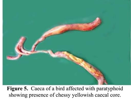
Ascites could be seen. Serous or fibrinous pericarditis was also seen. The ceca may contain caseous cores, swollen joint contains yellow viscous fluid.
Ascites could be seen. Serous or fibrinous pericarditis was also seen. The ceca may contain caseous cores, swollen joint contains yellow viscous fluid.
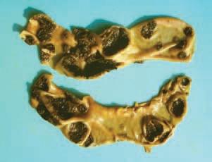
Adult chickens: lesion found mostly in chronic carrier hens with PD and FT are a few misshapen, discolored cytic or nodular ova among normal ovules. The involved ova may contain oily and caseous material enclosed in a thickened capsule. There degenerative follicles may be closely attached to the ovary, but frequently they are pedunculated and may detached from the ovary to the peritoneal cavity lead to extensive peritonitis.
Oviduct contains caseous exudate in the lumen. Fibrinous peritonitis and perihepatits with or without involrment of the reproductive tract could be seen. Hydropericadium with pericarditis were common. Occasionally cysts containing amber, caseous material may be found embedded in the abdominal fat or attached to the venticulus and intestine. Frequently, the pancreas may have white foci or nodules. In male, test may have white foci or nodules. Occasionally, caseous granuloma can be found in lungs and air sacs.
Adult chickens: lesion found mostly in chronic carrier hens with PD and FT are a few misshapen, discolored cytic or nodular ova among normal ovules. The involved ova may contain oily and caseous material enclosed in a thickened capsule. There degenerative follicles may be closely attached to the ovary, but frequently they are pedunculated and may detached from the ovary to the peritoneal cavity lead to extensive peritonitis. Oviduct contains caseous exudate in the lumen. Fibrinous peritonitis and perihepatits with or without involrment of the reproductive tract could be seen. Hydropericadium with pericarditis were common. Occasionally cysts containing amber, caseous material may be found embedded in the abdominal fat or attached to the venticulus and intestine. Frequently, the pancreas may have white foci or nodules. In male, test may have white foci or nodules. Occasionally, caseous granuloma can be found in lungs and air sacs.
Histopathology:
Histopathology:
Peracute cases of PD and FT, only severe vascular congestion in various organs.
Peracute cases of PD and FT, only severe vascular congestion in various organs.
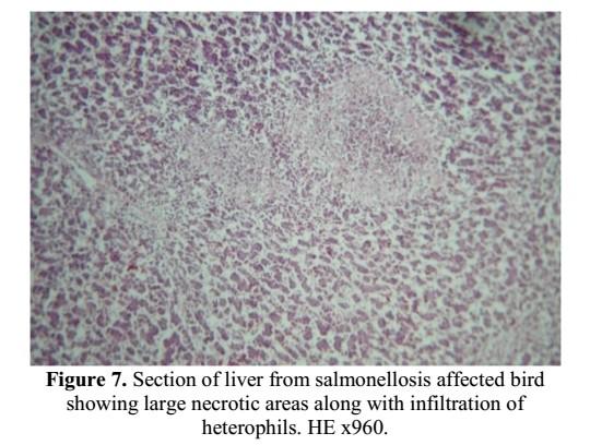
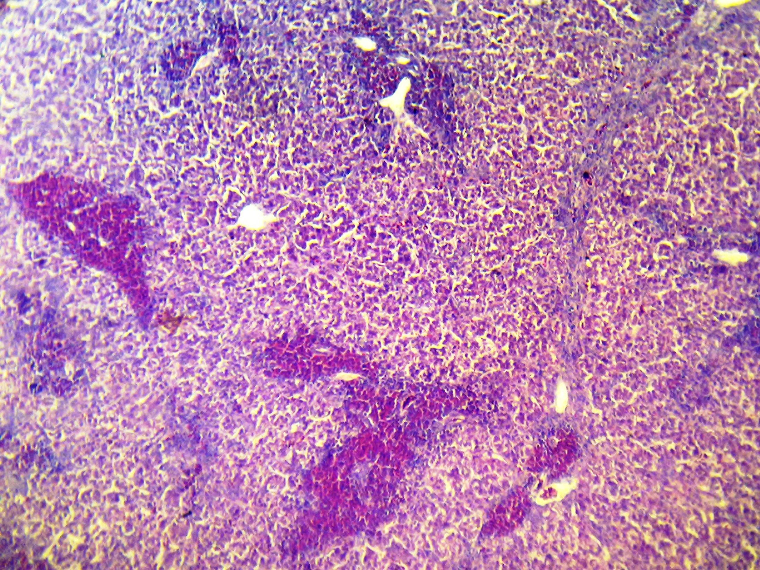
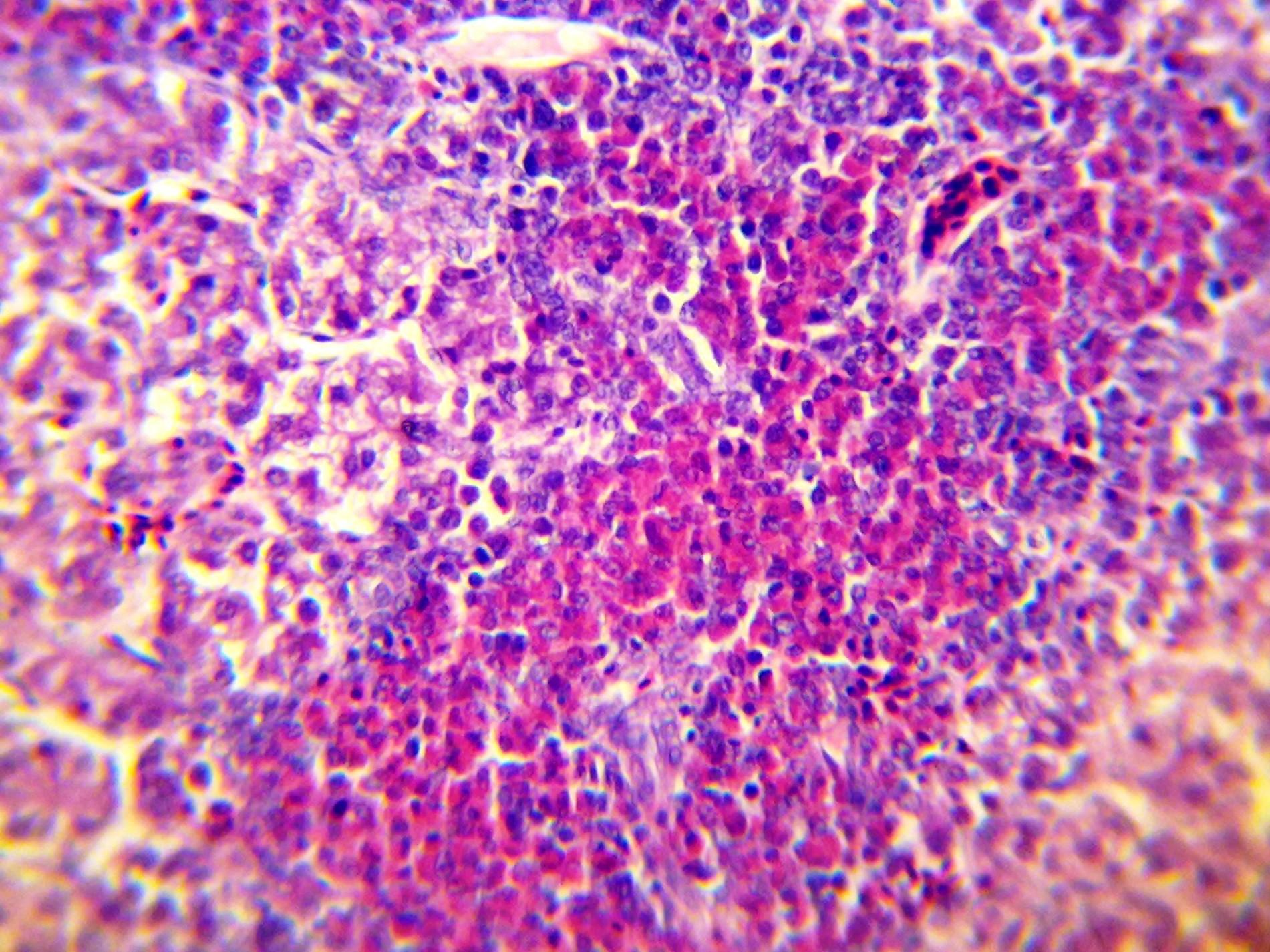
Acute and subacute cases, multifocyal necrosis of hepatocytes with accumulation of fibrin and infiltration of heterophils, periportal infiltration of heterophils and a few lymphocytes and plasma cells.
Acute and subacute cases, multifocyal necrosis of hepatocytes with accumulation of fibrin and infiltration of heterophils, periportal infiltration of heterophils and a few lymphocytes and plasma cells.
Spleen has severed congestion or fibrin exudats in acute cases and hyperplastic mononuclear phagocytic system in later stages.
Spleen has severed congestion or fibrin exudats in acute cases and hyperplastic mononuclear phagocytic system in later stages.
The ceca in young chicks may have extensive necrosis of mucosa and submucosal with accumulation of necrotic debris with fibrin and heterophils in the lumen.
The ceca in young chicks may have extensive necrosis of mucosa and submucosal with accumulation of necrotic debris with fibrin and heterophils in the lumen.
The characteristic microscopic lesions n the heart , proventriculus and pancreas represented by necrosis infiltrated with heterophils mixed with lymphocytes and plasma cells. Later on these cells are replaced by massive numbers of uniform histiocytes (large cells with irregular vesicular nuclei and faintly staining foam eosinophilia cytoplasm). They may be arranged in solid sheets forming nodules
The characteristic microscopic lesions n the heart , proventriculus and pancreas represented by necrosis infiltrated with heterophils mixed with lymphocytes and plasma cells. Later on these cells are replaced by massive numbers of uniform histiocytes (large cells with irregular vesicular nuclei and faintly staining foam eosinophilia cytoplasm). They may be arranged in solid sheets forming nodules
Serositis of various organs (pericardium, synovoium and serosa of the intestine) characterized by fibtin and heterophils and in late stage only lymphiocytes, plasma cells and histiocytes.
Serositis of various organs (pericardium, synovoium and serosa of the intestine) characterized by fibtin and heterophils and in late stage only lymphiocytes, plasma cells and histiocytes.
Ovarian lesions varied from acute fibrinosuppurative inflammation to severe pygoranuloma.
Ovarian lesions varied from acute fibrinosuppurative inflammation to severe pygoranuloma.
Less common lesions as catarrhal bronchitis, catarrhal enteritis and interstitial inflammation of lungs and kidneys.
Less common lesions as catarrhal bronchitis, catarrhal enteritis and interstitial inflammation of lungs and kidneys.
Immunohistochemical findings
Immunohistochemical findings
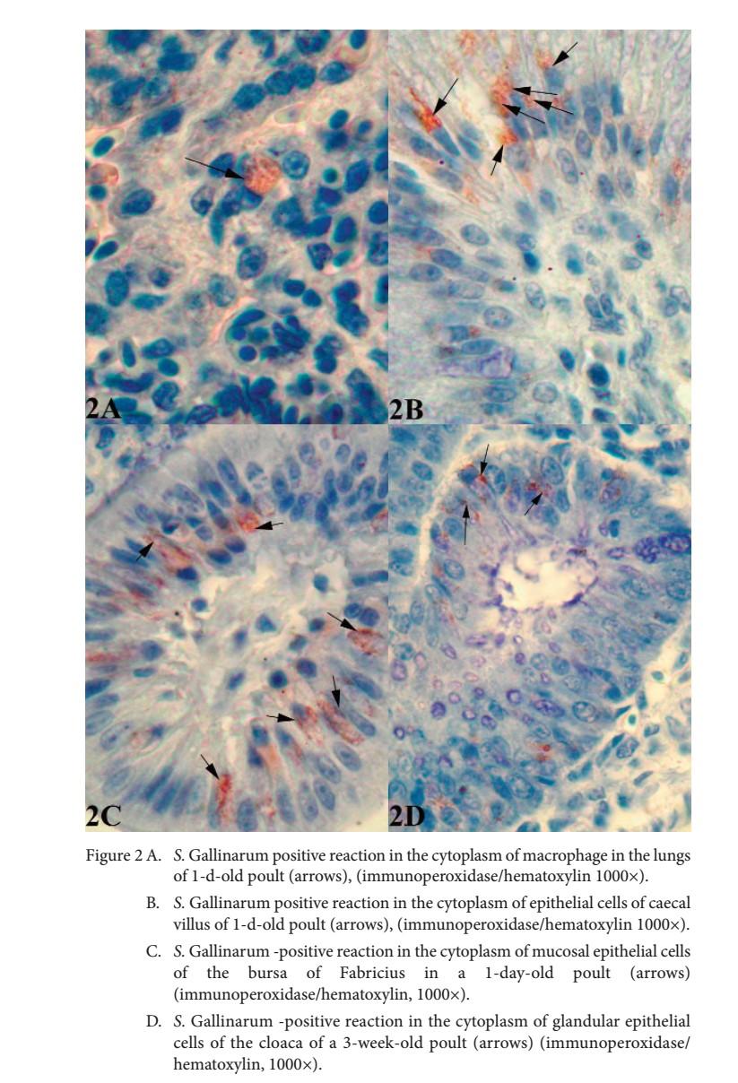
The bacterial antigen of S. Gallinarum was detected in the macrophages of the propria mucosa of the bronchi and in the epithelium of the mesobronchi
Strong immunoreactivity was noted in the cytoplasm of epithelial cells of the mucosa of the ileum, caecum and bursa of Fabricius, while immunolocalization was weak inside and outside of necrotic cells located in the propria mucosa of these tissue samples on days 4-10.
The bacterial antigen of S. Gallinarum was detected in the macrophages of the propria mucosa of the bronchi and in the epithelium of the mesobronchi Strong immunoreactivity was noted in the cytoplasm of epithelial cells of the mucosa of the ileum, caecum and bursa of Fabricius, while immunolocalization was weak inside and outside of necrotic cells located in the propria mucosa of these tissue samples on days 4-10.
PULLORUM DISEASE
susceptible species
Chicken and turkeys, S. pullorum is a specific bacteria but it can affect other bird species.
Distribution
Worldwide, nowadays it seems to be absent from the Pacific region.
Clinical signs
•The incubation period is usually a few days. The disease affects almost exclusively young birds which exhibit: Peracute infection with sudden death,
•Acute infection in first few days:
• Weakness,
• Somnolence,
• Anorexia,
• Poor growth,
• Pasting of vent with chalky white excreta,
• Death.
•In older birds:
• Lethargy,
• Huddling under brooders,
• Wing droop,
• Dyspnoea.
•Growth retardation and poor feathering of survivors.
Post-mortem findings
•Gross lesions may be seen in chronic disease, but are usually absent in peracute disease. When present the following may be seen: Enlargement and congestion of liver, spleen and kidneys,
•Yolk sac retention, with yolk appearing creamy or caseous,.
•Lung and heart may have white nodules, pericardium may be thickened, with yellow or fibrinous exudate,
•Gastro-intestinal tract - may have white nodules on the gizzard, caeca, large intestinal wall.
•Caseous cores may be seen in the caeca.
•Joints may be swollen with yellow viscous fluid.
Differential diagnosis
•Fowl typhoid,
•Fowl cholera,
•Erysipelas
Transmission
From infected birds, their faeces and their eggs. Ingestion of contaminated food, water or bedding, and contact transmission; also mechanical spread by humans, wild birds, mammals, flies, and on trucks, feed sacks. May occur in newly-hatched birds due to trans-ovarial transmission.
Risk of introduction
Pullorum could be introduced by importation of live infected chicken, hatching eggs. The bacteria can also be found in poultry meat but contamination of poultry flocks through this route is at low risk.
Control / vaccines
Live and inactivated vaccines are available for fowl typhoid in some countries. If introduced control should focus on eradication of the disease through isolation and destruction of contaminated flocks, proper disposal of carcasses and disinfection of fomites.
Colibacillosis
Colibacillosis
Definition:
Definition:
It refers to any localized or systemic infection caused entirely or partly by avian pathogenic E-Coli (APEC).
It refers to any localized or systemic infection caused entirely or partly by avian pathogenic E-Coli (APEC).
Etiology:
Etiology:
APEC and other infectious agents and other predisposing factors.
APEC and other infectious agents and other predisposing factors.
Two additional species are included E-fergusonii and E- albertii
Two additional species are included E-fergusonii and E- albertii
Natural hosts:
Natural hosts:
All avian species are susceptible to colibacillosis (chickens, turkey, ducks, quails, pheasants, pigeons, ostriches,……..).
All avian species are susceptible to colibacillosis (chickens, turkey, ducks, quails, pheasants, pigeons, ostriches,……..).
Transmission:
Transmission:
Direct or indirect contact with infected birds or their drooping's (feces) are the source of infection. Mechanical transmissions by adult house fly or beet flies are considered
Direct or indirect contact with infected birds or their drooping's (feces) are the source of infection. Mechanical transmissions by adult house fly or beet flies are considered
Ovotransmission follow oophortis, salpingitis or contaminated shell by infected feces.
Ovotransmission follow oophortis, salpingitis or contaminated shell by infected feces.
Incubation period:
Incubation period:
Generally 5-7 days in natural outbreak, and 1-3 day in experimental infection.
Generally 5-7 days in natural outbreak, and 1-3 day in experimental infection.
Localised or systemic infections and syndromes caused by avian pathogenic E. coli (Nolan et al., 2013)
Localised infections
Systemic infections
Colisepticaemia sequelae
Coliform omphalitis / yolk sac infection
Coliform cellulitis (inflammatory process)
Swollen head syndrome
Diarrhoeal disease
Venereal colibacillosis (acute vaginitis
Coliform salpingitis / perotonitis
Coliform orchitis / epididymitis
Colisepticaemia
Haemorrhagic septicaemia
Coligranuloma (Hjarre's disease)
Meningitis
Encephalitis
Panphthalmitis
Osteomyelitis
Synovitis
Clinical signs:
Clinical signs:
Clinical signs vary from inapparent to total unresponsiveness just prior to death depending on the specific type of disease produced by E. coli.
Clinical signs vary from inapparent to total unresponsiveness just prior to death depending on the specific type of disease produced by E. coli.
Localized infections result in fewer and milder clinical signs than systemic diseases. Also retained yolk sac with inspissated exudate could be seen.
Localized infections result in fewer and milder clinical signs than systemic diseases. Also retained yolk sac with inspissated exudate could be seen.
Morbidity and mortality:
Morbidity and mortality:
Both are highly variable depending on the type of the disease produced by E. coli.
Both are highly variable depending on the type of the disease produced by E. coli.
Pathology
Pathology
Several forms (diseases) of colibacillosis either localized or systemic infections are included.
Several forms (diseases) of colibacillosis either localized or systemic infections are included.
I-Localized forms of coli bacillosis
I-Localized forms of coli bacillosis
1-Coliform omphalitis (yolk sac infection)
1-Coliform omphalitis (yolk sac infection)
Due to fecal contamination of unhealed navel with APEC.
Due to fecal contamination of unhealed navel with APEC. It characterized by omphilits and yolk saculitis (mushy chicken disease)
It characterized by omphilits and yolk saculitis (mushy chicken disease)
Some embryos may die before hatching, particularly late in incubation. Wheres others die at or shortly after hatching.
Some embryos may die before hatching, particularly late in incubation. Wheres others die at or shortly after hatching.
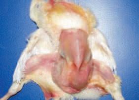
The ambilicus show swelling, edema, redness and possibly small abscesses (acute inflammation of navel) .The abdomen is often distended with hyperemic blood vessels.
In severe cases, the body wall and overlying skin undergo lysis and are wet and dirty (mushy chicks or poultry)
The ambilicus show swelling, edema, redness and possibly small abscesses (acute inflammation of navel) .The abdomen is often distended with hyperemic blood vessels. In severe cases, the body wall and overlying skin undergo lysis and are wet and dirty (mushy chicks or poultry)
Nonspecific lesions as dehydration, visceral gout, emaciation, vent pasting and enlarged gall bladder may be seen. Yolk sac is distended (unabsorbed yolk) with abnormal color, consistency and smell. Infected birds and those living more than 4 days also may have peritonitis, pericarditis or per hepatitis indicating local and systemic spread forms the yolk sac.
Nonspecific lesions as dehydration, visceral gout, emaciation, vent pasting and enlarged gall bladder may be seen. Yolk sac is distended (unabsorbed yolk) with abnormal color, consistency and smell. Infected birds and those living more than 4 days also may have peritonitis, pericarditis or per hepatitis indicating local and systemic spread forms the yolk sac.
Microscopic picture:
Microscopic picture:
The well of the infected yolk sac is edematous with mild inflammation. There is an outer C.T zone adjacent to a layer of inflammatory cells containing heterophils and macrophages, a layer of giant cells, a zone of necrotic heterophils and mass of bacteria.
The well of the infected yolk sac is edematous with mild inflammation. There is an outer C.T zone adjacent to a layer of inflammatory cells containing heterophils and macrophages, a layer of giant cells, a zone of necrotic heterophils and mass of bacteria.
2- Coliform cellulitis:
2- Coliform cellulitis:
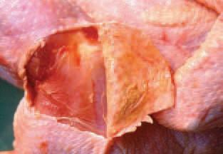
It characterized by sheets of serosanguinous to caseated, fibrinoheterophilic exudation in subcutinous tissues.
It characterized by sheets of serosanguinous to caseated, fibrinoheterophilic exudation in subcutinous tissues.
These plaques lesions are located in the skin over the abdomen or between the thigh and midline.
These plaques lesions are located in the skin over the abdomen or between the thigh and midline.
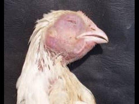
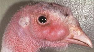
3- Swollen head syndrome (SHS):
3- Swollen head syndrome (SHS):
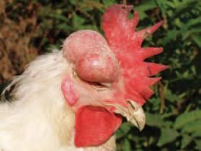
An acute or subacute cellulitis involving the periorbital and adjacent subcutaneous tissues of the head.
An acute or subacute cellulitis involving the periorbital and adjacent subcutaneous tissues of the head.
Swelling of the head is caused by inflammatory exudate beneath the skin.
Swelling of the head is caused by inflammatory exudate beneath the skin.
Microscopic lesions:
Microscopic lesions:
They include fibrino heterophilic inflammation and heterophilic granuloma in the spaces of the cranial bones, middle ear and facial skin. Lymphoplasmacyt conjunctivitis and tracheitis with formation of germinal centers also have been observed.
They include fibrino heterophilic inflammation and heterophilic granuloma in the spaces of the cranial bones, middle ear and facial skin. Lymphoplasmacyt conjunctivitis and tracheitis with formation of germinal centers also have been observed.
4- Diarrheal disease: primary enteritis due to E coli is in poultry.
4- Diarrheal disease: primary enteritis due to E coli is in poultry.
5- Venereal coli bacillosis (acute vaginitis): it is an acute and frequnitly fatal vaginitis that affects turkey breeder hens shortly after they are first inseminated. It characterized by vaginitis, cloacal and intestinal prolapse, peritonitis, egg binding and internal laying.
5- Venereal coli bacillosis (acute vaginitis): it is an acute and frequnitly fatal vaginitis that affects turkey breeder hens shortly after they are first inseminated. It characterized by vaginitis, cloacal and intestinal prolapse, peritonitis, egg binding and internal laying.
Egg binding is a common reproductive problem that causes the bird to retain the egg in the reproductive tract, unable to expel it naturally. The affected mucosa is markedly thickened, ulcerated and covered with a diphtheritic (Caseo necrotic) membrane, which causes obstruction of the lower reproductive tract. The upper oviduct appears normal.
Egg binding is a common reproductive problem that causes the bird to retain the egg in the reproductive tract, unable to expel it naturally. The affected mucosa is markedly thickened, ulcerated and covered with a diphtheritic (Caseo necrotic) membrane, which causes obstruction of the lower reproductive tract. The upper oviduct appears normal.
6- Coliform salpingitis / peritonitis (adult)
6- Coliform salpingitis / peritonitis (adult)
It is one of causes of mortality and decrease egg production in layers and breeders following ascending infection, coagulated yolk inside the body cavity (yolk or egg peritonitis) a companied by marked exudation and extensive inflammation.Salpingitis and egg binding may occur (obstructive mass within the oviduct).
It is one of causes of mortality and decrease egg production in layers and breeders following ascending infection, coagulated yolk inside the body cavity (yolk or egg peritonitis) a companied by marked exudation and extensive inflammation.Salpingitis and egg binding may occur (obstructive mass within the oviduct).
7- Coliform orchitis / epididymitis:
7- Coliform orchitis / epididymitis:
An ascending infection of male reproductive tract by E- coli. Testicles are swollen, firm, inflamed, irregularly shaped and have a mosaic of necrotic and viable tissue when opened.
An ascending infection of male reproductive tract by E- coli. Testicles are swollen, firm, inflamed, irregularly shaped and have a mosaic of necrotic and viable tissue when opened.
II)- Systemic forms of colibacellosis:
II)- Systemic forms of colibacellosis:
Colisepticemia: It progresses through the following stages: Acute septicemia ,Subacute polyserositis .Chronic granulomation inflammation
Colisepticemia: It progresses through the following stages: Acute septicemia ,Subacute polyserositis .Chronic granulomation inflammation
Acute septicemia:
Acute septicemia:
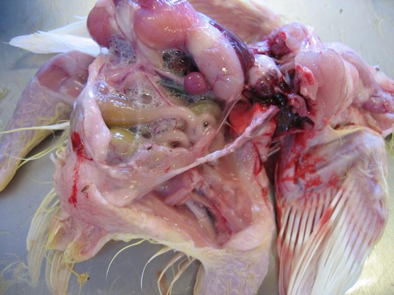
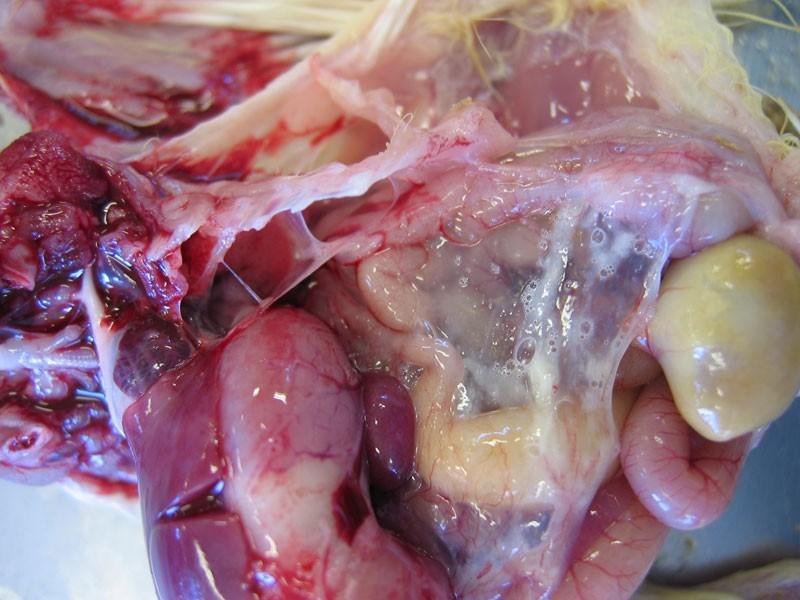
The characteristic features include a green discoloration of the liver following exposure to air and a characteristic odor (indol product by bacteria). Pericarditis is a characteristic lesions associated with myocarditis (with adhesions)
The characteristic features include a green discoloration of the liver following exposure to air and a characteristic odor (indol product by bacteria). Pericarditis is a characteristic lesions associated with myocarditis (with adhesions)
1-Respiratory- organ colisepticemia: it follows upper respiratory viral infection or vaccination or mycoplasma and ammonia (predisposing factors)
1-Respiratory- organ colisepticemia: it follows upper respiratory viral infection or vaccination or mycoplasma and ammonia (predisposing factors)
- It characterized by air sac disease or CRD -Lesions are prominent in respiratory tissues (trachea, lungs and air sacs),
pericardial sac and peritoneal cavities (subacute polyserositis). Infected air sacs thickened by caseous exudate and pluropnemonia.
- It characterized by air sac disease or CRD -Lesions are prominent in respiratory tissues (trachea, lungs and air sacs), pericardial sac and peritoneal cavities (subacute polyserositis). Infected air sacs thickened by caseous exudate and pluropnemonia.
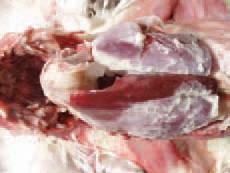
2-Enteric-organ colisepticemia: it is common in turkeys. lesions are typical of acute septicemic stage .The most characteristic lesions are: green discoloration of liver, enlarged and congested spleen and congested muscle.
2-Enteric-organ colisepticemia: it is common in turkeys. lesions are typical of acute septicemic stage .The most characteristic lesions are: green discoloration of liver, enlarged and congested spleen and congested muscle.
3-Haemorrhagic septicemia: it occurs in turkeys, it characterized by circulatory disorder, generalized necrosis and enlarged liner spleen, kidneys.
3-Haemorrhagic septicemia: it occurs in turkeys, it characterized by circulatory disorder, generalized necrosis and enlarged liner spleen, kidneys.
4-Neonatal colisepticemia:
4-Neonatal colisepticemia: Baby chicks after hatching ,Initial lesions (congested lungs, edematous serous membranes, splenomegaly). Lateron, proventriculus and lungs develop a dark color.
Baby chicks after hatching ,Initial lesions (congested lungs, edematous serous membranes, splenomegaly). Lateron, proventriculus and lungs develop a dark color.
5-Layer colisepticemia:
5-Layer colisepticemia:
It is usually a disease of young birds but outbreaks occurs in mature chickens and turkeys
It is usually a disease of young birds but outbreaks occurs in mature chickens and turkeys
It characterized by (sudden death, poly serositis or perihepatitis/ pericarditis) and peritonitis with free yolk in the peritoneal cavity.
It characterized by (sudden death, poly serositis or perihepatitis/ pericarditis) and peritonitis with free yolk in the peritoneal cavity.
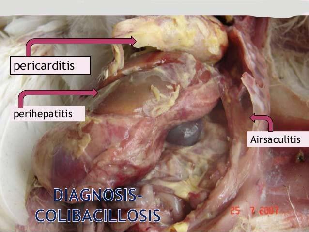
6-Coliform septicemia: It characterized by Pericarditis, perihepatits and air saculaitis.
6-Coliform septicemia: It characterized by Pericarditis, perihepatits and air saculaitis.
A- characterisitic odor at necropsy ,Swollen liver and spleen
A- characterisitic odor at necropsy ,Swollen liver and spleen
B. Meningoencephalitis: it is uncommon
B. Meningoencephalitis: it is uncommon
C. Panophthalmitis: it is un common
C. Panophthalmitis: it is un common
d. Osteoarthritis and synovitis: it is a common sequel of colisepticemia. It characterized by (osteomyelitis and polyarthritis). It results in lameness and poor growth.
d. Osteoarthritis and synovitis: it is a common sequel of colisepticemia. It characterized by (osteomyelitis and polyarthritis). It results in lameness and poor growth.
E. Coligranuloma (Hjarres disease): it is common in chickens and turkeys. It characterized by multiple granulomas in liver, ceca, duodenum and mesentery but not spleen.
E. Coligranuloma (Hjarres disease): it is common in chickens and turkeys. It characterized by multiple granulomas in liver, ceca, duodenum and mesentery but not spleen.
Microscopically:
Microscopically:
Heterophilic granuloma is present in the affected tissue (pyogranulomatous typhlitis and hepatitis).
Heterophilic granuloma is present in the affected tissue (pyogranulomatous typhlitis and hepatitis).
The lesions that develop in the articular spaces of thoracolumbar
The lesions that develop in the articular spaces of thoracolumbar
vertebrae result in spondylitis (spondylosis) and after that, in progressive paresis and paralysis.
vertebrae result in spondylitis (spondylosis) and after that, in progressive paresis and paralysis.
Arthritis, Osteomyelitis and Osteonecrosis (inflammation of joints, bone marrow and bone necrosis, respectively). The lesions are a common sequel to E. coli septicaemia. Clinically, lameness, prolonged lying down, dehydration and retarded growth rate are observed. The coxofemoral joints, the femur and tibiotarsal joints are most commonly affected. The bacteria colonize the physes of growing bones and provoke an inflammatory response that is further causing osteomyelitis. Pathoanatomically, fractures of the femoral head are usually discovered.
Arthritis, Osteomyelitis and Osteonecrosis (inflammation of joints, bone marrow and bone necrosis, respectively). The lesions are a common sequel to E. coli septicaemia. Clinically, lameness, prolonged lying down, dehydration and retarded growth rate are observed. The coxofemoral joints, the femur and tibiotarsal joints are most commonly affected. The bacteria colonize the physes of growing bones and provoke an inflammatory response that is further causing osteomyelitis. Pathoanatomically, fractures of the femoral head are usually discovered.
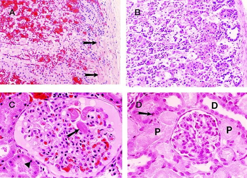
prominent microthrombus formation in the subcapsular vessels (arrows) of the adrenals (A) and also in the renal glomeruli (arrow) (C). Note the extensivecorticalhemorrhagein the adrenal gland (A) and the early ischemic/necrobiotic changes (karyopyknosis) in the proximal tubular epithelial cells (triangle) of the kidney (C). In B and D, the animals that had relatively long survival, widespread necrotic foci were apparent with associated polymorphonuclear leukocyte infiltrates in the adrenal cortex (B) and severe acute tubular necrosis involving the proximal tubules (P) of the kidney (D).
prominent microthrombus formation in the subcapsular vessels (arrows) of the adrenals (A) and also in the renal glomeruli (arrow) (C). Note the extensivecorticalhemorrhagein the adrenal gland (A) and the early ischemic/necrobiotic changes (karyopyknosis) in the proximal tubular epithelial cells (triangle) of the kidney (C). In B and D, the animals that had relatively long survival, widespread necrotic foci were apparent with associated polymorphonuclear leukocyte infiltrates in the adrenal cortex (B) and severe acute tubular necrosis involving the proximal tubules (P) of the kidney (D).
Thedistaltubules(D)werewellpreserved. Occasional mitotic figures of the proximal tubular epithelial cells are also seen as featuresof regeneration(arrow) (D). Note the lack of thrombi and hemorrhage in the “late” group(BandD).
Thedistaltubules(D)werewellpreserved. Occasional mitotic figures of the proximal tubular epithelial cells are also seen as featuresof regeneration(arrow) (D). Note the lack of thrombi and hemorrhage in the “late” group(BandD).
Colibacillosis, Colisepticemia
Colibacillosis, Colisepticemia
Introduction
Introduction
Colisepticaemia is the commonest infectious disease of farmed poultry. It is most commonly seen following upper respiratory disease (such as Infectious Bronchitis) or Mycoplasmosis. It is frequently associated with immunosuppressive diseases such as Infectious Bursal Disease Virus (Gumboro Disease) in chickens or Haemorrhagic Enteritis in turkeys, or in young birds that are immunologically immature. It is caused by the bacterium Escherichia coli and is seen worldwide in chickens, turkeys, etc.
Colisepticaemia is the commonest infectious disease of farmed poultry. It is most commonly seen following upper respiratory disease (such as Infectious Bronchitis) or Mycoplasmosis. It is frequently associated with immunosuppressive diseases such as Infectious Bursal Disease Virus (Gumboro Disease) in chickens or Haemorrhagic Enteritis in turkeys, or in young birds that are immunologically immature. It is caused by the bacterium Escherichia coli and is seen worldwide in chickens, turkeys, etc.
Morbidity varies, mortality is 5-20%. The infectious agent is moderately resistant in the environment, but is susceptible to disinfectants and to temperatures of 80°C. Infection is by the oral or inhalation routes, and via shell membranes/yolk/navel, water, fomites, with an incubation period of 3-5 days. Poor navel healing, mucosal damage due to viral infections and immunosuppression are predisposing factors. Signs
Morbidity varies, mortality is 5-20%. The infectious agent is moderately resistant in the environment, but is susceptible to disinfectants and to temperatures of 80°C. Infection is by the oral or inhalation routes, and via shell membranes/yolk/navel, water, fomites, with an incubation period of 3-5 days. Poor navel healing, mucosal damage due to viral infections and immunosuppression are predisposing factors. Signs
•Respiratory signs, coughing, sneezing.
•Respiratory signs, coughing, sneezing.
•Snick.
•Snick.
•Dejection.
•Dejection.
•Reduced appetite.
•Reduced appetite.
•Poor growth.
•Poor growth.
•Omphalitis.
•Omphalitis.
post-mortem lesions
post-mortem lesions
Airsacculitis.
Airsacculitis.
Pericarditis.
Pericarditis.
Perihepatitis.
Perihepatitis.
Swollen liver and spleen.
Swollen liver and spleen.
Peritonitis.
Peritonitis.
Salpingitis.
Salpingitis.
Omphalitis.
Omphalitis.
Synovitis.
Synovitis.
Arthritis.
Arthritis.
Enteritis.
Enteritis.
Granulomata in liver and spleen.
Granulomata in liver and spleen.
Cellulitis over the abdomen or in the leg.
Cellulitis over the abdomen or in the leg.
Lesions vary from acute to chronic in the various forms of the disease.
Lesions vary from acute to chronic in the various forms of the disease.
Diagnosis
Diagnosis
Isolation, sero-typing, pathology. Aerobic culture yields colonies of 2-5mm on both blood and McConkey agar after 18 hours - most strains are rapidly lactose-fermenting producing brickred colonies on McConkey agar.
Isolation, sero-typing, pathology. Aerobic culture yields colonies of 2-5mm on both blood and McConkey agar after 18 hours - most strains are rapidly lactose-fermenting producing brickred colonies on McConkey agar.
Differentiate from acute and chronic infections with Salmonella spp, other enterobacteria such as Proteus, as well as Pseudomonas, Staphylococcus spp. etc.
Differentiate from acute and chronic infections with Salmonella spp, other enterobacteria such as Proteus, as well as Pseudomonas, Staphylococcus spp. etc.
Treatment
Treatment
Amoxycillin, tetracyclines, neomycin (intestinal activity only), gentamycin or ceftiofur (where hatchery borne), potentiated sulphonamide, flouroquinolones.
Amoxycillin, tetracyclines, neomycin (intestinal activity only), gentamycin or ceftiofur (where hatchery borne), potentiated sulphonamide, flouroquinolones.
Escherichia coli, a gram-negative bacillus that is part of the intestinal flora, is also an important opportunistic pathogen, causing diarrhea and dysentery, urinary tract infection, pneumonia and neonatal meningitis. Escherichia coli causes various patterns of human enteric diseases caused by different varieties of Escherichia coli. Some of these are - Enterotoxigenic ; Enteroinvasive ;
Enteroadherent ; Enterohemorrhagic. Enterotoxigenic Escherichia coli causes diarrheal disease by elaborating two plasmid-mediated enterotoxins.
Escherichia coli, a gram-negative bacillus that is part of the intestinal flora, is also an important opportunistic pathogen, causing diarrhea and dysentery, urinary tract infection, pneumonia and neonatal meningitis. Escherichia coli causes various patterns of human enteric diseases caused by different varieties of Escherichia coli. Some of these are - Enterotoxigenic ; Enteroinvasive ; Enteroadherent ; Enterohemorrhagic. Enterotoxigenic Escherichia coli causes diarrheal disease by elaborating two plasmid-mediated enterotoxins.
The heat-labile toxin is antigenically, structurally and functionally related to the cholera toxin, although the toxin of Escherichia coli is less potent than that of cholera. The heat-labile toxin also binds to DM1 ganglioside on the intestinal epithelial cells. As in cholera, resulting activation of adenyl cyclase produces a hypersecretory diarrhea.The heat-stable toxin of Escherichia coli is different from cholera toxin and apparently acts to impair sodium and chloride absorption and to reduce the motility of small intestine through a mechanism dependent on cyclic GMP. Dehydration and electrolyte imbalance is a significant cause of morbidity and mortality when appropriate rehydration is lacking.
The heat-labile toxin is antigenically, structurally and functionally related to the cholera toxin, although the toxin of Escherichia coli is less potent than that of cholera. The heat-labile toxin also binds to DM1 ganglioside on the intestinal epithelial cells. As in cholera, resulting activation of adenyl cyclase produces a hypersecretory diarrhea.The heat-stable toxin of Escherichia coli is different from cholera toxin and apparently acts to impair sodium and chloride absorption and to reduce the motility of small intestine through a mechanism dependent on cyclic GMP. Dehydration and electrolyte imbalance is a significant cause of morbidity and mortality when appropriate rehydration is lacking.
Enterotoxigenic Escherichia coli is also responsible for 50% of traveller’s diarrhea.
Enterotoxigenic Escherichia coli is also responsible for 50% of traveller’s diarrhea.
Enteroinvasive Escherichia coli produces a dysentery-like disease resembling shigellosis , although it is less severe and requires a much larger infecting dose of organisms. Enteroinvasive Escherichia coli invades the intestinal mucosa and causes local tissue destruction and sloughing of necrotic mucosa. Bloody mucoid stools contain neutrophils.
Enteroinvasive Escherichia coli produces a dysentery-like disease resembling shigellosis , although it is less severe and requires a much larger infecting dose of organisms. Enteroinvasive Escherichia coli invades the intestinal mucosa and causes local tissue destruction and sloughing of necrotic mucosa. Bloody mucoid stools contain neutrophils.
Enteroadhesive Escherichia coli has been associated with diarrheal diseases.
Enteroadhesiveness is plasmid-dependent and is apparently mediated by pili, which bind tightly to receptors of the intestinal epithelial cells.
Enteroadhesive Escherichia coli has been associated with diarrheal diseases. Enteroadhesiveness is plasmid-dependent and is apparently mediated by pili, which bind tightly to receptors of the intestinal epithelial cells.
The mechanism of diarrhea is unknown. The most common strain of enterohemorrhagic Escherichia coli is 0157:H7. This organism adheres to intestinal epithelial cells and produces a cytotoxin similar to that produced by Shigella dysenteriae.
The mechanism of diarrhea is unknown. The most common strain of enterohemorrhagic Escherichia coli is 0157:H7. This organism adheres to intestinal epithelial cells and produces a cytotoxin similar to that produced by Shigella dysenteriae.
Contaminated meat is the most frequent mode of transmission, but infection may also occur through contaminated water, milk, produce, and person-to-person contact.
The symptoms include bloody diarrhea with severe abdominal cramps and mild or no fever. Non bloody, watery diarrhea may occur. Some patients have fecal leukocytes.
Contaminated meat is the most frequent mode of transmission, but infection may also occur through contaminated water, milk, produce, and person-to-person contact. The symptoms include bloody diarrhea with severe abdominal cramps and mild or no fever. Non bloody, watery diarrhea may occur. Some patients have fecal leukocytes.
Patients are at risk for the hemolytic-uremic syndrome or thrombotic thrombocytopenic purpura. Children and elderly patients are at particular risk of serious illness.
Patients are at risk for the hemolytic-uremic syndrome or thrombotic thrombocytopenic purpura. Children and elderly patients are at particular risk of serious illness.
Patients may have colonic edema, erosions, ulcers, and hemorrhage, and the right colon is usually more severely affected. The lamina propria and submucosa contain marked edema and hemorrhage, with associated mucosal acute inflammation, cryptitis, crypt abscesses, ulceration, and necrosis. Microthrombi may be seen within small vessels, and pseudomembranes are occasionally present. About 80% of all infections of the urinary tract in humans, ranging from mild cystitis to fatal pyelonephritis, are caused by Escherichia coli. In addition, Escherichia coli is the etiologic agent in many caes of nosocomial pneumonia, most often in elderly patients with underlying chronic disease. Aspirates of endogenous oral flora containing Escherichia coli appear to be the cause of this bronchopneumonia , although in bacteremic patients pneumonia may result from seeding by septic emboli. Empyema is a common complication, especially in patients with disease lasting more than a week. Only rarely does Escherichia coli cause meningitis in adults, but it is a major cause of neonatal meningitis.
Patients may have colonic edema, erosions, ulcers, and hemorrhage, and the right colon is usually more severely affected. The lamina propria and submucosa contain marked edema and hemorrhage, with associated mucosal acute inflammation, cryptitis, crypt abscesses, ulceration, and necrosis. Microthrombi may be seen within small vessels, and pseudomembranes are occasionally present. About 80% of all infections of the urinary tract in humans, ranging from mild cystitis to fatal pyelonephritis, are caused by Escherichia coli. In addition, Escherichia coli is the etiologic agent in many caes of nosocomial pneumonia, most often in elderly patients with underlying chronic disease. Aspirates of endogenous oral flora containing Escherichia coli appear to be the cause of this bronchopneumonia , although in bacteremic patients pneumonia may result from seeding by septic emboli. Empyema is a common complication, especially in patients with disease lasting more than a week. Only rarely does Escherichia coli cause meningitis in adults, but it is a major cause of neonatal meningitis.
Between 40% and 80% of infants with Escherichia coli meningitis die, and the survivors frequently suffer from neurologic or developmental anomalies.
Between 40% and 80% of infants with Escherichia coli meningitis die, and the survivors frequently suffer from neurologic or developmental anomalies.
Microscopic and immuno-h istochemical findings: In the microscopic examination, the presence of necrosis or hyperplasia in the epithelium of the bronchi was seen. The presence of heterophils, lymphocytes, macrophages, desquamated epithelial cells, cellular deposits composed of erythrocytes and mucus in the lumen was seen. Also, similar cells were seen in the interlobular septa and the pleura. Furthermore, lymphoid hyperplasia was noticed in the periphery of blood vessels and in secondary & tertiary bronchi (Fig.1a, b). In the liver, marked degenerative alterations of the hepatocytes were seen, while some were observed as necrotic. Furthermore, similar inflammatory cells were present in parenchyma (Fig. 2c).
Microscopic and immuno-h istochemical findings: In the microscopic examination, the presence of necrosis or hyperplasia in the epithelium of the bronchi was seen. The presence of heterophils, lymphocytes, macrophages, desquamated epithelial cells, cellular deposits composed of erythrocytes and mucus in the lumen was seen. Also, similar cells were seen in the interlobular septa and the pleura. Furthermore, lymphoid hyperplasia was noticed in the periphery of blood vessels and in secondary & tertiary bronchi (Fig.1a, b). In the liver, marked degenerative alterations of the hepatocytes were seen, while some were observed as necrotic. Furthermore, similar inflammatory cells were present in parenchyma (Fig. 2c).
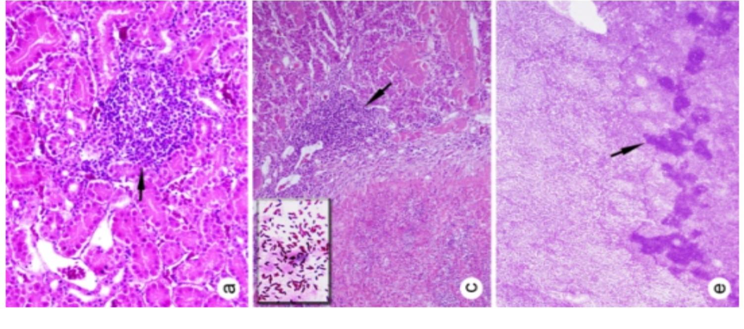
In the kidney, blood vessels were hyperemic, tubular epithelial cells were characterized by degenerative alterations and some were observed necrotic. In some birds, heterophils, lymphocytes and macrophages were seen in the interstitial area (Fig. 2a). The heart, blood vessels were hyperemic and heart muscle cells were degenerative. In a few cases, multifocal coagulation necrosis was present in the spleen (Fig. 2e). In addition, in only three birds, granulomatous foci were localized in the liver and lung.
In the kidney, blood vessels were hyperemic, tubular epithelial cells were characterized by degenerative alterations and some were observed necrotic. In some birds, heterophils, lymphocytes and macrophages were seen in the interstitial area (Fig. 2a). The heart, blood vessels were hyperemic and heart muscle cells were degenerative. In a few cases, multifocal coagulation necrosis was present in the spleen (Fig. 2e). In addition, in only three birds, granulomatous foci were localized in the liver and lung.
Apoptotic cells were seen in all of the tissues. The number of apoptotic cells increased as the tissue damage increased.
Apoptotic cells were seen in all of the tissues. The number of apoptotic cells increased as the tissue damage increased.
Apoptosis was observed in all tissues including bronchial epithelial cells in lungs (Fig.1c, d), hepatocytes in liver, (Fig. 2d), tubular epithelial cells and glomerulus in kidneys (Fig. 2b), myocytes in heart, white and red pulp in spleen (Fig. 2f), endothelial cells in vessels, and all inflammatory cells in varying proportions
Apoptosis was observed in all tissues including bronchial epithelial cells in lungs (Fig.1c, d), hepatocytes in liver, (Fig. 2d), tubular epithelial cells and glomerulus in kidneys (Fig. 2b), myocytes in heart, white and red pulp in spleen (Fig. 2f), endothelial cells in vessels, and all inflammatory cells in varying proportions
Fig. 1: Photomicrograph of lungs of broilers suffering from colibacillosis. a) Lymphoid hyperplasia and inflammation in bronchi (arrow), b) Inflammatory cells in the interlobular septa (arrow) and c-d) Apoptosis in bronchial epithelial cells (arrow). Stain: a & b: H&E; c & d: TUNEL; X20.
Fig. 2: Photomicrograph of various organs of broilers suffering from colibacillosis. a)
Inflammatory cells in the interstitial area (arrow), b) Apoptosis in tubular epithelial cells and glomerulus (arrow), c) Necrosis and inflammatory cells in the liver (arrow).
Above small plate: E coli (Gram stain, X 100), d) Apoptosis in the liver (arrow), e)
Necrosis in the spleen (arrow) and f) Apoptosis in the spleen (arrow). Stain: a, c & e: H&E; b, d & f: TUNEL; X20.
Pasteurellosis
Pasteurellosis
Fowl cholera
Fowl cholera
It is a contagious disease affecting domesticated and wild birds. It usually appears as a septicemic disease associated with high morbidity and mortality but chronic or benign conditions often occur.
It is a contagious disease affecting domesticated and wild birds. It usually appears as a septicemic disease associated with high morbidity and mortality but chronic or benign conditions often occur.
Etiology:
Etiology:
Pasteurella multocida (Pasteurella aviseptica) Endotoxins are produced by all P. multocida both virulent and nonvirulent. It is more prevalent in late summer, fall and winter.
Pasteurella multocida (Pasteurella aviseptica) Endotoxins are produced by all P. multocida both virulent and nonvirulent. It is more prevalent in late summer, fall and winter.
Natural hosts:
Natural hosts:
Outbreaks of FC are common in chickens, turkeys, ducks and geese. Also game birds, companion birds, birds in zoo and wild birds are affected.
Outbreaks of FC are common in chickens, turkeys, ducks and geese. Also game birds, companion birds, birds in zoo and wild birds are affected.
Transmission:
Transmission:
Horizontally infected birds are considered to be a major source of infection.
Horizontally infected birds are considered to be a major source of infection.
Free-flying birds have a role of dissemination of infection.
Free-flying birds have a role of dissemination of infection.
Clinical signs:
Clinical signs:
A-Acute:
A-Acute:
Sudden deaths.Some birds may develop fever, anorexia, ruffled feathers, mucus discharge from the mouth, diarrhea and increase respiratory rate. Cyanosis of unfeathers areas of the head (comb and wattles) immediately prior to death.
Sudden deaths.Some birds may develop fever, anorexia, ruffled feathers, mucus discharge from the mouth, diarrhea and increase respiratory rate. Cyanosis of unfeathers areas of the head (comb and wattles) immediately prior to death.
Feces initially watery and whitish in color later became greenish and contain mucus
Feces initially watery and whitish in color later became greenish and contain mucus
Some birds survived and become emaciated, dehydrated and become chronically infected.
Some birds survived and become emaciated, dehydrated and become chronically infected.
Fowl cholera is an infectious disease in domestic fowl, waterfowl and other avian species. It is manifested either in acute septicaemic form with a high morbidity and death rates or as chronic local forms (independently or secondary to acute ones). Acute fowl cholera. The sudden and unexpected death could be the first sign of the disease. In this form, the lesions are predominantly related to vascular injuries.
ommonly observed signs are anorexia, ruffled feathers, oral and nasal mucus discharge, cyanosis and white or greenish watery mucoid diarrhoea.
Frequently, subserous petechial or ecchymosed haemorrhages in the anterior part of the small intestine, the gizzard or the abdominal fat are discovered.
Chronic fowl cholera. It is characterized by local inflammations. The periorbital sinuses are frequently affected by a serofibrinous inflammation.
Another local form is the injury of wattles that are strongly distended because of their filling with fibrinous caseous content. The flocks that recuperated ( تفاعت)from fowl cholera continue to carry and shed Pasteurella multocida. The carriers store the organism in nasal choanas and contaminate the forage, water and the environment with oral discharges. Wild birds and some mammals (swine) could also carry the agent and introduce it into poultry flocks. Cannibalism is an essential route of spreading the infection.
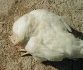
possibly be spread from sinuses to adjacent air-filled skull bones with subsequent necrosis and onset of neurological signs (opisthotonus and torticolis). The diagnosis is made on the basis of disease history, clinical signs, the lesions and the results of bacteriological studies. Fowl cholera should be differentiated from acute E. coli septicaemia, erysipeloid, fowl typhoid etc. The immunization of birds at the age of 8 -12 weeks gives very promising results. Many antibiotics and sulfonamides could lower death rate, but at discontinuation of the treatment, the disease could recur. Sulfonamides are appropriate for treatment, but they inhibit egglaying.
B-Chronic:
B-Chronic:
Signs related to localized infections. Wattles, sinuses, leg or wing joints, foot pads and sternal bursae often become swollen. Exudative conjunctival and pharyngeal lesions may be observed and torticollis Tracheal rales and dyspnea may also seen.
Signs related to localized infections. Wattles, sinuses, leg or wing joints, foot pads and sternal bursae often become swollen. Exudative conjunctival and pharyngeal lesions may be observed and torticollis Tracheal rales and dyspnea may also seen.
Pathology
Pathology
Gross and microscopic:
Gross and microscopic:
Acute disease:
Acute disease:
Hyperemia and bacteremia in all veins of the abdominal viscera but pronounced in small vessels of the duodenal mucosa.
Hyperemia and bacteremia in all veins of the abdominal viscera but pronounced in small vessels of the duodenal mucosa.
Petechial and ecchymotic hemorrhages on serous membranes ,lungs and abdominal fat and intestinal mucosa. Hydro pericardium and hydroperitonum may occur. Disseminated intravascular clotting or fibrinous thrombosis. Livers of acutely affected birds May swollen and contain multiple areas of coagulative necrosis and heterophilic infiltration. Pneumonias is severe in turkeys than chickens.
Petechial and ecchymotic hemorrhages on serous membranes ,lungs and abdominal fat and intestinal mucosa. Hydro pericardium and hydroperitonum may occur. Disseminated intravascular clotting or fibrinous thrombosis. Livers of acutely affected birds May swollen and contain multiple areas of coagulative necrosis and heterophilic infiltration. Pneumonias is severe in turkeys than chickens.
Large amounts of viscid mucus in digestive tract particularly in the pharynx, crop and intestine Ovaries of laying hens contain flaccid follicles with presence of yolk
peritonitis( acute serofibrinous oophoritis)
Large amounts of viscid mucus in digestive tract particularly in the pharynx, crop and intestine Ovaries of laying hens contain flaccid follicles with presence of yolk peritonitis( acute serofibrinous oophoritis)
Chronic disease:
Chronic disease:
Localized suppurative lesions distributed in the affected birds mainly in sinuses and pneumatic bones. Pneumonia is common mainly in turkeys .Conjunctivitis and facial edema may be observed. Localized infections mainly in hock joints, foot pads, peritoneal cavity and oviduct, middle ear, cranial bones, meninges (yellowish caseous exudate in air spaces of the calvaria & bones of turkeys.Calvaria( superior portion of frontal and occipital bone)
Localized suppurative lesions distributed in the affected birds mainly in sinuses and pneumatic bones. Pneumonia is common mainly in turkeys .Conjunctivitis and facial edema may be observed. Localized infections mainly in hock joints, foot pads, peritoneal cavity and oviduct, middle ear, cranial bones, meninges (yellowish caseous exudate in air spaces of the calvaria & bones of turkeys.Calvaria( superior portion of frontal and occipital bone)
Heterophilic infiltration and fibrin were observed in air spaces, middle ear and meninges beside multinucleated giant cells and necrotic masses of heterophils. Reduction egg production (10-40%)
Heterophilic infiltration and fibrin were observed in air spaces, middle ear and meninges beside multinucleated giant cells and necrotic masses of heterophils. Reduction egg production (10-40%)
A foul old or in chronic cases which complicated by other bacteria.
A foul old or in chronic cases which complicated by other bacteria.
Pumble foot of chickens and rabbits due to staph, psudo. Or E,coli.
Infections coryza
Infections coryza
It is an acute respiratory disease of chickens caused by the bacterium known as Hemophilus paragallinarum.
It is an acute respiratory disease of chickens caused by the bacterium known as Hemophilus paragallinarum.
Etiology:
Etiology:
H. paragallinarum (avibacterium gallinarum)
H. paragallinarum (avibacterium gallinarum)
There are 9 serovars for this bacterium A( A1, A2, A3, A4) B1 and C (C1, C2, C3,C4)
There are 9 serovars for this bacterium A( A1, A2, A3, A4) B1 and C (C1, C2, C3,C4)
Natural hosts: the chicken is the natural hosts.
Natural hosts: the chicken is the natural hosts.
Transmission:
Transmission:
Chronic or healthy carrier birds have long been recognized as the main reservoir of IC infection.
Chronic or healthy carrier birds have long been recognized as the main reservoir of IC infection.
Incubation period:
Incubation period:
Short incubation period (1-3 days) with a course of about 2-3 weeks.
Short incubation period (1-3 days) with a course of about 2-3 weeks.
Clinical signs:
Clinical signs:
Acute respiratory signs in outbreaks mainly coughing and sneezing
Acute respiratory signs in outbreaks mainly coughing and sneezing
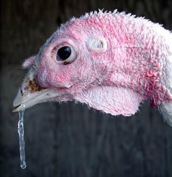
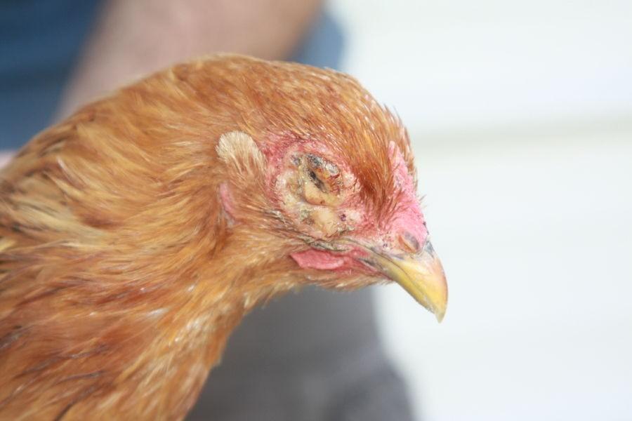
Serous to mucoiod nasal discharge .Facial edema and conjunctivitis .Swollen wattles mainly in males (swollen head syndrome) .Rales may be heard in birds.Arthritis and septicemia in broilers and layer flocks. Diarrhea, decrease feed and water in turkeys.
Serous to mucoiod nasal discharge .Facial edema and conjunctivitis .Swollen wattles mainly in males (swollen head syndrome) .Rales may be heard in birds.Arthritis and septicemia in broilers and layer flocks. Diarrhea, decrease feed and water in turkeys.
Clinically,The disease has a low mortality rate but leads to a drop in egg production up to 40 % in layer hens and increased culling in broilers and thus poses significant financial liability to chicken farmers.The disease is caused by gram negative bacteria H. paragallinarum(Avibacterium paragallinurum). The disease is characterized by rapid onset and high morbidity in flock, decreased feed consumption, decreased egg production, oculonasal conjunctivitis, edema of the face, respiratory noise, swollen infraorbital sinus, and exudates in the conjuncivital sac . Respiratory sign of infectious coryza persists for a few weeks if complicated by fowl pox, Mycoplasma gallisepticum, Newcastle disease, infectious bronchitis, pasteurellosis and infectious laryngotracheitis . So, certainly it has a huge negative impact in poultry industry.The gross lesions) includ mucous in nasal passage, conjunctivitis, swelling of sinuses and face and congested lungs.Microscopically , acanthosis and congested blood vessels of nasal passage and pneumonic lesion of lung are usually seen.
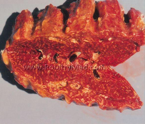
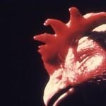
Turkey Coryza (Bordetellosis): Caused by Bordetella avium
It is upper respiratory tract infection primarily of young turkey poults; swollen sinus, collapsed trachea, watery discharge from eyes, tracheitis: deciliation, squamous metaplasia, and lymphoplasmacytic inflammation.
NB.: B. avium can be a significant pathogen in young broiler chickens, ratites(Ostrich), passerines(
) and psittacines(Parrots) (lock jaw)
Mycoplasma gallisepticum infection (MG)
Mycoplasma gallisepticum infection (MG)
It is associated with chronic respiratory disease (CRD) of chickens and infectious sinusitis of turkey.
It is associated with chronic respiratory disease (CRD) of chickens and infectious sinusitis of turkey.
Transmission:
Transmission:
Horizontal transmission occurs readily by direct or indirect infected birds with clinical or sub clinical infected birds mainly via upper respiratory tract or conjunctiva.
Horizontal transmission occurs readily by direct or indirect infected birds with clinical or sub clinical infected birds mainly via upper respiratory tract or conjunctiva.
Vertical transmission (in ovo transmission) by eggs laid from naturally infected hens chickens or turkeys.
Vertical transmission (in ovo transmission) by eggs laid from naturally infected hens chickens or turkeys.
Incubation period: it various from 6-12 days.
Incubation period: it various from 6-12 days.
Clinical signs:
Clinical signs:
Chickens
Chickens
The characteristic signs of natural infection included rales, nasal discharge and coughing
The characteristic signs of natural infection included rales, nasal discharge and coughing
Reticulation in feed consumption and weight loss beside drop in egg production in layers
Reticulation in feed consumption and weight loss beside drop in egg production in layers
High morbidity and mortality due to concurrent infections and environmental factors.
High morbidity and mortality due to concurrent infections and environmental factors.
Turkeys:
Turkeys:
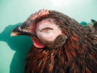
Respiratory signs severe sinusitis (particular closed eyes)
Respiratory signs severe sinusitis (particular closed eyes)
General signs as chickens.
General signs as chickens.
Pathology:
Pathology:
Gross lesions:
Gross lesions:
Primarily, mucosal congestion and catarrhal exudate in nasal and paranasal passages, trachea, bronchi and air sacs
Primarily, mucosal congestion and catarrhal exudate in nasal and paranasal passages, trachea, bronchi and air sacs
Sinusitis with Caseous exudate (focal or multifocal or diffuse) mainly in turkey.
Sinusitis with Caseous exudate (focal or multifocal or diffuse) mainly in turkey.
Some degrees of pneumonia
Some degrees of pneumonia
In severe cases, air sac disease in chicken and turkey (Caseous air saculitis) with fibrinous perihepatiis and pericarditis.
In severe cases, air sac disease in chicken and turkey (Caseous air saculitis) with fibrinous perihepatiis and pericarditis.
Keratoconjunctivitis (edema in facial subcutis, and eyelids with occasional corneal opacity in layers. Also salpingitis could be seen.
Keratoconjunctivitis (edema in facial subcutis, and eyelids with occasional corneal opacity in layers. Also salpingitis could be seen.
Microscopically:
Microscopically:
Chicken and turkey:
Chicken and turkey:
Marked thickening of respiratory tract and sinus, mucous membrane by mononuclear cells and lymphoid aggregation in mucosal lamina propria.
Loss of cilia, swollen epithelial or complete epithelial destruction.
Marked thickening of respiratory tract and sinus, mucous membrane by mononuclear cells and lymphoid aggregation in mucosal lamina propria. Loss of cilia, swollen epithelial or complete epithelial destruction.
Lateron, accumulation of lymphocytic plasma cells, macrophages and some heterophils in the lamina propria.
Lateron, accumulation of lymphocytic plasma cells, macrophages and some heterophils in the lamina propria.
Pneumonia areas in lungs (lymphofolliculuare changes) and granuloma.
Pneumonia areas in lungs (lymphofolliculuare changes) and granuloma.
Air saculitis (fibrino heterophilic or lymphoplastic infeltrations)
Air saculitis (fibrino heterophilic or lymphoplastic infeltrations)
Keratoconjunctivitis in layer chickens.
Keratoconjunctivitis in layer chickens.
Salpingitis in layer chickens
Salpingitis in layer chickens
Encephalitis in turkey's brain.
Encephalitis in turkey's brain.
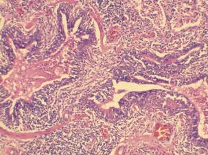
Most consistent histopathological alterations were observed in trachea and lungs. The tracheal epithelium was necrosed and swollen. Epithelial cells and cilia in some areas were covered with thick layer of mucous. Epithelial and sub mucosal infiltration with leukocytes was observed. Hyperplasia of epithelial mucous glands, thickness was increased due to cellular infiltration and oedema. Sloughing of mucosa and hemorrhages of variable degree was also noted, Lungs showed congestion, haemorrhages, focal necrosis and leukocytic infiltration (lymphocytes & polymorphs). Emphysema and exudates in alveoli was also recorded. Grey hepatization of some lungs was also observed. Giant cells, miliary granuloma in lungs were observed In complicated cases, mostly with E. coli. , liver and heart showed congestion and leukocyt ic infiltration (Lymphocytic & Polymorphonuclear). Haemorrhages, degeneration and necrosis of hepatocytes were recorded in some sections. Degeneration of cardiac muscles and pericarditis was also seen .
Mycoplasma meleagridais infection
Mycoplasma meleagridais infection
It is a specific pathogen of turkey It characterized by:
It is a specific pathogen of turkey It characterized by:
Air saculitis in the progeny (egg transmission) Skeletal abnormalities as sternal bursitis , synovitis and ascites & poor growth. Decreased hatchability.
Air saculitis in the progeny (egg transmission) Skeletal abnormalities as sternal bursitis , synovitis and ascites & poor growth. Decreased hatchability.
Mycoplasma synoviae infection:
Mycoplasma synoviae infection:
It is common in chickens and turyes .The main lesions
It is common in chickens and turyes .The main lesions
include: Synovitis of joints and teandoon sheaths and keel bursa (hock shoulder) ,Hepatosplenomegaly and
include: Synovitis of joints and teandoon sheaths and keel bursa (hock shoulder) ,Hepatosplenomegaly and
Air saculitis may be present,No gross lesions in upper respirstory tract could be seen.
Air saculitis may be present,No gross lesions in upper respirstory tract could be seen.
Clostridial disease
Clostridial disease
Ulcerative enteritis:
Ulcerative enteritis:
It is found in a wide range of avian hosts but quail were among the most susceptible species.
It is found in a wide range of avian hosts but quail were among the most susceptible species.
Incubation period:
Incubation period:
It ranges from 1-3 days and the course of the disease 3 weeks
It ranges from 1-3 days and the course of the disease 3 weeks
Clinical signs:
Clinical signs:
Sudden deaths from acute disease
Sudden deaths from acute disease
Sometimes, watery, white droppings.
Sometimes, watery, white droppings.
When the disease progress, infected birds become restless and humped up with partly closed eyes and dull feather
When the disease progress, infected birds become restless and humped up with partly closed eyes and dull feather
Notable emaciations and atrophy of pectoral mucsels.
Notable emaciations and atrophy of pectoral mucsels.
Gross lesions:
Gross lesions:
Acute form:
Acute form:
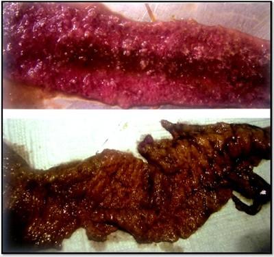
Marked hemorrhagic duodenitis
Marked hemorrhagic duodenitis
Small punctate mucosal ulcers surrounded by hemorrhagic hale. (visible from the serosa )
Small punctate mucosal ulcers surrounded by hemorrhagic hale. (visible from the serosa )
Ulceration may results in perforation of the intestinal wall and leads to peritonitis with adhesion.
Ulceration may results in perforation of the intestinal wall and leads to peritonitis with adhesion.
Subacute or chronic:
Subacute or chronic:
Multiple large, roundish yellow ulcers surrounded by hemorrhages in any part of small or large intestine
Multiple large, roundish yellow ulcers surrounded by hemorrhages in any part of small or large intestine
Diphtheritic membrainous may be observed on the intesitineal wall.
Diphtheritic membrainous may be observed on the intesitineal wall.
Blood may be found in the gut
Blood may be found in the gut
Ulcers may be seen in the mucosa with raised borders.
Ulcers may be seen in the mucosa with raised borders.
Ulcers in ceca may have central depression filled with strongly attached, dark-staining material.
Ulcers in ceca may have central depression filled with strongly attached, dark-staining material.
Liver from light yellow mottling to multiple large irregular, gray or yellow circumscribed foci.
Liver from light yellow mottling to multiple large irregular, gray or yellow circumscribed foci.
Congested, enlarged and hemorrhagic spleen
Congested, enlarged and hemorrhagic spleen
Necrotic enteritis
Necrotic enteritis
It is a disease of primarily young chickens caused by infection with and toxin production ny closteridium perfringens, type A and type C.
It is a disease of primarily young chickens caused by infection with and toxin production ny closteridium perfringens, type A and type C.
Clinical signs:
Clinical signs:
Depression and birds relactant to more and have ruffled feathers
Depression and birds relactant to more and have ruffled feathers
Diarrhea, inappetance and dehydration were seen.
Diarrhea, inappetance and dehydration were seen.
Peracute deaths with high mortality
Peracute deaths with high mortality
Gross lesions:
Gross lesions:
Lesion are usually jejunal and ileial but may be extended to duodenal and cecal portus
Lesion are usually jejunal and ileial but may be extended to duodenal and cecal portus
The small intestine is distended and friable
The small intestine is distended and friable
The mucosa may have Turkish towel appearance or may have specific lesion
The mucosa may have Turkish towel appearance or may have specific lesion
As focal to confluent psudomembrane and petechial hemorrhages
As focal to confluent psudomembrane and petechial hemorrhages
Liver pale fecal have necrotic areas
Liver pale fecal have necrotic areas
Gall bladder distended with inspissated material
Gall bladder distended with inspissated material
Microscopic lesions:
Microscopic lesions:
Extensive villous necrosis and cellular degeneration which to may extend to submucosal and even the muscularies mucosa.
Extensive villous necrosis and cellular degeneration which to may extend to submucosal and even the muscularies mucosa.
Coagulative necrosis of villous tips.
Coagulative necrosis of villous tips.
Large gram positive rods present
Large gram positive rods present
Cholengiohepatits
Cholengiohepatits
Chiken coccidiosis
Compylobacteriosis
Compylobacteriosis
It is a disease of domestic poultry including chickens, turkeys, ducks and geese
It is a disease of domestic poultry including chickens, turkeys, ducks and geese
Fibrino-necrotic epatitis has been reported in laying hens and ostriches causing high morbidity and mortality
Fibrino-necrotic epatitis has been reported in laying hens and ostriches causing high morbidity and mortality
Etiology:
Etiology:
Compylobacter species:
Compylobacter species:
Campylobacter jejunic
Campylobacter jejunic
Campylobacter coli
Campylobacter coli
Transmission:
Transmission:
Horizontal transmissions: from environment to poultry hens.
Horizontal transmissions: from environment to poultry hens.
Vertical transmission: very rarly on poultry farms.
Vertical transmission: very rarly on poultry farms.
Incubation period: it ranged from 2-5 days
Incubation period: it ranged from 2-5 days
Clinical signs and pathology:
Clinical signs and pathology:
No clinical signs in natural infection
No clinical signs in natural infection
Sometimes, infection of young chickens result in watery / mucous or /bloody diarrhea, weight loss and a few mortality may occure
Sometimes, infection of young chickens result in watery / mucous or /bloody diarrhea, weight loss and a few mortality may occure
Gross lesions:
Gross lesions:
It confined to the gastrointestinal trade
It confined to the gastrointestinal trade
Distended intestine with fluid or excess mucus (watery foamy material in ceca)
Distended intestine with fluid or excess mucus (watery foamy material in ceca)
Blood and mucous in the lumen of small intestine
Blood and mucous in the lumen of small intestine
Petechial hemorrhages in serous and mucous membranes of small intestine & large intestine (ceca )
Petechial hemorrhages in serous and mucous membranes of small intestine & large intestine (ceca )
Necrotic lesions in liver
Necrotic lesions in liver
Microscopic lesions:
Microscopic lesions:
Necrosis of intestinal epithelium
Necrosis of intestinal epithelium
Mild edema in lamina propria and submucosal mainly in ceca
Mild edema in lamina propria and submucosal mainly in ceca
Villous atrophy
Villous atrophy
Mononuclear cells infiltration
Mononuclear cells infiltration
