5 minute read
Will robots replace humans in oral pathology?
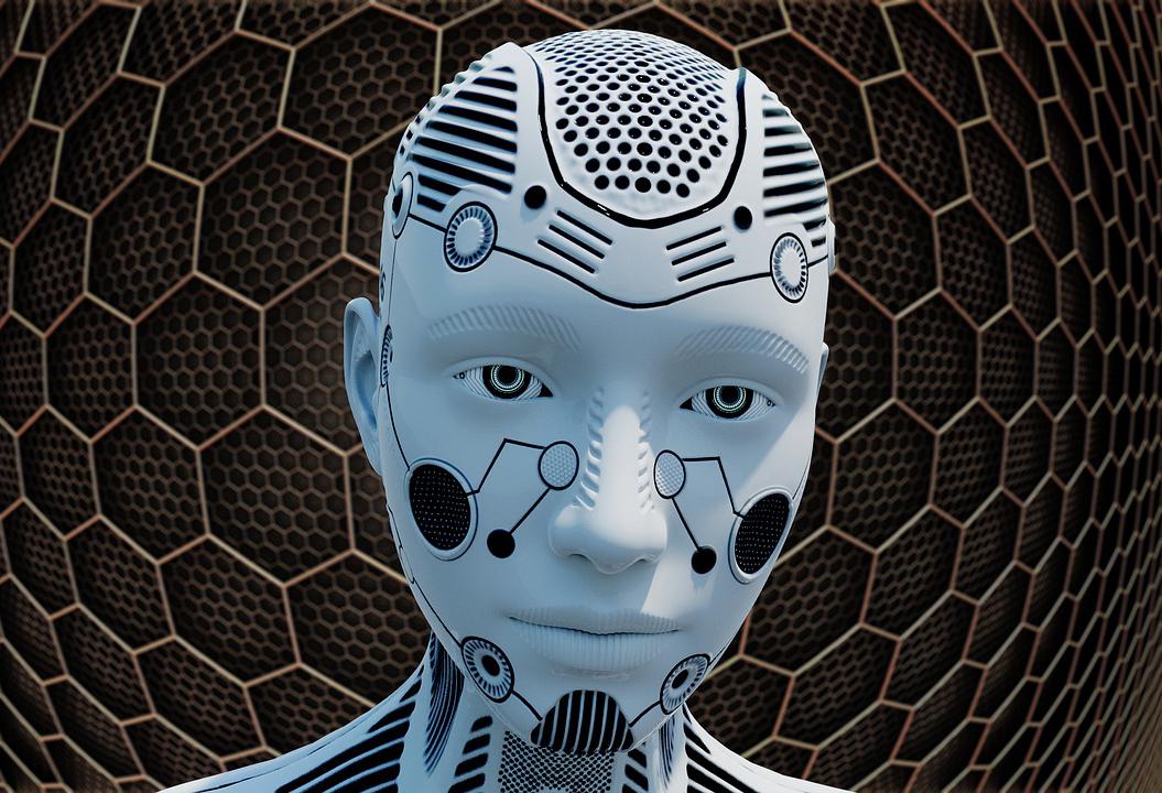
Artificial Intelligence is improving at an exponential rate. Technology has started to replace many previously human-based activities and continues to threaten the positions of humans due to their objectivity, accuracy and low maintenance demands. Will this proceed into the realms of dentistry? Will robots really end up replacing humans?
Tanaka Kadiyo, United Kingdom
Advertisement
World-renowned physicist Stephen Hawking offered stark, timeless words of warning (BBC, 2014) amid the rapid development of artificial intelligence that even facilitated his ability to communicate, having to depend on advanced technology following the progression of his amyotrophic lateral sclerosis (ALS). Are humans, as Professor Hawking proposed, bound to be ‘superseded’ by machines of their own concoction? And, how can we expect such matters to unfold as it relates to pathology in Dentistry? How will the continuous and rapid development of artificial intelligence shape the dynamics of Dentistry; the way people are diagnosed, treated and managed? Should we, as dental professionals, academics, or even patients, be worried or excited? Will it be for the better, or for worse?
A pathologist? Before answering any questions, the most important component that underpins this discussion is defining pathology itself. Originating from the words ‘pathos’ meaning disease, and ‘logos’ pertaining to ‘study of’, oral pathologists are typically dentally qualified professionals that study the causes and effects of diseases (RCPath, 2020). The weight of the work is highly localised in the analysis of laboratory samples of oral tissue for diagnostic purposes. Specialist pathologists play an integral role in healthcare through referrals and the treatment pathway for an incredible volume of patients. Pathology is central to the work of general dentists too, and indeed it is difficult to diagnose and organise the management of any condition without a grasp of the underlying disease process and a basic understanding of pathology. The author of New York Times Bestseller ‘The High Mountains of Portugal’ poetically describes the life of a pathologist by expressing that: “Under the pathologist’s microscope, life and death fight in an illuminated circle in a sort of cellular bullfight. The pathologist’s job is to find the bull among the matador cells” (Martel, 2016). Oral and Maxillofacial Pathology is one of the thirteen recognised dental specialties. With less than thirty specialists in the United Kingdom, very few dentists will ever consider this specialty and very few will have had the opportunity to work alongside one (BSOMP, 2020). The oral pathologist practises an assortment of laboratory techniques to scrutinise human tissue samples of the oral cavity, jaws and salivary glands. These techniques can be labelled as macroscopic, microscopic and molecular. Macroscopic investigation symbolises gross sections of tissue, such as the appearance of white confluent patches found in Oral Hairy Leukoplakia (OHL) (Cho et al., 2010 ). Microscopy includes the subspecialties of histopathology, cytology and immunohistochemistry. The histopathologist can see cellular features such as hyperkeratosis, acanthosis and “balloon cells” (Greenspan et al., 2016) - Pathology is full of strange descriptions, from ‘orange peel’ and ‘soap bubble’ to ‘snowstorm’.
The case for robots Manual interpretation of medical images is very time-consuming, requires considerable specialist expertise, and is prone to inaccu-
racy with questionable consistency. For this reason, in the early 1980s, computer-aided diagnosis (CAD) systems were developed to improve the efficiency of medical image interpretation (Bengio, 2009).
As humans, we are subject to what is widely known as “human error”. This phenomenon is subdivided into either a “skillbased error” or “mistake”. A skillbased error is unawareness, in the moment, of the correct protocol and can be via a slip of action or memory lapse. A mistake occurs whereby the individual commits an action that is generally accidental and can be rule-based or knowledge-based. Even if we controlled all of the knowledge in the world - all the possible causes and outcomes - it would be impossible to sustain 100% accuracy in all of our work (Henriksen and Brady, 2013). As artificial intelligence, cognitive computing and machine learning systems become better than humans at medicine and cost less, it might even become unethical not to replace people. In manual microscopy, it is standard practice for a pathologist to “take a look” at a histological slide under a microscope. To start with, there are many issues with microscopy alone. For example, most microscopes are not US Food and Drug Administration (FDA) cleared medical devices (they pre-date the FDA, but this is starting to change), different microscopes have different optics and sources of light (e.g. blue vs. orange light), and the microscopes in use are often not properly calibrated (Evans et al., 2018). The next task now facing the pathologist, is to assess anywhere between 500,000 to 1,000,000 cells - which may have substantial heterogeneity - and condense all the data into a simplified diagnosis. A situation where robots may be advantageous over humans is in the analysis of results, particularly when using a method such as immunohistochemistry. For example, it may be required that a pathologist dyes a potentially cancerous cell with a staining chemical (e.g., DAB – 3,3’-Diaminobenzidine). This frequently occurs with cells from a suspected oral melanoma. The pathologist then has to determine the percentage of cells that appear stained and compare this against a threshold (>10%) to see if this cell is cancerous (Lilyquist et al., 2017). This is an unbelievably challenging computational task, and without surprise, leads to abnormal levels of intra- and inter-pathologist variation. Conversely, a robotic system is unlikely to have this same level of error. Early diagnosis is the most important determinant of oral and oropharyngeal squamous cell carcinoma (OPSCC) survival outcomes, and yet most of these cancers are detected late, resulting in poorer outcomes (Ford and Farah, 2013). A potentially positive screening outcome at a routine dental check-up leads to referral to a specialist for biopsy and histopathological analysis. Artificial Intelligence (AI) systems now have the ability to combine multifactorial data streams into powerful integrated diagnostic and predictive systems spanning divergent data streams from sources such as images, genomics, pathology, electronic health records, and even social networks (Langlotz, 2019). A recent survey of the literature reported a 15% to 20% improvement in the accuracy of cancer prediction outcomes in clinical practice using AI techniques (Cruz and Wishart, 2006). To date, there are few publications on the application of these techniques to imaging in the oral cavity. In a recent study, the performance of a deep learning algorithm for detecting oral cancer from hyperspectral images of patients with oral cancer was evaluated. The investigators reported a classification accuracy of 94.5% for differentiating between images of malignant and healthy oral tissues (Jeyaraj and Samuel Nadar, 2019).
In defence of pathologists It is often easy to forget the breadth and depth of knowledge a pathologist possesses. Defini-


