8 minute read
Factors affecting the osseointegration of dental implants
tive diagnoses are only one tool in the pathologist’s toolbox. A basal cell carcinoma, for example, is a fairly simple diagnosis to deduce, even on clinical examination only. However, a pathologist’s report may include further information on the staging and progression of a cancerous lesion (Dourmishev et al., 2013). A pathologist needs to be ready to investigate anything suspicious (including incipient findings) by synthesising chart review information, holding discussions with clinicians, consulting old reports and concurrent labs, judging patient-specific factors and conducting additional special investigations. At the end of the day, the pathologist is the focal spokesperson who uses their wealth of experience to set the baseline for treatment planning. This is why it is difficult to believe artificial intelligence will inherit a greater role beyond screening, but decision-support is going to make our pathologists much more efficient, accurate and consistent. The mounting demand for and the current dearth of pathologists could stabilise more once the technology matures and develops.
Conclusion “Could pathologists be replaced by robots” is unnecessarily alarmist, and the wrong question to be addressing. Perhaps a more useful question is “How can Artificial Intelligence enhance the work of pathologists?”. AI approaches can rapidly analyse complex images to provide decision-making guidance. Additional studies are needed to identify optimal imaging approaches for each clinical need and to finalise the configuration and clinical guidance outcomes of AI-based algorithms. It is an exciting field with many pioneers leading the effort, but not one where humans are at risk of being ‘superseded’ any time soon.
Advertisement
An introduction to modern day implant osseointegration.
Christianna Iris Papadopoulou, Dimitrios Angelakis, Greece
Dental implants have become a more common treatment for replacing missing teeth. In clinical dentistry, the goal of dental implants is to increase patient satisfaction in terms of improved chewing efficiency, physical health, and esthetics. In 1965, Brånemark introduced the term “osseointegration” to describe the process of bone growing right up to the implant surface, with no soft tissue in between. When osseointegration occurs, the implant is held so tightly in place by the bone, that there is no relative movement between the two. The process typically takes four to six months to occur. Kieswetter et al. described this process in his article in 1996 in more detail. Some days after implantation, bone regeneration is regulated by several growth and differentiation factors that are released around the implant. In 2003 Davies stated that the process of bone regeneration is formed either on the implant surface or from the surrounding bone towards the implant surface. Finally, bone remodeling occurs by replacing immature with mature bone at the implant site, providing biological (mechanical) stability. To be considered successful, an osseointegrated oral implant has to meet certain criteria. As Karthik et al. described in 2013, these basic criteria are immobility, absence of peri-implant radiolucency, adequate width of
the attached gingiva, and absence of infection. A wider implant has a better long-term success than a narrow implant. Co-existing medical conditions and smoking also play an important role in evaluating the success of an implant. Additional criteria are: bone loss less than 0.2mm annually after the first year postoperative (Albrektsson et al., 1986), and no bleeding pockets exceeding 5mm of probing depth (Mombelli et al., 1994). According to Esposito et al. (1998), failures of osseointegration can be divided into biological failures, i.e., the inadequacy of the host tissue to establish or to maintain osseointegration, mechanical failures of the components of the oral implant, iatrogenic failures (malpositioning) and inadequate patient adaptation (psychological, esthetic and phonetic problems). Biological failures can further be divided according to chronological criteria in early and late failures. Early (primary) failure is the failure to establish osseointegration, i.e., an interference with the healing process. Late (secondary) failure is the failure to maintain the established osseointegration, i.e., processes involving a breakdown of osseointegration. Many factors affect the osseointegration of a dental implant. This article describes the most important.
Biocompatibility of the implant material In his 1987 Dictionary of Biomaterials, Williams defines biocompatibility as “the ability of a material to perform with an appropriate host response in a specific application”. One of the most significant factors for assessing biocompatibility is corrosion behaviour due to the adverse effects (e.g. allergic reaction, increased toxicity) that metal ions can generate both systemically and in the immediately related tissue. Titanium is one of the most widely accepted materials used to manufacture dental implants since 1981. According to a recent article (Nicholson, 2020), the most used titanium alloys are cpTi and Ti-6A1-4V, which have a 99% of success over a period of 10 years. Other dental implant materials include ceramic metals, stainless steel, chromium-nickel-vanadium and tantalum. Although ceramics are gradually becoming more and more popular, the fact that titanium is the most biocompatible material remains undisputed. Titanium has a low electrical conductivity and its electrochemical oxidation leads to formation of a thin passive oxide layer which makes it highly resistant to corrosion. Titanium has an oxide isoelectric point of 5-6 and the protective, passive layer is retained at the same pH levels as the human body. Finally, in aqueous environments Ti and its oxides have low reactivity with macromolecules due to their low ion-formation Implant design A significant function of dental implants is force transfer (Steigegna, 2003). Thus biting forces and occlusal overload are two main factors to be considered when designing implants. Implant shape impacts the distribution of stress to the surrounding tissues greatly. In particular, cylindrical and screw-shaped implants produce less stress than conical or stepped ones.
To assess the quality of osseointegration related to implant design, three tests can be applied: pull-out, push-out, and torque measurement. The first two evaluate the shear strength of the implant whereas the third refers to the resistance of loosening of the implant. According to a recent research (Kayabasi, 2020), bone to implant contact (BIC) is essential in osseointegration and more important than implant length. Due to the difficulty of performing implant tests in vivo, a variety of mathematical models is needed to ensure efficient implant design.
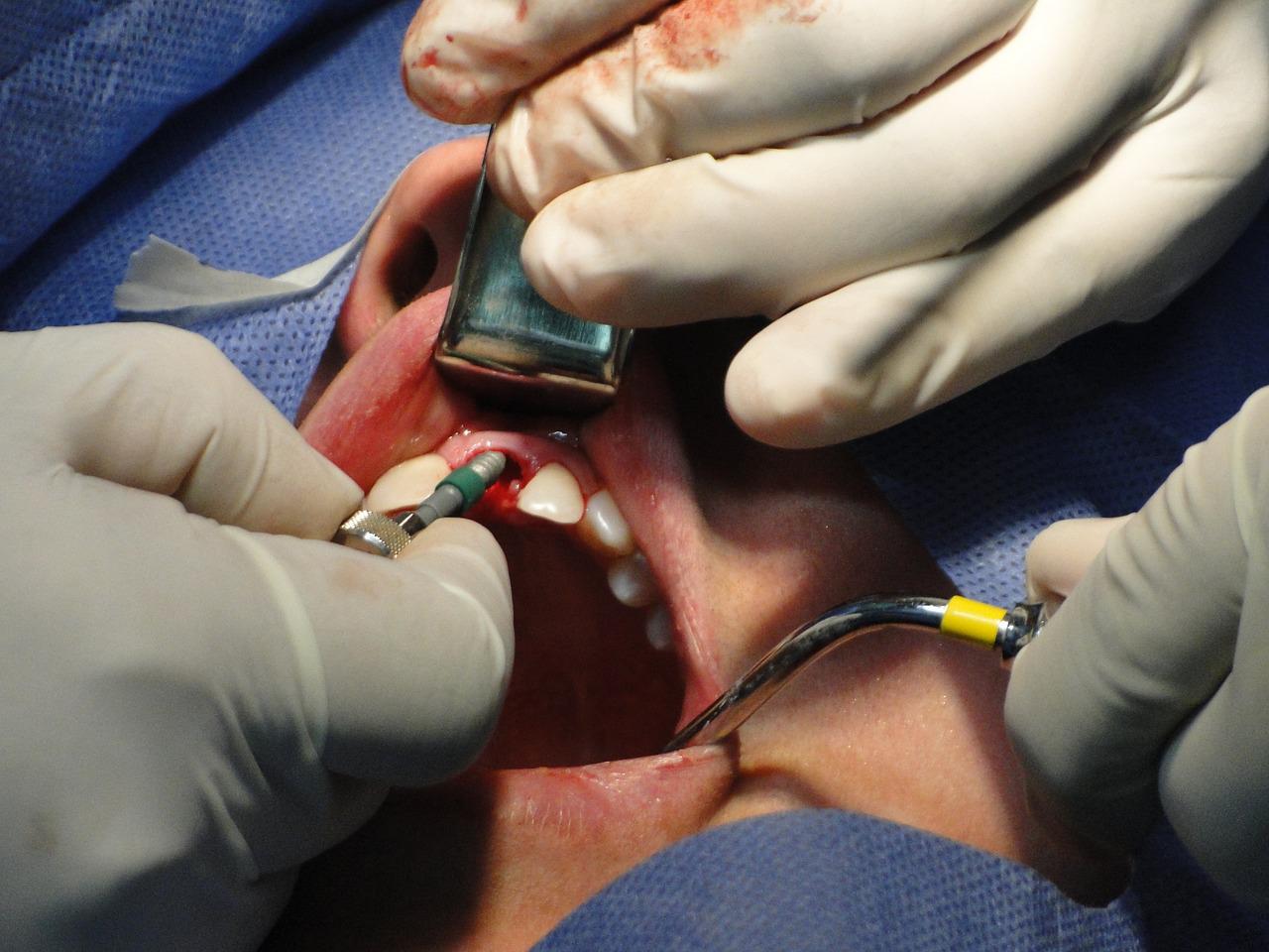
Implant surgical technique and training of the operator The experience of the operator plays a great role in accurately placing dental implants with a bone supported stereolithographic surgical template (Cushen, 2013). It is undisputed that the skills of the dental specialist are vital to the success of the procedure and the subsequent osseointegration of dental implants. Additionally, basic surgical rules need to be followed to ensure successful osseointegration. These include, among others, cautious surgical handling, cooling of the drills in order to avoid overheating and prevention of contaminating the dental implants via exposure to oral fluids (Vrotsos et al., 2016).
Subsequent prosthetic design The subsequent prosthetic design is essential to the success of osseointegration. Prosthetic planning prior to the surgical procedure is of utmost importance since it will determine the most effective position for the dental implant. Each case requires a different approach. Dentists can choose between screw-retained prosthesis, which is easier for maintenance, and cement-retained prosthesis - both can be equally effective. The main goal of the dentist should be to avoid excessive forces placed on implants as this will undermine osseointegration. Furthermore, occlusal adjustments after prosthetic placement are vital because shearing forces can overload the entire prosthetic set, leading to screw fracture, porcelain fracture, screw loosening and implant fracture (Gustavo, 2018).
Loading condition Esposito et al. (2013) established three main protocols for implant loading: immediate loading (within one week of the implant placement), early loading (between one week and two months) and conventional loading (after two months). When the Brånemark implants were first introduced in 1965, the trending notion was that loading should be implemented 3–6 months after implant placement. Brånemark and other researchers concluded that earlier loading would lead to failure of osseointegration. Despite that, in 1979, Ledermann implemented the immediate loading protocol using Titanium implants with a larger surface area. Today, the immediate loading protocol is becoming more and more popular due to its problem-solving nature for both dentists and patients. For this protocol to be successful, some guidelines should be followed: placement of an acceptable number of implants (4–10), proper distribution of implants in the bone, initial implant stability that exceeds 40Ncm and normal occlusion.
Bone quality The success rate obtained with dental implants depends to a great extent on the volume and quality of the surrounding bone. According to Lekholm and Zarb (1985), sufficient bone density and volume are crucial factors for ensuring implant success. Bone quality is broken down into four groups according to the proportion and structure of compact and trabecular bone tissue: Type I to IV (Bone Quality Index-BQI). Clinical reports have indicated that areas with Type III or IV bone, often found in the posterior maxilla, have a higher chance of failure compared to areas with Type II bone. When compared to the maxilla, there was a higher survival rate for dental implants in the mandible, particularly in the anterior region of the mandible, which has been associated with better volume and density of the bone (Turkyilmaz et al., 2008). Type I bone also requires a great deal of caution because its preparation may cause overheating and necrosis of the bone, which leads to early failure.
Periodontal health and infection control The success of implant osseointegration, like in any other treatment plan, depends on periodontal health and presence/absence of infection. Patient’s history of periodontal and/or endodontic disease represents one of many risk factors contributing to the failure of dental implants. Bacteria and inflammatory elements in the area (cytokines, inflammatory mediators and inflammatory cells) potentially progress deeper into the bone and undermine the osseointegration process. To avoid early failure of implant osseointegration, the dentist must make sure the area of the implant and the surrounding periodontal tissues are clear of infection.


