Oar feet and opal teeth: about copepods and copepodologists 1st edition charles b. miller all chapte
and Opal
Charles B. Miller
Visit to download the full and correct content document: https://ebookmass.com/product/oar-feet-and-opal-teeth-about-copepods-and-copepo dologists-1st-edition-charles-b-miller/
More products digital (pdf, epub, mobi) instant download maybe you interests ...
Oar Feet and Opal Teeth: About Copepods and Copepodologists 1st Edition Charles B. Miller
Oxford University Press is a department of the University of Oxford. It furthers the University’s objective of excellence in research, scholarship, and education by publishing worldwide. Oxford is a registered trade mark of Oxford University Press in the UK and certain other countries.
Published in the United States of America by Oxford University Press 198 Madison Avenue, New York, NY 10016, United States of America.
All rights reserved. No part of this publication may be reproduced, stored in a retrieval system, or transmitted, in any form or by any means, without the prior permission in writing of Oxford University Press, or as expressly permitted by law, by license, or under terms agreed with the appropriate reproduction rights organization. Inquiries concerning reproduction outside the scope of the above should be sent to the Rights Department, Oxford University Press, at the address above.
You must not circulate this work in any other form and you must impose this same condition on any acquirer.
Library of Congress Cataloging-in-Publication Data
Names: Miller, Charles B., 1940- editor.
Title: Oar feet and opal teeth : about copepods and copepodologists / [edited by] Charles B. Miller.
Description: New York, NY : Oxford University Press, [2023] | Includes bibliographical references.
Identifiers: LCCN 2022039027 (print) | LCCN 2022039028 (ebook) | ISBN 9780197637326 (hardback) | ISBN 9780197637340 (epub)
LC record available at https://lccn.loc.gov/2022039027
LC ebook record available at https://lccn.loc.gov/2022039028
DOI: 10.1093/oso/9780197637326.001.0001
Printed by Integrated Books International, United States of America
Preface
In the early 1960s I started sampling zooplankton from oceans and estuaries. Almost consistently the most numerous and most readily named animals in the catches have been copepods, members of a large and diverse class of Crustacea. I learned about some of them, those living their whole lives swimming in the sea. My interests came to focus on their life histories: spawning, hatching, molting through distinctive phases, forming their teeth, migrating, maturing, mating, and again, spawning. By 1970, I had a university job teaching biological oceanography and generating research projects. Teaching about zooplankton required learning quite generally about their biology, about things I did not work on myself, like modes of feeding. Copepods dominate the zooplankton, so that learning was mostly about them. Most of my research also connected me to them.
A recurring experience for people who study copepods—copepodologists—is that most people we meet have never heard of copepods: our neighbors, our dentist, even our children’s biology teacher. They have no idea copepods exist, what they are like, or that they are key components of all wet habitats (except perhaps clouds). Unlike insects, no images spring to mind. So in 2017, I decided to write a book to introduce them more widely. This is it. Many chapters include some biological background to make their topics accessible. There are quasi-literary asides in places to leaven the loaf.
In 2017 I attended the 13th International Conference on Copepoda, held at a hotel in Los Angeles. Those meetings are organized by the World Association of Copepodologists. Yes, that exists, and such meetings are held somewhere about every three years (delayed lately by the COVID-19 epidemic). There, I met with and interviewed some of the leading copepod biologists. For other interviews I traveled to meet the subjects, or used online “facetime.” Those interviews provided biographical materials for characterizing the life histories of people doing this work at a professional level. The selection is much narrower than I would like. Most, but not all, of the stories are about people I have worked with or followed closely because of my professional connections (e.g., people trained by Bruce Frost). A majority are from the United States or Canada, but specimens of about ten other nationalities are considered. There are several thousand important copepodologists active now or recently (thinking in decades), and the sampling here is quite parochial. If you are a copepodologist who feels left out of the book, I’m sorry. You almost certainly deserved to be in it.
The subjects covered are listed in the table of contents. Far from everything known about copepods is covered. As stated in the first chapter, the subject is the free-living copepods of lakes and oceans. Parasitic and meiofaunal groups are only mentioned, and somebody else should write semipopular books about them. The focus is on the animals themselves, on aspects of their individual lives, much more so than on their place in the ecology of their habitats. There are good books about those ecosystem dynamics; just not this one. Later in the book the focus shifts to the methods and results of molecular genetics applied to copepods. I have reached for an understandable level of explanation. Knowing DNA sequences in detail has provided a new lens for discerning the long evolutionary history of copepods and their phylogeny. The tentative genetic conclusions have also been guided by recent and very refined morphological comparisons. Those back chapters tell how that cross fertilization has been accomplished. Extraordinary insights have already emerged from the combined efforts, and more are already in lab notebooks. We start to see how the orders and families of copepods have diverged through time.
I hope you enjoy reading Oar Feet and Opal Teeth, and I hope you feel like recommending it to others you think would enjoy it as well.
Acknowledgments
I thank all the copepodologists whom I interviewed for the personal insights they offered, for their photographs, and for checking what I wrote about them and their scientific contributions. Reading on, you will meet them. Among those copepodologists I especially thank Russ Hopcroft, for sharing so many of his beautiful copepod photos with me and the anticipated readers. Thanks also to all the other photographers credited in figure captions. All were generous with permission. Thank you Jeremy Lewis and Michelle Kelley at Oxford University Press for taking on this project and seeing it through. Thank you Martha Clemons, my wife, and for some years a valued coauthor contributing to life history studies of Neocalanus. Thanks to her for staying the course with me through the multiyear adventure that writing Oar Feet and Opal Teeth has been. Thanks to everyone who has taught me important things, and special thanks to John McGowan, my graduate school advisor. Thanks for the honest arguments and good company to the crews of my scientific colleagues at Oregon State University, Scripps, Woods Hole, Plymouth Marine Lab, National Institute of Water and Atmospheric Research in New Zealand, and the universities of Washington, Tokyo, Hiroshima, and Tromsø. Indeed, I am grateful for my many scientific and personal connections with marine and copepod scientists around the world. Thanks everyone.
Finally, warm thanks to copy editor Anne Sanow for hundreds of “serial commas,” for cooling my outbursts of enthusiasm (and cynicism), for slogging through every detail of the references.
Charlie Miller // Biddeford, Maine // 29 November 2022
1 Planktonic Copepods Have Those Oar Feet
Defining Plankton
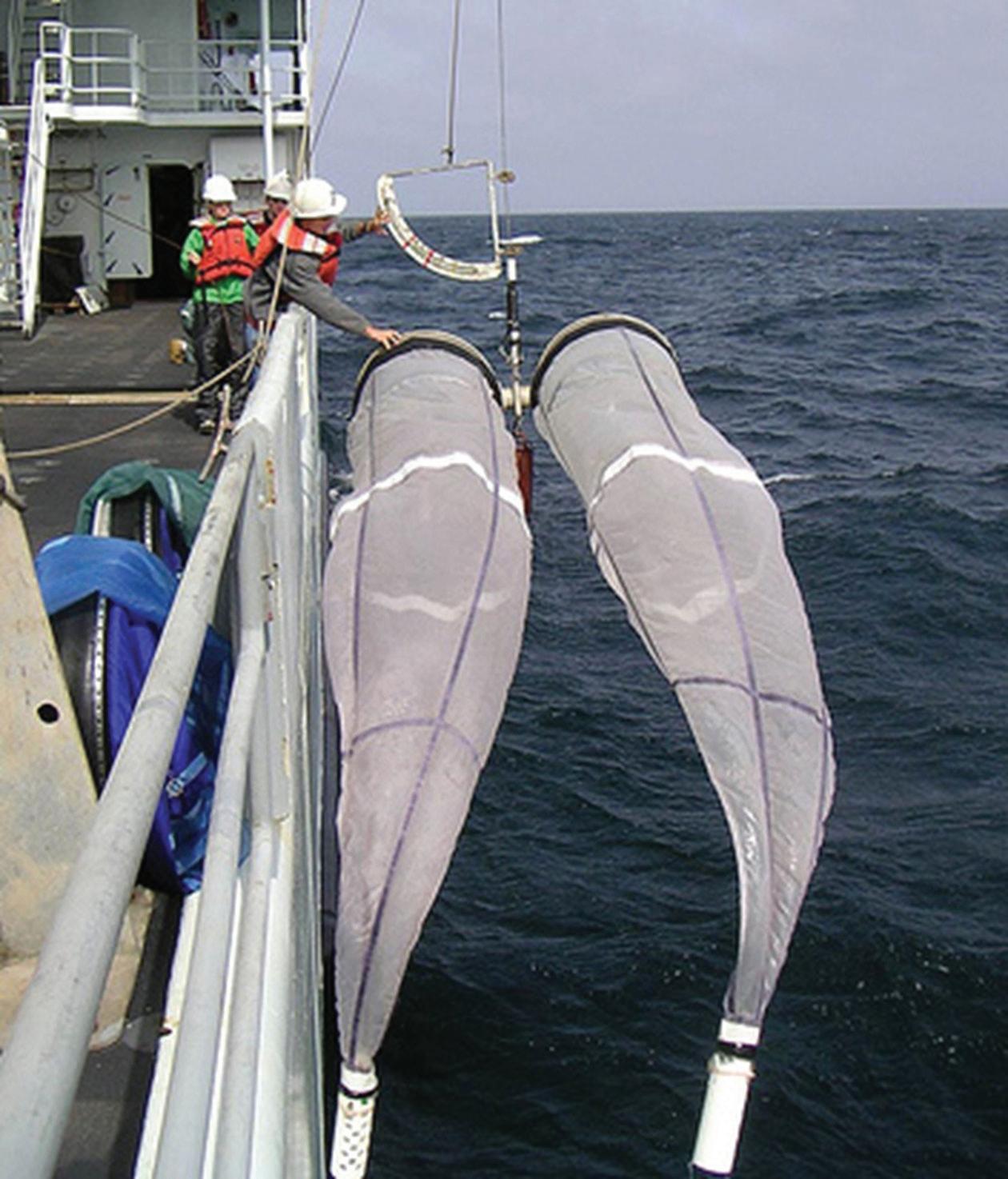
Many of the organisms living in water are grouped as plankton. The word was taken from the ancient Greek word πλαγκτοσ (planktos). It is a beautiful word meaning “that which must wander or drift,” things like clouds. Oversimplifying current classifications considerably, the plankton includes bacteria, algae (the phytoplankton), and a wide range of animal types, the zooplankton. Scientists who call themselves planktologists sample these organisms in several ways. The larger zooplankton (0.1 mm to 10 cm) are most commonly sampled with fine-mesh nets (example in Figure 1.1) that are hauled through the water like large, conical kitchen strainers, usually with mouth openings from about 15 cm to 2 meters. Pumps and water-sampling bottles are also used. When nets with mesh holes a millimeter or less across are pulled through ocean water, the catch most often will be dominated by small, torpedo-shaped crustaceans of the subclass Copepoda, familiarly called copepods.
Because copepods make up half or more of such catches, especially in numbers, many limnologists, oceanographers, and zoologists who take an interest in plankton find themselves intensely engaged with the life and times of copepods. Results of their studies are the subject of this book, along with some notes on the lives and times of these copepodologists. In some places I will emphasize my own experiences of discoveries, few as those seem from the vantage point of age. However, I can speak to those from the distorted memory of having been there. I have also interviewed a number of the significant students of copepods, and I will represent some of their experiences as they told them to me.
Again, copepods are among the most abundant and most diverse multicellular (metazoan) animals in the ocean. They are also abundant in lakes and are found in underground streams; in the water around tropical, epiphytic bromeliads; even as very small versions living in the interstitial water among the grains of sand in beaches, seafloors, and lakebeds. Some, like salmon lice, are parasites, and it is likely that the commensal and parasitic forms exceed all the others in species diversity. They will need a book of their own (by somebody else). The free-living forms are beautiful, so study of them is an aesthetic as well as scientific pleasure.
Figure 1.1 A paired “Bongo” net system ready to tow with the ship underway, obliquely down and back up. Suspension from the central axle avoids having towing bridles in front of the net mouths. Two mesh sizes can be used. Catches are taken by unlatching the white “cod ends” from the tails. The initial bongo designs were drawn in the mid-1960s by John McGowan of Scripps Institution of Oceanography. Photo taken by Andrew King on a cruise for a California Current Ecosystem Research project.
Copepods have an evolutionary history dating way back, likely to the Cambrian period (541 to 485 million years ago), the time when metazoans generally began to leave a fossil record. According to paleontologists, some tiny Cambrian fossils look like the toothed edges of copepod jaws,1 though not all copepodologists think those bits could only be from copepods. Unlike the much later emerging insects, which molecular genetic data2 suggest derived from crustacean ancestors, copepods are known all but exclusively to those copepodologists and ecologists who study them. However, they are fascinating and well worth learning about, hence this book. Copepodologists? Really? Yes, there are some, and there is even a World Association of Copepodologists with more than 500 members.
Like the term “plankton,” the name copepod derives from Greek words: “cope” (κουπι) for oar and “pod” (ποδι) for foot: oar-foot. According to David Damkaer,3 the premier historian of copepod studies, the name was first applied by Henri Milne Edwards in 1830. He was a French carcinologist at a time when reasonably accurate relationships among crustacean groups were first emerging into view. In those days essentially every well-educated person knew something of both Greek and Latin, and they combined Greek or Latin roots to name almost everything they felt needed naming. We still do that today, though very few of us actually know either ancient language.
Copepods are surprisingly unfamiliar to most people, surprising because of their abundance and nearly universal presence in natural waters. Well, they are small. They go unnoticed by human swimmers, though small fish can spot them and then eat them. There is virtually no mention of them in nonscientific literature. Long ago I asked a friend, J. Dennis Evans, then an English Instructor at Oregon State University, whether there were any poetic or other references to copepods in English literature. As it happened, he was writing a thesis4 on the works of the American poet A. R. Ammons. In “Hymn IV,” Ammons5 is cursing God (or some omnipotent entity, addressed as “you”) for all the evils from disease to “life’s all-clustered grief.” Here (with permission) are those three stanzas:
I hold you responsible for every womb’s neck clogged with killing growth
I keep you existent at least as a ghost crab moon-extinguished his crisp walk silenced on broken shells
answering at least as the squiggling copepod for the birthing and aging of life’s all-clustered grief
Ammons at the time (1957 or 1958) seems to have run out of full stops and commas. Why “the squiggling copepod” should answer for any of life’s grief he did not explain, though apparently he was only keeping some God existent because he/she/it was as trivial as a copepod for explaining the problem of evil (that is, why does evil exist, if there is a big-G God?). At least (to borrow Ammons’s recurring limit to the minimal), he clearly knew copepods exist, he mentions
them elsewhere. He majored in biology at college and worked for decades at Cornell University, where a limnologist maybe mentioned them during lunch at the Faculty Soup Club. Unlike insects, lobsters, crabs, shrimp, and crayfish, all of which have wider poetic representation, a squiggling specimen seems to be it for copepods, at least (again), in literary English. Copepodologists constantly meet people not familiar with copepods. Reading on will remove you from that crowd.
Meet One of the Larger Copepod Species
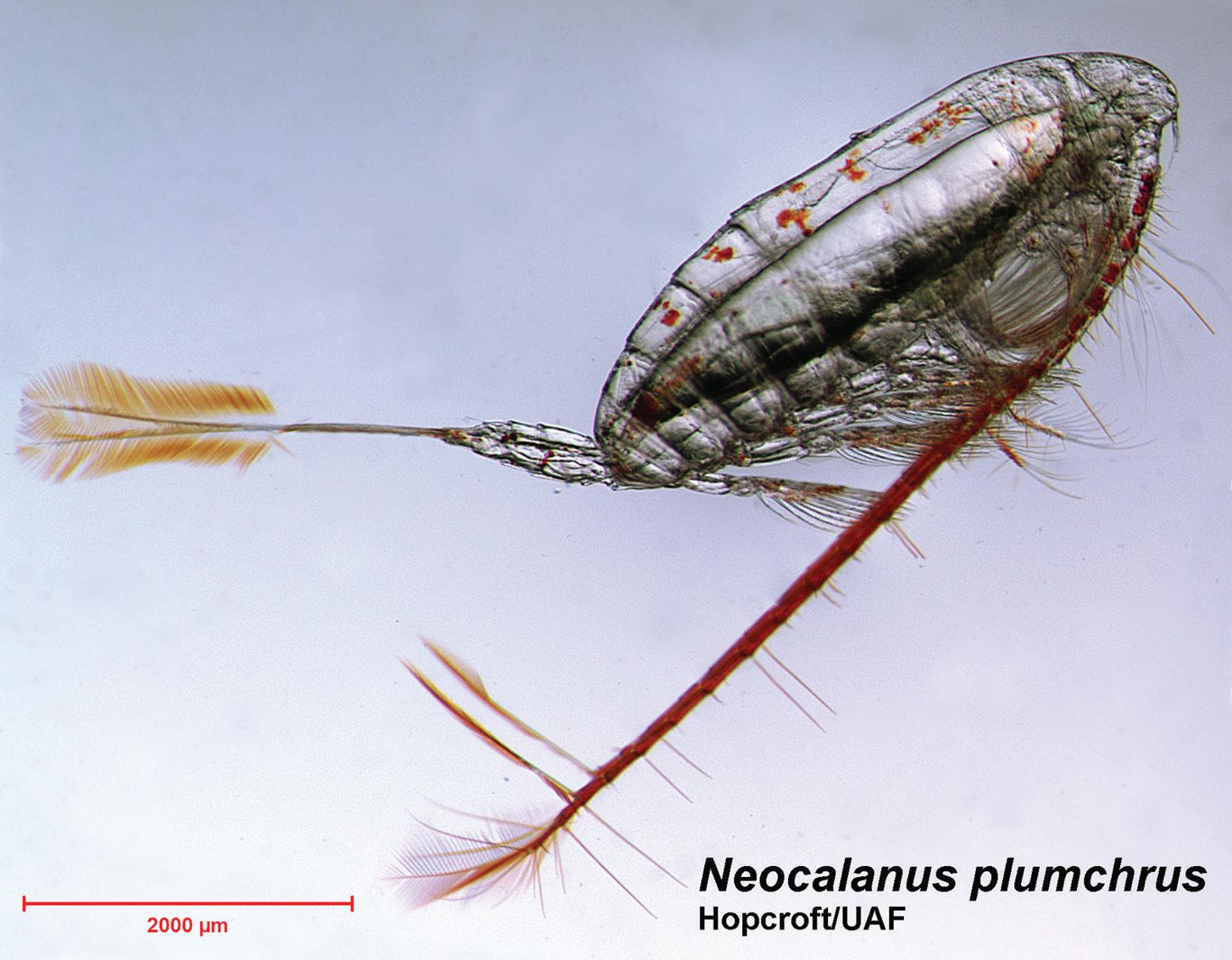
Figure 1.2 is a photo of the last larval stage, the fifth “copepodite,” of Neocalanus plumchrus, a freely swimming copepod that lives in the subarctic reaches of the Pacific, waters north of 45°. It has none of the primary or secondary sexual features of an adult copepod, so it shows well the more basic features of this crustacean class. Copepods have the general features of that class, so like all arthropods they have plastic-like, chitin sheaths as outer skeletons. Chitin serves as armor and as other interfaces with the outside world, even in some species as eye lenses. Implications of this crop up in every aspect of their lives. Chitin is a starch-like polymer of glucose molecules, but each of those includes an amine (NH3) group, which allows crosslinking into pliable sheets as well as chains.
Figure 1.2 The fifth copepodite (last larval stage) of a relatively large calanoid copepod: not counting the long tail setae, the total length is around 5 mm (2,000 μm is 2 mm). Photo by Russell Hopcroft (University of Alaska, Fairbanks), one of the outstanding photographers of planktonic organisms. With his kind permission.
As depicted (Figure 1.2), the head end is at the right, the tail at the left. The main body, called the fore-body or prosome, has the drag-reducing shape and smooth surface of a somewhat bulky torpedo. The carapace or shell over the head segments is fused into a continuous skin of chitin. Each prosome segment bears a pair of limbs on the ventral side. Five pairs of limbs at the back are the oar feet attached below the thorax, so they are termed “thoracic legs.” Chitin over the first thoracic segment (T1) is continuous with the fused carapace of the head (also known as the cephalosome, “head body”). The last four thoracic segments are articulated with each other and the cephalosome, so the thorax can bend a little.
Limbs beneath the head are specialized in different ways: for sensing odor and motion in the water, for slow swimming, and for feeding. Much of the specialization between genera and species is located in the leading antennules (the long tubes turned below the body in Figure 1.2) and among the next five pairs of appendages, the mouth parts, each adapted for a specific aspect of feeding. At the left is the urosome, a tubular tail section terminated by two short, pad-like appendages, the caudal furcae. Those are tipped by the two sides of a fan of setae, which in a few genera includes a very long medial pair of setae. In N. plumchrus those are orange, with ribbon-like setules or side whiskers. Once biologists have written out a genus name, as in Neocalanus plumchrus, they allow themselves to abbreviate it to one or two letters.
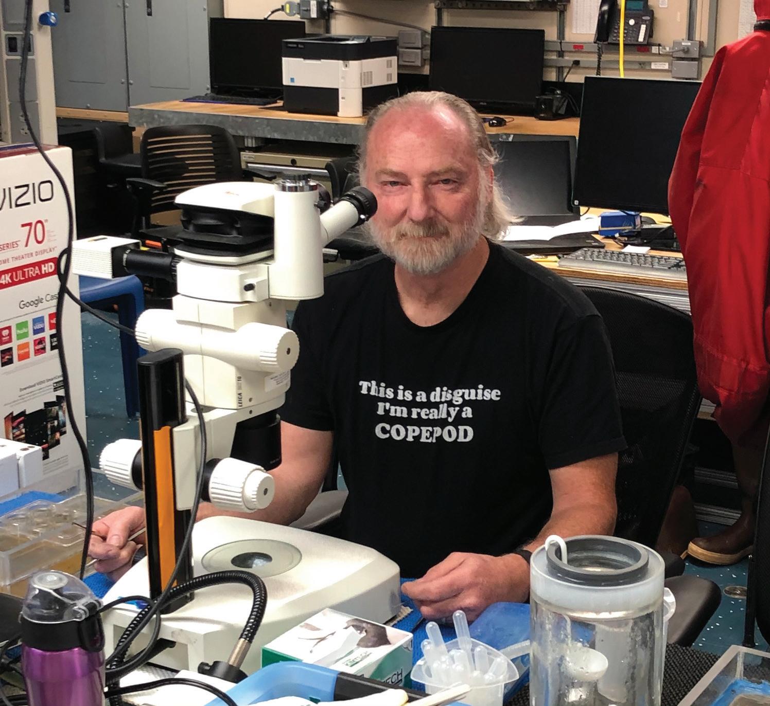
A First Example of a Copepodologist, Russell Hopcroft
The meaning of “copepodologist” is obvious: somebody who studies copepods. Copepods vary greatly in where and how they live, though only in wet places. They also vary in their sizes and shapes, especially the shapes of parasitic forms. Thanks to all that variation, copepodologists come in many versions: basic taxonomists, evolution specialists (phylogeneticists), biomechanicians, limnologists, oceanographers, parasitologists (including fisheries experts), and recently molecular biologists. I will introduce some of those. Russell Hopcroft (Figure 1.3) is an ocean ecologist and zooplankton photographer, who lives in Fairbanks, Alaska, teaching at the branch there of the state university. He studies zooplankton all around that huge peninsula from Prince William Sound and along the southern coast, up through the Bering and Chukchi Seas, even spreading out around the entire Arctic Ocean. He did not start with such high latitude interests. Russ was born in Exeter, Ontario, a town of about 500, and moved at age five to live near his grandparents in a town of about 6,000. He went through most of his early schooling there, and then the family followed his father as he shifted among several jobs. They moved to Toronto and later west to near Hamilton. He recalls early interest in the bugs and tadpoles of nearby freshwater, often catching
Figure 1.3 Russell Hopcroft at his microphotography station, with a Leica stereoscope with megapixel digital cameras. Both Russ Hopcroft and the scopecamera were aboard the University of Alaska ship RV Sikuliaq, preparing for a cruise from Seward across the Gulf of Alaska shelf. His t-shirt indicates a majority interest in respect to his planktonic friends. Photo by the author.
and watching them in jars and wading pools. He thinks an influence may have come from Jacques Cousteau’s television programs about oceans and ocean life. He knew entering high school that marine biology would be his life interest and he prepared for college accordingly, as well as learning dive skills. Included was a gap year to think about life’s possibilities. College, starting in 1978, was at the University of Guelph, a public college offering specialization in the subject.
During his senior year of college Russ started to work with Professor John Roff. Their first joint publication in 1985 reported the respiration of a rockclinging sea cucumber6 measured during a study trip to the Huntsman Centre on Passamaquoddy Bay in New Brunswick. Roff also gave him access to an early desktop computer and some video-microscopy gear; the challenge was to develop a length-measuring system for tiny animals like copepod nauplii. Russ had a major part in that, and the system is still in use.7 At the time Roff was engaged in an extended series of plankton studies in and near Kingston Harbor, Jamaica, a partnership between University of Guelph and the marine labs of the University of the West Indies. He took on Russ as grad student, who took him on
as an advisor, and they worked together on tropical plankton until about 1997, though more papers came out later. It wasn’t all copepods; there were also studies of phytoplankton and sedimenting particles that were reported in his master’s thesis.
It is a badly kept secret that marine labs powerfully foster romance among their human occupants, as well as allowing study of exotic aspects of mating among snails, whales, and, of course, copepods. Another of Roff’s students was Cheryl Clarke, who worked mostly on components of the plankton other than copepods.8 She and Russ had met at the Huntsman Centre. Interest continued in Guelph and Jamaica. They completed their Jamaica-based MS degrees in 1990 and married in 1991. Cheryl pushed more than Russ (according to him), that he continue for his doctorate. He did, turning to the population dynamics and production rates of copepods. “Production” in ecology refers to how much organic matter accumulates in an organism or population, a central interest for Roff and so for his students. Tropical nearshore plankton are predominantly small, including copepods like Oithona, Euterpina, and Corycaeus that mature at about 1 mm. So Russ became a refined microscopist in order to study aspects of growth and production among these small crustaceans, emphasizing the effects of body size on growth rates. He finished in 1997.9 Some of his results10 concerned growth rates and fecundity of small copepod varieties that carry their eggs to hatching in sacs attached to their urosomes.
As Dr. Hopcroft, Russ moved with Cheryl to Moss Landing, California, where he had been offered a postdoctoral position, limited by contract to two years, at the Monterey Bay Aquarium Research Institute (MBARI). Their first child, a daughter, was born there, and their twin boys were born in Alaska in 2000. Cheryl’s training in zooplankton research was not set aside, and she now works with Russ as his lead technician at the University of Alaska in Fairbanks (UAF), appearing in bylines as C. Clarke. At MBARI, Russ worked with Bruce Robison, who largely led the institute’s submersible and remotely operated vehicle (ROV) studies of deep-sea animals. Using an ROV, they photographed, collected, and later described four previously unknown but relatively large (0.5 to 1 cm) deepliving larvaceans, gelatinous zooplankters that feed by spreading elaborate filtering structures made of mucous around themselves.11 (You could learn about those by looking up the paper; they are not copepods, so we’re not going into more detail here.)
Back on the job market, Russ applied for an opening at UAF that came about because the lead zooplankton worker there retired. As he puts it, most of the smaller oceanographic institutes in the United States and Canada have one or only a couple of positions in each subject matter “slot.” He got an offer to fill the UAF zooplankton slot, and he took it. There are not many zooplankton slots in the United States or Canada, and Russ is still in the one at Fairbanks. Siting an
oceanography department over 600 kilometers from the sea in a city with brutally cold winters (including rare days below -50°C) surely once had a political logic. No other explanation serves. The university, with about 10,000 students, is one of the city’s principal industries. Russ says that apart from the cold and the mosquito swarms in spring, it is a good place to live and raise a family. He and Cheryl have done that; the twins are now in college.
The cover of a recent novel, pH, by Nancy Lord, offers a picture by Russ of a planktonic snail, Limacina. That was chosen because ocean acidification (lower pH) threatens erosion of their shells. The novel has two principal characters. Jackson Oakley, an ocean-acidification chemist, works against his discipline in return for oil company payoffs, and Ray Berringer, who is rather like Russ in several respects, leads numerous ocean cruises, trains graduate students, and works with talented postdocs (many of whom are women), is recognized for excellent plankton photography, and has a supportive family. It’s a good yarn, though the fictional Ray Berringer probably had more struggles with university administration than the real-life Russ does. In the novel the struggles came about because of the payoffs and other sick deals connected to Oakley. Possibly Dr. Hopcroft likes beer as much as did Dr. Berringer.
When we met at Seward aboard UAF’s R/V Sikuliaq for an interview in early July of 2019, Russ was just back from one of the regular transect cruises for a Long-Term Ecosystem Research (LTER) program, sampling at stations from the mouth of Resurrection Bay across the shelf to deep water over the Aleutian Trench. The goal is a long record of changes in the ecosystems of the Alaskan coastal currents and the northern Gulf of Alaska. In two days he would be sailing again, leading a cruise involving an ROV study of the fauna on and above a sea mount, helping a student sample large jellyfish with a specially designed net, and sampling with Petra Lenz and Vittoria Roncalli (of the University of Hawaii) to obtain some Neocalanus flemingeri from depths below 1,000 m for study of gene activity related to their summer resting stage (see Chapter 16). In the years since 2000, when he started at UAF, Russ has spent long weeks and months in northern waters. Many good papers have come from those cruises, many of them about copepods, most coauthored with eminent copepodologists (e.g., Ksenia Kosobokova of Shirshov Institute of Oceanology, Moscow) or his students.
Drawings of Copepods
Color photographs are lovely and informative, especially those by Russ. However, the traditions for depicting animals developed before cameras, and drawings remain the medium of exchange among copepod taxonomists. Like
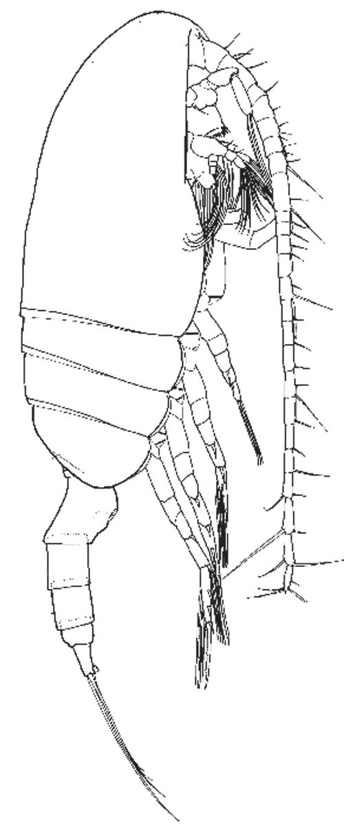
the photographs, they strongly flatten rounded shapes into a two-dimensional projection, but in the case of taxonomic work they are usually just outlines with the segment edges drawn in. For the whole animal, such a drawing (Figure 1.4) is termed a habitus drawing, often a side view. Copepods in a preserved plankton sample mostly lie on their sides, so the habitus is what you generally see through a stereomicroscope before your probes toss an animal into other postures. This drawing, by Gayle Heron from a paper by Bruce Frost,12 is unusually complete. Often the aim is just to show the shape of the body, the prosome and urosome, so the legs and setae are left out. Getting the body shape about right is easy, especially by using a camera lucida (or by tracing a photograph). Adding the realism of legs, and particularly to include setae, takes a real artist. We have some among us yet, though Heron has retired. Rony Huys of the British National Museum and several others keep great drawings coming.
Figure 1.4 Pseudocalanus moultoni, female, from Frost,12 a habitus drawing by Gayle Heron. The total length of this specimen, not including the tail setae (at left), was 1.65 mm. Permission, Canadian Journal of Zoology
Oar Feet
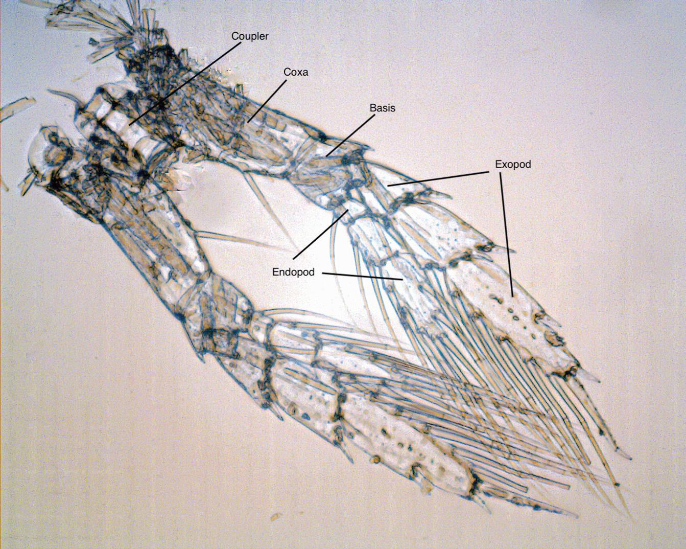
Let’s look at more construction details of a copepod starting with the thoracic legs, those oars. The paired left and right thoracic legs are also known as pereiopods: walking legs, from shrimp terminology, often abbreviated as P1 to P5. A photograph of the P4, or fourth from the front, from a specimen of N. plumchrus, is labeled in Figure 1.5. The paired left and right legs are mirror images in shape and are attached to each other below the body by a coupler plate (formally, an “intercoxal sclerite”). It forces them to move together. These legs are used for amazingly rapid escape or prey-chase swimming. When a predator is sensed moving nearby, certainly by tiny water motions sensed by the long antennules, the legs swing backward in sequence from back (P5) to front (P1). Each leg, acting as an oar, accelerates the copepod forward, which with some luck is away from an approaching predator or toward potential prey. Nothing is foolproof for protecting prey from predators. For example, a fish can actually
Figure 1.5 Neocalanus plumchrus, fifth copepodite: fourth thoracic leg (P4) from a preserved specimen. Segment names are given. Dissection and micrograph graciously provided by Atsushi Yamaguchi (University of Hokkaido). Some setae are broken, which is common for net-caught copepods.
take advantage by approaching the head, sucking in a little, and the copepod “escapes” right into its mouth.
When all of the legs have pushed back against the water and point aft (as in Figure 1.4), they swing forward (as in Figure 1.2) for another series of thrusts. The elaborate jointing of the legs facilitates that return. The muscles for the return stroke pull the coupler forward, and the leg is feathered back to reduce water resistance, or drag. Human rowers rotate their paddles on the back stroke, “feathering” them, to slip them forward through waves. On copepod legs, all the segments bend backward at their joints to present less surface to the water ahead. The joints can bend because the hard casings of the segments are joined around their edges by thin and flexible connectors (arthrodial membranes) with folds inside the articulations. The joints do not bend forward on the power strokes, because there are hard stops on their forward edges to prevent it. Tiny muscles in the segments closer to the body (termed proximal), visible in the micrograph as pale tan stripes, attach to the inner surface and reach through the joint to just inside next (more distal) segment, straightening the leg at the end of the return stroke. The main pressure against the water of a whole leg in the power stroke comes from large, fan-shaped muscles converging from the upper wall of its thoracic segment to attachments in the most proximal joint in the leg (the coxa). There are, of course, retractor muscles to help the leg back forward, but probably some of that return force comes from springiness of the exoskeleton.
Indeed, to the uninitiated there is a sort of mumbo- jumbo tone to the terminology (cephalosome, coxa, arthrodial, proximal, distal). All science can be like that, offering many odd terms. You might think the vocabulary is a sort of code designed to turn away all but the cognoscenti. And you might be right! In legal practice (habeas corpus) that is a major purpose. I introduce terms here so you can start to sense what the chatter is like at a conference of copepodologists.
A few more things about these oar feet; they have two branches (that is, they are biramous), a feature of some limbs in other crustacean groups. In copepods the outer branch (exopod) of each leg is longer than the inner branch (endopod). In many copepod species both branches have three segments, but in others some segments are fused; the legs have lost one or more articulations. The branches of some thoracic legs in different species have evolved various specialized shapes or tools. The P4 shown in the photo is close to the primitive original, or at least we (all those copepodologists) think so. The outer tips of the exopod segments have articulated spines. How those function is uncertain, but possibly they enable shedding of tiny eddies during the power strokes.
The medial sides of the exopods and both sides of the endopods bear many setae, long bristles that are part of the exoskeleton, and those bristles have short,
paired side branches called setules. The arrangement is a little like the barbs and barbules on bird feathers. As the fan of setae is spread by drag from the water during the power stroke, the array becomes a sort of webbed extension of the oar. An aspect of fluid flow, surprising to most people, is that flow along a solid surface is restrained by viscosity such that layers right at that surface do not move. There is no “slip.” That was initially proved for flowing water by the German fluid dynamicist Ludwig Prandtl,13 who published his results in 1904. Relative velocity accelerates progressively from zero at the surface out into the main flow as a boundary layer.
Mimi Koehl and Rudi Strickler14 suggested much later (in 1981) that the boundary layers around setae, and more so around setules, are close enough together to overlap and let very little water through, even as a limb is moving rapidly. Thus, the setules effectively create an almost solid surface, like oar blades, and their flexibility lets them bend to return forward by sliding almost lengthwise through the water. A little (only that) more explanation of the different sources of fluid drag is offered in a box at the end of the chapter.
The Tail Fan
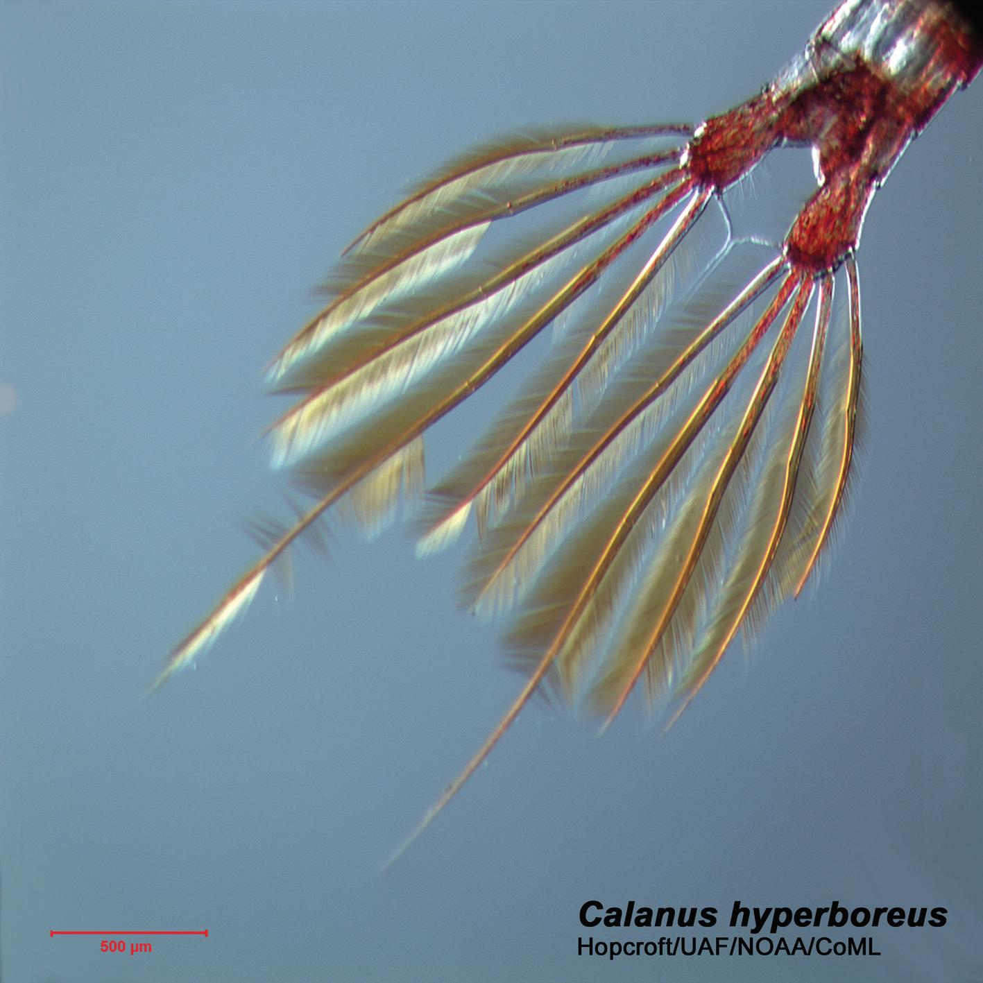
In the side view of the N. plumchrus copepodite (Figure 1.2) above, the tail fan is just a line. A dorsal view (Figure 1.6) shows that the urosome carries a fan of setae spreading from two blobs, the caudal furcae, that extend from the last segment of the urosome (location of the anus, and thus the “anal segment”—What, not Greek or Latin? Well, it is from Latin). The long muscles in the prosome and urosome can flip this fan like a fish swishing its tail. If the copepod were still this would have little effect, the viscosity of the water would hold the body almost in place, but once the thoracic legs have the whole body moving, the final swish of the fan can accelerate it further. Again, at the smaller scale of the setae and setules, the boundary layers would make it an almost solid sculling blade.
In the early 1990s, two biological oceanographers who did not usually work directly with copepods, Victor Smetacek from Germany and the late Peter Verity from the United States, were nevertheless talking about them.15 They must have asked each other why so many variations have evolved in the anterior feeding limbs and in the life ways of planktonic copepods, species varying in adult size by about a hundred-fold, when there are only minor variations in the pattern of smoothly tapered prosomes, oar-like thoracic legs cocked forward when resting and a tail fan at the rear. Their answer was that all of them have the need in common for extremely accelerated escape jumps. Surely they were right.
Figure 1.6 Calanus hyperboreus, just the anal segment, two pad-like extensions (the caudal furcae) and five stiff setae on each side. Two smaller midline setae bend together and touch along the midline. The lateral five setae have dense lines of long setules on each side that fill the plane as a tail fan. The scale in red is hard to read here; it is 500 μm (0.5 mm). Photo by Russell Hopcroft.
Escape: How Far, How Fast?
So, how fast exactly (or approximately) do copepods move when they feel something approach? Ed Busky and colleagues16 used high frame-frequency TV recordings to study jumps of several species of Acartia (about 1.0 mm long) and found speeds up to 500 body-lengths per second, or around 50 cm per second. They did not jump very far, about 6–8 mm, using 1 to 9 thrust sequences, but probably that is far enough and fast enough to provide substantially improved survival (otherwise they would evolve so as not to waste the necessary energy). They also mentioned what happens in all species: the initial acceleration swings the antennules back into the position shown in Figure 1.2.
I am fearlessly using metric units here. All scientists use them, and it becomes very hard for us to think, at least for scientific subjects, in what are now almost exclusively “American” units: inches, feet, cubic feet, and so on. Metric units are
better because of the ten-fold ratios between units adjacent with differing names (1 cm = 10 mm; 1 liter = 1,000 milliliters), and because they give Americans a language for measurements in common with the rest of the world. American readers, stick with me in this; it isn’t difficult.
Many workers beside Ed Busky have taken a turn at estimating the speed and power of copepod escape jumps, with an early entry from William Vlymen,17 who examined the drag encountered by Labidocera trispinosa as it decelerated to rest after a leap. In succeeding years and decades movies and video cameras with better and better slow-motion capability have been used to follow copepod movement in aquaria that are thin from front to back (to keep the copepods almost in focus). Leonid Svetlichny, working in Crimea, provided one of the best studies.18 He enlarged vertical and horizontal dimensions of his thin aquarium enough to see how many thrust cycles his copepods applied in rushing away from alarms. Thirty years ago he had a movie camera that could record up to 3,500 frames per second. You can buy one of those SKS-1M cameras on eBay, maybe even film for it.
Later Svetlichny worked with Thomas Kiørboe19 of Denmark (see Chapter 11) using smaller aquaria and sufficiently motion-slowing video (1,200 frames per second) to characterize the cycles of thoracic leg power strokes in several species. A graph of their results (Figure 1.7) for Calanus helgolandicus shows its speed pulses, velocity rising as the legs swing back, falling off as they are feathered forward for the next cycle. Notice the very short durations of each cycle, rising up to about 80 cm/second, then falling to 20 or 40 in about 10 milliseconds (10
Figure 1.7 Calanus helgolandicus. Initial velocity cycles of an escape jump. The x-axis is time in seconds. The black bars are the intervals in which the thoracic legs thrust back in sequence (P5, P4, . . . , P1). During the gaps between bars the legs feather into cocked position forward. From Svetlichny et al.19 Permission, Journal of Experimental Biology.
ms = 0.01 seconds). You cannot blink that fast (~100 ms at ~20 cm/second). The peak speeds of successive cycles slow slightly. I added up the distances covered as the speeds varied: 2.7 cm in 40 milliseconds. In his earlier work Svetlichny18 showed that C. helgolandicus (~3.2 mm) can sustain that rush for a full second, though the peak velocities of successive thrust cycles fall off, as they started to do in the graphed data, decreasing to approximately 40 cm/second before thrusts ceased. The final distance when again at rest can be nearly a meter, maybe 300 body lengths.
Petra Lenz and colleagues,20 also using video, measured top speeds of Calanus finmarchicus (~3 mm prosome) in the same range, around 80 cm per second. So an adult Calanus arrives almost a meter away from an attacking fish, when viscosity drags it again to a complete stop. Visual paths are short in water, and shorter for nearly transparent (Figure 1.2) and tiny objects, so the jump provides a substantial gain in safety. Just as important, Lenz showed that an escape response to a new vibrational stimulus initiates in about two milliseconds for Calanus, faster than the proverbial greased lightning. That quickness comes from very rapid transmission of motor impulses from the antennules along “giant” neurons in the ventral nerve cord. All of the invertebrates with their main along-body nerve below the gut (annelids, molluscs, arthropods) have these giant neurons carrying the news of danger to the muscles in order to initiate immediate movement. In copepods those nerves are also short. Repeating the cycling requires extremely fast recharge of muscles with fuel, with the adenosine-triphospate (ATP) molecules that are the energy currency of all animals. Lots of mental exercise has come from calculating the power output capacity of copepod muscles, showing that it is probably the greatest (per gram!) in the animal kingdom.
There are refinements. Petra Lenz and colleagues discovered in the late 1990s21 that some copepods (e.g., Calanus, Bestiolina, and Parvocalanus) have myelinated axons, whereas some other copepods do not (e.g., Acartia, Eurytemora, and Centropages). Axons with myelin are likely, based on data from animals generally, to have much faster impulse conduction than bare nerve fibers. More recently, Lenz, with Ed Busky and other colleages,22 conducted experimental observations with high-speed cameras by applying pulsed and directional hydrodynamic disturbances to copepods from both groups. Copepodites of “amyelinate” species escaped in the direction they were already headed when the pulse hit them. The danger of that, noted above, is that a signal from in front can cause them to swim toward an attacker. Copepodites of two myelinate species, Bestiolina and Parvocalanus, swam away from the stimulus source regardless of where it hit them. The myelin evidently provides more time for processing the signals from setal sensors around the body in order to leap away.
Resting
Finally, moving all the way back, the center of the tail fan of N. plumchrus has those two amazing setae, termed the II bristles, with orange, ribbon-like setules (Figure 1.2). Most of the time copepods are not escaping; they are hanging nearly motionless in the water. Mark Benfield23 measured the orientations of 152 individual older copepodites and adults of Calanus finmarchicus (a familylevel relative of N. plumchrus) from pictures of them captured by high-speed video cameras towed through the ocean while recording. It is possible for such cameras to record from far enough ahead that the pressure wave from their approach does not obviously disturb animals until after their images are recorded. A strong majority of specimens were oriented head up, tail down, with the antennules extending to either side. That is also how live specimens orient in aquaria when eating (or, for all we know, just thinking). If a preserved specimen of N. plumchrus with intact II bristles is released tail first into water, the ribbons spread out into a sort of three-dimensional feather (much as in Figure 1.2), a sort of sea anchor resisting sinking. If you pull that specimen forward with forceps through the water, the ribbons fold back along the bristle, reducing their drag. That is surely to accommodate the need for high escape velocity. Similar vertical sea anchors are found in other families, notably the Euchaetidae (Euchaeta and Paraeuchaeta). You will meet them further on.
Enough about the back end; let’s move now to the front.
Box 1 About Drag
Motion of an animal through water is resisted by drag of two kinds. Both require work to overcome the resistance. One, the water has mass, so it must be accelerated out from in front of the animal, and then accelerated in the opposite direction to fill the space the animal has just left. Basically, the latter is done by accelerating the body forward, pushing against the inertia of the water. Work must be done to impart new momentum to the water in the way, force motion where there was none before. The rule is Newton’s Second Law: Force needed = Mass × Acceleration
Applying that to fluid motion is more complicated than calculating the speed of a ball falling through a vertical vacuum, but it can be done. The amount of work needed to overcome this inertial drag depends on how far the water must move, farther if a beast is bigger and on how fast it is moving. For a given shape, inertial drag increases as the square of the velocity. It also
depends on the density of the water. Heavier water that is colder or saltier takes more work than does warmer, fresher and thus lighter water.
Two, water is sticky: the molecules are attracted to each other and to the animal’s surfaces. So, moving through them requires work against viscous drag, work to stretch and rearrange those attractive forces. The amount of that work is greater if the intermolecular forces are greater: stronger in cold water than warm. Intermolecular forces are always present, and the faster an animal swims the greater the rate of rearrangements, the resulting drag increasing proportionally with velocity.
The difference in velocity’s effect on inertial and viscous drag means that viscous drag becomes relatively less significant as a copepod accelerates. But as it slows, not expending propulsive power, viscous drag quite rapidly hauls it to a stop. Copepods usually are a little denser than water and tend to sink, but slowly, and viscous drag dominates. So, when resting, viscosity tends to hold them almost in place. They are in exactly the right size range for this to work well. They are also the right size for inertial drag to make their thoracic legs effective oars, their tail fan effective for sculling.
When swimming, people experience drag dominated by inertial effects, including long glide paths. A viscosity-dominated regime has seemingly counterintuitive effects, such as miniscule glide paths, essentially none. Another key effect in respect to swimming is that the viscous drag along the length of a moving seta (or any cylinder) is half that of flow around it. So, small crustaceans push setae side-on through water during power strokes and slide them through lengthwise on return strokes. That is also why protozoan flagellae, at sizes smaller than setae, can act as propellers. More effects of viscosity will come up in respect to feeding mechanics (in Chapter 7).
References
1. Harvey, T. H. P., & B. E. Pedder (2014) Copepod mandible palynomorphs from the Nolichucky shale (Cambrian, Tennessee): implications for the taphonomy and recovery of small carbonaceous fossils. Palaios 28(5): 278–284.
2. Regier, J. C., J. W. Shultz, A. Zwick, A. Hussey, B. Ball, R. Wetzer, J. W. Martin, & J. W. Cunningham (2010) Arthropod relationships revealed by phylogenomic analysis of nuclear protein-coding sequences. Nature 463: 1079–1083.
3. Damkaer, D. (2002) The Copepodologist’s Cabinet, A Biographical and Bibliographical History. Two volumes to date. American Philosophical Society, Philadelphia.
4. Evans, J. Dennis (1978) The Book of Laws Founded Against Itself: The Poetry of A.R. Ammons, 1951–1976. PhD dissertation, University of California, Berkeley.
5. West, Robert M., ed. (2017) “Hymn IV.” In The Complete Poems of A. R. Ammons, Vol. 1, 1955–1977. W.W. Norton & Co., New York.
6. Hopcroft, R. R., D. B. Ward, & R. C. Roff (1985) The relative significance of body surface and cloacal respiration in Polus fabricii (Holothuroidea: Dendrochirotida). Canadian Journal of Zoology 64: 2878–2881.
7. Roff, J. C., & R. R. Hopcroft (1986) High precision microcomputer based measuring system for ecological research. Canadian Journal of Fisheries and Aquatic Sciences 43: 2044–2048.
8. Clarke, C., & R. C. Roff (1990) Abundance and biomass of herbivorous zooplankton off Kingston, Jamaica, with estimates of their annual production. Estuarine, Coastal and Shelf Science 31: 423–437.
9. Hopcroft, R. R. (1997) Size-Related Patterns in Growth Rate and Production of Tropical Marine Planktonic Communities Along a Trophic Gradient. PhD dissertation, University of Guelph.
10. Hopcroft, R. R., & J. C. Roff (1996) Zooplankton growth rates: diel egg production in the copepods Oithona, Euterpina and Corycaeus from tropical waters. Journal of Plankton Research 18: 789–803.
11. Hopcroft, R. R., & B. H. Robison (2005) New mesopelagic larvaceans in the genus Fritillaria from Monterey Bay, California. Journal of the Marine Biological Association, U.K 85: 665–678.
12. Frost, B.W. (1989) A taxonomy of the marine calanoid copepod genus Pseudocalanus. Canadian Journal of Zoology 67: 525–551.
13. Anderson, John D. Jr. (2005) Ludwig Prandtl’s boundary layer. Physics Today 58: 42–48.
14. Koehl, M., & J. R. Strickler (1981) Copepod feeding currents: food capture at low Reynolds number. Limnology and Oceanography 26: 1062–1073.
15. Verity, P., & V. Smetacek (1996) Organism life cycles, predation and the structure of marine ecosystems. Marine Ecology Progress Series 130: 277–293.
16. Buskey, E. J., P. H. Lenz, & D. K. Hartline (2002) Escape behavior of planktonic copepods in response to hydrodynamic disturbances: high speed video analysis. Marine Ecology Progress Series 235: 135–146.
17. Vlymen, W. (1970) Energy expenditure of swimming copepods. Limnology and Oceanography 15(3): 348–356.
18. Svetlichny, L. (1987) Speed, force and energy expenditure in the movement of copepods. Oceanology 27: 497–502.
19. Svetlichny, L., P. S. Larson, & T. Kiørboe (2018) Swim and fly: escape strategy in neustonic and planktonic copepods. Journal of Experimental Biology 221: doi:10.1242/ jeb.167262.
20. Lenz, P. H., A. E. Hower, & D. K. Hartline (2004) Force production during pereiopod power strokes in Calanus finmarchicus. Journal of Marine Systems 49: 133–144.
21. Davis, A. D., T. M. Weatherby, D. K. Hartline, & P. H. Lenz. (1999) Myelin-like sheaths in copepod axons. Nature 398: 571.
22. Buskey, E. J., J. R. Strickler, C. J. Bradley, D. K. Hartline, & P. H. Lenz (2017) Escapes in copepods: comparison between myelinate and amyelinate species. Journal of Experimental Biology 220: 754–758.
23. Benfield, M., C. S. Davis, & S. M. Gallager (2000) Estimating the in-situ orientation of Calanus finmarchicus on Georges Bank using the Video Plankton Recorder. Plankton Biology and Ecology 47: 69–72.
2
The Front End
Olfaction, Motion Sensors, Feeding Limbs Teeth
The head end, or cephalosome (“head body”) of a generalized copepod, one much like Calanus helgolandicus (~2.5 mm) that is common in British waters and south to Spain, was drawn side view (Figure 2.1) and published in 1928 by H. Graham Cannon.1 His picture shows the general layout of the complex tangle of limbs for dealing with aspects of copepod life other than escape and reproduction. Things could get technical here, and I will go there later. Instead, let’s skip to the mandible (labeled in Figure 2.1) to finish explaining the book title.
Barbara Sullivan Finds Opals in Copepod Mouths
The most usual format of the mandible (Figure 2.22) includes the coxa, articulating with the copepod’s ventral surface, and a palp. The mandibular palp is two basis segments and two segmented branches: exopod and endopod. It extends below and alongside the body (Figure 1.2) and can swing forward and back, moving water toward the mouth or away from it. That is part of feedingcurrent generation. The coxae bear relatively long side branches (edites), known as the mandibular gnathobases, ending in chewing surfaces that are the copepod’s jaws. Left and right gnathobases extend under the labrum (a fat front lip) and meet inside the mouth. They are tilted enough (Figure 2.3) that the little spine, shown at the bottom in Figure 2.2, is termed the dorsal seta.
Barbara K. Sullivan (Figure 2.4), a graduate student working with me at Oregon State University (OSU) in the early 1970s, discovered in the course of studying the gut contents of arrow worms (chaetognaths) that teeth of many copepods have opal crowns,3 which led to the title of this book. Discoveries are not always planned; often enough they just happen along the way. Arriving at OSU after my undistinguished postdoctoral year in New Zealand (neither my advisor there nor I had any clear idea what I should do as a postdoc), I needed to find research projects. That went quickly enough, but I was expected also to begin advising graduate students. There is no training whatever for that, except having been one. It was also expected that I would generate stipends for students