I would like to present another edition that is older and wiser with the help of your comments and suggestions. Even after minor surgery, a few transplants, and some trimming and plumping, the fifth edition of The Human Body in Health and Illness has emerged with the same sense of humor!
The Human Body in Health and Illness tells the story of the human body with all its parts and the way these parts work together. It is a story that we have told many times in our classes. It is also a story that gets better with each telling as the body continues to reveal its mysteries and how marvelously it has been created. I hope that you enjoy telling the story as much as I do.
The Human Body in Health and Illness is a basic anatomy and physiology text addressed to the student preparing for a career in the health professions. It is written for students with minimal preparation in the sciences; no prior knowledge of biology, chemistry, or physics is required. The text provides all the background science information needed for an understanding of anatomy and physiology.
The basic principles of chemistry and biochemistry are presented in Chapters 2 and 4, and they set the stage for an understanding of cellular function, fluid and electrolyte balance, endocrine function, and digestion. Chapter 5, Microbiology Basics, presents clinically relevant microbiological topics. Check out the stories “Rick, Nick, and the Sick Tick” and “Dr. Semmelweis Screams: ‘Wash Those Mitts!’ ” The latter is an amusing presentation of a sad tale in the history of medicine and corresponds to the current emphasis on hand hygiene and healthcare-associated infection.
The anatomy and physiology content is presented in a traditional order, from simple to complex. The text begins with a description of a single cell and progresses through the various organ systems. There are two key themes that run throughout the text: (1) the relationship between structure and function—the student must understand that an organ is anatomically designed to perform a specific physiological function, and (2) homeostasis—the role that each organ system plays in sustaining life and what happens when that delicate balance is disturbed.
The text addresses two concerns about the selection of content. The first has to do with the amount of content. The field of anatomy and physiology is huge; therefore, there must be a selection of content that can be mastered in the short period of time that a
semester (or even two) allows. This text focuses on the physiology that is basic and most clinically relevant. Pathophysiology is introduced primarily to clarify physiological function. For instance, the different types of anemias illustrate the various steps in the making of the red blood cell. A second concern has to do with the recognition that we are not preparing physiologists; instead, we want the student to be able to use the physiology to understand clinically relevant content such as pathophysiology, physical assessment, diagnostics, and pharmacology. An understanding of physiology is crucial for advancement in the medically related sciences.
TEXTBOOK STRENGTHS
• Anatomy and physiology are clearly and simply explained. A meticulously prepared set of illustrations—complete with amusing cartoons— supports the text. In fact, the story of the body is told as much through the art as through the written word.
• The text truly integrates pathophysiology; it is not merely boxed in or tacked on at the end. The integrated pathophysiology is used primarily to amplify the normal anatomy and physiology. The expanded Medical Terminology and Disorders tables and frequent references to common medical terminology allow the text to be used for an introductory course in pathophysiology and medical terminology.
• In addition to the pathophysiology, other topics are liberally integrated throughout the text. These include common diagnostic procedures such as blood count, lumbar puncture, urinalysis, and electrocardiography. Pharmacological topics are also introduced and, like the pathophysiology, are used to amplify the normal anatomy and physiology. For instance, the discussion of the neuromuscular junction is enhanced by a description of the effects of the neuromuscular-blocking agents. Because of the effort of the text to make clinical correlations, it sets the stage for the more advanced health science courses, including pharmacology and medicalsurgical nursing.
• Re-Think boxes are liberally distributed throughout each chapter and encourage students to master that content before progressing through the chapter. New Ramp It Up boxes develop selected clinically
relevant topics that are simply too advanced to be included in the text as critical information. These boxed features contain new or advanced content commonly used in the clinical setting and allow instructors to scale their coverage in a manner appropriate to the course. They offer students the chance to make further connections between the text and their future careers. (See the To the Student preface on page ix for descriptions and examples of each of the chapter features.)
• Medical terminology is introduced, defined, and used throughout the text. Common clinical terms such as hyperkalemia, vasodilation, hypertension, and diagnosis are defined and reused so that the student gradually builds up a substantial medical vocabulary. The new and expanded Medical Terminology and Disorders tables were deliberately constructed to maximize the use of common medical terms and disorders. To help foster broader understanding of medical terminology, word parts and their meanings are included for nearly every term presented. Repetition of these helps students gain greater ground in understanding the very specific medical language they will be learning to use for a future in the health professions. A description is also provided, which gives the definition or other pertinent information on the topic.
• The Review Your Knowledge section has been expanded to include questions that require an analytical response. The Go Figure questions are based on the story told by the artwork. The questions can only be answered by analyzing the art and/or the information presented in the tables. This exercise encourages the student to see beyond the “pretty pictures” and realize that a picture is truly worth a thousand words.
• The text is supported by many activities, exercises, puzzles, and games (e.g., Body Bingo) on Evolve (http://evolve.elsevier.com/Herlihy). These activities emphasize the focus of this text—clinically relevant anatomy and physiology.
• Last, the text incorporates many amusing anecdotes from the history of medicine. Although the human body is perfectly logical and predictable, we humans think, do, and say some strange things. Tales from the medical crypt provide some good laughs and much humility.
CLASSROOM RESOURCES
Materials from the Study Guide and Evolve Instructor Learning Resources can be used to:
1. Remediate students who are having difficulty in grasping the content
2. Remediate students who have missed class(es)
3. Review students engaged in pathophysiology and pharmacology whose memories need to be refreshed in the physiology
STUDY GUIDE
The Study Guide for The Human Body in Health and Illness offers something for students at all levels of learning and is a ready-made resource for instructors looking for homework assignments. Each chapter includes three parts: Part I, Mastering the Basics, with matching, labeling, and coloring exercises; Part II, Putting It All Together, containing multiple-choice practice quizzes, completion exercises, and case studies; and Part III, Challenge Yourself!, which has grouping exercises and word puzzles. Textbook page references are included with the questions, and the answer key is available on the Evolve website, only to instructors.
EVOLVE INSTRUCTOR LEARNING RESOURCES
The Evolve website for The Human Body in Health and Illness (http://evolve.elsevier.com/Herlihy) includes all of the Student Resources (see the inside back cover), as well as the following Instructor Resources:
• Answer Key for the Study Guide, Audience Response System questions, Image Collection, Teaching Tips, and ExamView Test Banks that include over 2300 questions—more than 600 questions have been added!
• Instructor’s Chapter Exams, Classroom Activities— including Bingo, Line ‘Em Up, Sorting, and Word Puzzles—and the TEACH Instructor Resource.
TEACH Instructor Resource on Evolve
Instructors who adopt the textbook will also receive access to the TEACH Instructor Resource, which links all parts of the Herlihy educational package with customizable Lesson Plans based on objectives drawn from the text. The TEACH Lesson Plans are based on the chapter-by-chapter organization of The Human Body in Health and Illness and can be modified or combined to meet your curriculum’s scheduling and teaching needs.
TEACH has been completely updated and revised for this edition. The TEACH Lesson Plans help instructors prepare for class and make full use of the rich array of ancillaries and resources that come with the textbook. The content covered in each textbook chapter is divided across one or more lesson plans, each designed to occupy 50 minutes of class time. Lesson plans are organized into easily understandable sections that are each tied to the chapter learning objectives:
• Instructor Preparation This section provides a checklist of all the things you need to do to prepare for class, including a list of all the items you need to bring to class to perform any activity or demonstration included in the lesson plan, and all pertinent key terms covered in that lesson.
• Student Preparation Textbook readings, study guide exercises, online activities, and other applicable
homework assignments for each lesson are provided here along with an overall estimated completion time.
• The 50-Minute Lesson Plan A lecture outline that reflects the chapter lecture slides that come as part of TEACH is included, as well as classroom activities and online activities, one or more critical thinking questions, and time estimates for the classroom lecture and activities.
• Assessment Plan To ensure that your students have mastered all the objectives, the new TEACH includes a separate Assessment Plan section. An easy-to-use table maps each assessment tool to the lesson plans and chapter objectives so you can see all your assessment options—by chapter, by lesson, and by objective—and choose accordingly.
To the Student
This book will take you on an amazing journey through the human body. You will learn many body parts, and more importantly, how they work in an integrated manner to keep you going. You will use this information in your clinical practice when patients become ill with disorders of those structures. The following special features were created to help make learning enjoyable and fun.
TEXTBOOK FEATURES
KEY TERMS
Key terms are listed at the beginning of each chapter along with a page reference. Each is (1) presented in the text in blue print, (2) accompanied by a pronunciation guide, (3) thoroughly explained within the chapter, and (4) defined in the glossary.
OBJECTIVES
Numbered objectives identify the goals for each chapter.
ILLUSTRATIONS
Original illustrations and full-color cartoons help you make sense of anatomy and physiology using humor, clarity, and insight.
MEDICAL TERMINOLGY AND DISORDERS TABLES
These tables describe medical terms and specific disorders related to individual body systems, with a focus on developing a strong working vocabulary, which is necessary for a career in the health professions.
END-OF-CHAPTER FEATURES
Summary Outline
A detailed outline at the end of each chapter summarizes key concepts and serves as an excellent review of the chapter content. Use it as a study tool to review your reading and prepare for exams.
Review Your Knowledge
The matching and multiple-choice questions in this section cover the major points of the chapter and allow you to test your comprehension.
Go Figure
This review section asks you to reflect on the figures in the chapter and reinforces the importance of the concepts presented.
ANSWERS TO REVIEW YOUR KNOWLEDGE AND GO FIGURE QUESTIONS
DO YOU KNOW…
Most of these boxed vignettes refer to clinical situations; others relate to interesting and amusing historical events related to anatomy and physiology.
RAMP IT UP!
These features challenge you with more advanced anatomy and physiology topics.
RE-THINK
Questions included cover key information found in the chapter and help reinforce important concepts.
AS YOU AGE
These boxed features contain numbered lists describing how human anatomy and physiology is affected by the aging process.
SUM IT UP!
These features appear regularly throughout the chapters and help the student synthesize key concepts.
The Appendix contains answers to all Review Your Knowledge and Go Figure questions found in the textbook.
GLOSSARY
The glossary includes a pronunciation guide and a brief definition of all key terms and many other words in the text.
STUDY GUIDE
Enhance your learning of the textbook content with the accompanying Study Guide for The Human Body in Health and Illness. The Study Guide has something to offer students at all levels of learning, from labeling and coloring exercises to multiple-choice practice tests and case studies.
1 Introduction to the Human Body, 1
Anatomy and Physiology: What They Are, 1
What’s It Mean?, 1
Why Do I Need to Know This?, 1
The Body’s Levels of Organization, 2
Major Organ Systems, 2
Homeostasis: Staying the Same, 6
Anatomical Terms: Talking About the Body, 6
Anatomical Position, 6
Relative Positions, 6
Planes of the Body, 7
Regional Terms, 8
Cavities of the Body, 9
Dorsal Cavity, 9
Ventral Cavity, 10
2 Basic Chemistr y, 15
Matter, Elements, and Atoms, 15
Matter, 15
Elements, 15
Atoms, 16
Chemical Bonds, 18
Ionic Bonds, 18
Covalent Bonds, 18
Hydrogen Bonds, 18
Ions, 20
Cations, Anions, and Electrolytes, 20
Ion Formation, 20
Ionization, 20
Molecules and Compounds, 20
Molecules, 20
Compounds, 21
Some Important Compounds and Molecules, 21
Chemical Reactions, 22
Acids and Bases, 22
Acids, 23
Bases, 23
Neutralization of Acids and Bases, 23
Measurement: The pH Scale, 23
Energy, 24
Forms of Energy, 24
Conversion of Energy, 25
Energy Transfer: The Role of Adenosine Triphosphate, 25
Mixtures, Solutions, Suspensions, and Precipitates, 25
3 Cells, 30
Typical Cell, 30
Cell Membrane, 30
Inside the Cell, 31
On the Cell Membrane, 35
Movement across the Cell Membrane, 36
Passive Transport Mechanisms, 36
Active Transport Mechanisms, 40
Cell Division, 41
Cell Cycle, 42
Cell Differentiation, 43
Stem Cells, 43
Order, Disorder, and Death, 44
4 Cell Metabolism, 48
Metabolism, 48
Carbohydrates, 48
Monosaccharides, 48
Disaccharides, 49
Polysaccharides, 49
Uses of Glucose, 50
The Breakdown of Glucose, 50
The Making of Glucose, 51
Lipids (Fats), 52
Uses of Lipids, 54
Metabolism of Lipids, 54
Proteins, 54
Amino Acids, 54
Uses of Proteins, 55
Protein Synthesis and DNA, 56
DNA Structure, 57
Steps in Protein Synthesis, 59
5 Microbiology Basics, 64
What Is Disease?, 64
Types of Pathogens, 64
Microorganisms (Microbes), 65
Other (Multicellular) Disease-Causing Organisms, 67
Laboratory Identification of Pathogens, 68
The Spread of Infection, 69
Portals of Entry and Exit, 69
Five Germ-Laden Stories, 70
Dr. Semmelweis Screams, “Wash Those Mitts!”, 70
Flora and Her Vaginal Itch, 71
Rick, Nick, and the Sick Tick, 71
Why Typhoid Mary Needed to Lose Her Gallbladder, 72
A Pox News Alert!, 72
6 Tissues and Membranes, 77
Epithelial Tissue, 77
Where Is It Found?, 77
What Does It Do?, 77
What Is It Like?, 77
Classification, 78
Connective Tissue, 81
Where Is It Found?, 81
What Does It Look Like?, 81
Types of Connective Tissue, 82
Nervous Tissue, 85
Neurons, 85
Muscle Tissue, 85
Skeletal Muscle, 86
Smooth Muscle, 86
Cardiac Muscle, 86
Tissue Repair, 86
Membranes, 88
Classification of Membranes, 88
7 Integumentar y System and Body
Temperature, 93
Functions of the Integumentary System, 93
Structure of the Skin, 94
Layers of the Skin, 94
Skin Color, 96
Accessory Structures of the Skin, 97
Hair, 97
Nails, 98
Glands, 99
Body Temperature, 101
Heat Production, 101
Heat Loss, 101
Regulation, 101 When Skin Is Burned, 103
A Note About Skin Care, 105
8 Skeletal System, 111
Arrangement and Functions of Bones, 111
The Skeletal System: What It Does, 111
Many Sizes and Shapes of Bones, 113
Bone Tissue and Bone Formation, 113
Compact and Spongy Bone, 114
Long Bones, 115
Ossification, 115 Growing Bones, 115
Divisions of the Skeletal System, 117
Axial Skeleton, 117
Appendicular Skeleton, 126
Joints (Articulations), 132
Joint Classification, 133
Naming Joints, 134
Moving Synovial Joints, 134
Clinically “Big” Synovial Joints, 135
9 Muscular System, 142
Types and Functions of Muscles, 142
Skeletal Muscle, 142
Smooth Muscle, 142
Cardiac Muscle, 143
Structure of the Whole Muscle, 143
Muscle, 143
Layers of Connective Tissue, 144
Muscle Attachments, 145
Structure and Function of a Single Muscle Fiber, 145
How Muscles Contract, 145
Sliding Filament Mechanism, 145
The Role of Calcium and Adenosine Triphoshate, 145
Skeletal Muscles and Nerves, 147
Somatic Motor Neuron, 147
The Motor Unit, 147
The Neuromuscular Junction, 147
Force of Muscle Contraction, 150
Contractile Force of a Single Muscle Fiber, 150
Contractile Force of a Whole Muscle, 151
Muscle Tone, 151
Energy Source for Muscle Contraction, 151
Muscle Fatigue, 152
Muscle Terms, 152
Origin and Insertion, 152
Prime Mover, Synergist, and Antagonist, 152
Muscle Overuse and Underuse Terms, 153
How Skeletal Muscles Are Named, 153
Muscles from Head to Toe, 154
Muscles of the Head, 154
Muscles of the Neck, 160
Muscles of the Trunk, 160
Muscles of the Shoulder (Pectoral) Girdle and Arm, 162
Muscles That Move the Forearm, 163
Muscles That Move the Wrist, Hand, and Fingers, 163
Muscles That Move the Thigh, Leg, and Foot, 164
10
Ner vous System: Ner vous Tissue and Brain, 172
The Nervous System: Structure and Function, 172
Divisions of the Nervous System, 172
Functions of the Nervous System, 172
Cells That Make Up the Nervous System, 173
Neuroglia, 173
Neuron, 173
White Matter Versus Gray Matter, 175
The Neuron Carrying Information, 175
The Nerve Impulse: What It Is, 175
The Nerve Impulse: What Causes It, 176
The Nerve Impulse: What Causes It to Move, 177
The Nerve Impulse: What Causes It to Move
Quickly, 178
Synapse across Neurons, 179
Parts of a Synapse, 179
Events at the Synapse, 180
Brain: Structure and Function, 182
Cerebrum, 182
Diencephalon, 186
Brain Stem, 187
Cerebellum, 188
Structures across Divisions of the Brain, 188
Protecting the Central Nervous System, 190
Bone: First Layer of Protection, 190
Meninges: Second Layer of Protection, 190
Cerebrospinal Fluid: Third Layer of Protection, 191
Blood–Brain Barrier: Fourth Layer of Protection, 193
11 Ner vous System: Spinal Cord and Peripheral Nerves, 200
What the Spinal Cord Is, 200
Location and Size, 200
Gray on the Inside, White on the Outside, 201
Spinal Nerves Attached to the Spinal Cord, 203
What the Spinal Cord Does, 203 Reflexes, 204
What Reflexes Are, 204
The Reflex Arc, 205
Many, Many Reflexes, 205
Peripheral Nervous System, 206 Nerves, 206
Classifying the Peripheral Nervous System, 206
12 Autonomic Ner vous System, 218
Autonomic (Visceral) Reflexes, 218
What They Do, 218
Pathway, 218
Organization and Function of the Autonomic Nervous System, 219
Division of the Autonomic Nervous System, 219
Autonomic Tone and Vasomotor Tone, 221
Autonomic Nervous System Neurons, 221
Numbers and Ganglia, 221
Neurons of the Sympathetic Nervous System, 221
Neurons of the Parasympathetic Nervous System, 222
Compatibility and Incompatibility of Blood Types, 300
Rh Classification System, 301
16 Anatomy of the
Heart
, 308
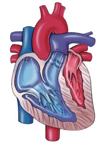
Function, Location, and Size of the Heart, 308
Layers and Covering of the Heart, 308
Endocardium, 308
Myocardium, 309
Epicardium, 309
Pericardium, 310
A Double Pump and Two Circulations, 310
The Heart’s Chambers and Great Vessels, 311
Right Atrium, 311
Right Ventricle, 312
Left Atrium, 312
Left Ventricle, 312
Great Vessels of the Heart, 313
Heart Valves, 313
Atrioventricular Valves, 313
Semilunar Valves, 314
Heart Sounds, 315
Pathway of Blood Flow through the Heart, 315
Blood Flow and Shunts, 315
Blood Supply to the Myocardium, 316
Ischemia and Infarction, 317
Cardiac Enzymes and Leaky Cells, 318
Cardiac Conduction System, 318
Parts of the Cardiac Conduction System, 318
Automaticity and Rhythmicity, 320
Electrocardiogram, 320
17 Function of the Heart, 325
The Coordinated and Adaptable Pump, 325
Cardiac Cycle, 325
Autonomic Control of the Heart, 326
Heart Talk, 330
Heart Talk: Clinical Terms, 330
Heart Talk: Receptor Language, 331
The Failing Heart: When the Heart Can’t
Pump, 332
Left Heart Failure, 332
Right Heart Failure, 333
18 Anatomy of the Blood Vessels, 340
Circles, Circuits, and Circulations, 340
Blood Vessels, 341
Naming the Blood Vessels, 341
Blood Vessel Walls: The Layered Look, 341
Blood Vessels: What They Do, 342
Major Arteries of the Systemic Circulation, 343
Aorta, 343
Branches of the Aorta, 344
Major Veins of the Systemic Circulation, 346
Venae Cavae, 346
Special Circulations, 348
Blood Supply to the Head and Brain, 348
Blood Supply to the Liver and the Hepatic Portal Circulation, 350
Fetal Circulation, 351 Pulse, 353
What Is a Pulse?, 353
What Can You Learn About a Patient by Feeling the Pulse?, 353
19 Functions of the Blood Vessels, 357
Blood Vessels Deliver, 357
Blood Vessels Regulate Blood Pressure, 357
Measurement of Blood Pressure, 357
Blood Pressure in Different Blood Vessels, 359
What Determines Blood Pressure?, 360
How Blood Pressure Stays within Normal Limits, 362
Blood Vessels Act as Exchange Vessels, 364
What Is an Exchange Vessel?, 364
Why Capillaries Are Good Exchange Vessels, 365
Capillary Forces: Exchange, 365
Blood Vessels Distribute Blood, 367
Blood Vessels Regulate Body Temperature, 367
20 Lymphatic System, 373
The Lymphatic System, 373
Lymph: What It Is, Where It Comes From, 373
Lymphatic Vessels, 373
Movement through the Lymphatic Vessels, 374
Lymphoid Organs, 374
Lymph Nodes, 375
Tonsils, 376
Thymus Gland, 376
Spleen, 378
21 Immune System, 382
Classification of the Immune System, 382
Nonspecific Immunity, 382
Specific Immunity: Third Line of Defense, 386
Types of Immunity, 390
Genetic Immunity, 391
Acquired Immunity, 391
Other Immune Responses, 392
Allergic Reactions, 392
Autoimmune Disease, 393
Organ Rejection, 393
22 Respirator y System
, 399
Structure: Organs of the Respiratory System, 399
Upper and Lower Respiratory Tracts, 399
Nose and Nasal Cavities, 399
Pharynx, 401
Larynx, 401
Trachea, 404
Bronchial Tree: Bronchi, Bronchioles, and Alveoli, 404
Lungs, 406
Pleural Membranes, 407
Collapsed and Expanded Lungs, 407
Why Lungs Collapse, 408
Why Lungs Expand, 409
Saying It Another Way: Compliance, 410
Respiratory Function, 411
Three Steps in Respiration, 411
Amounts of Air, 416
Control of Breathing, 418
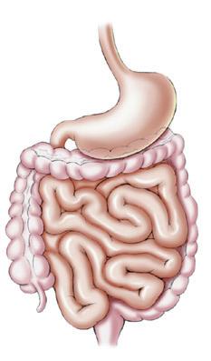
23 Digestive System, 428
Overview of the Digestive System, 428
Digestion and Absorption, 429
Layers, Nerves, and Membranes, 429
Structures and Organs, 431
Mouth, 431
Pharynx, 433
Esophagus, 434
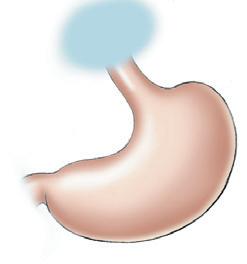
Stomach, 434
Small Intestine, 437
Large Intestine, 438
Accessory Digestive Organs, 443
Liver, 443
Gallbladder, 445
Pancreas, 445
When Accessory Digestive Organs Are Not Working Right, 446
Digestion and Absorption, 448
Carbohydrates and Carbohydrate-Splitting Enzymes, 448
Proteins and Protein-Splitting Enzymes, 449
Fats, Bile, and Fat-Splitting Enzymes, 449
Nutrition: Concepts to Know, 449
Carbohydrates, 450
Proteins, 450
Fats (Lipids), 451
Vitamins, 451
Minerals, 452
Health and a Balanced Diet, 452
Body Energy, 453
24 Urinar y System, 460
Excretion, 460
Organs of Excretion, 460
Urinary System Organs, 460
Urinary System Terms, 461
Kidneys, 461
Location, 461
Structure, 461
Blood Supply, 461
Nerve Supply, 461
Functions of the Kidneys, 461
Urine Making: The Nephron Unit, 462
Structures, 462
Urine Formation, 463
Hormones That Work on the Kidneys, 465
Aldosterone, 465
Antidiuretic Hormone, 466
Natriuretic Peptides, 467
Parathyroid Hormone, 467
Composition of Urine, 467
Uremia and Dialysis, 468
Your Plumbing, 469
Ureters, 470
Urinary Bladder, 470
Urination, 472
Urethra, 472
An Autonomic Moment, 473
25 Water, Electrolyte, and Acid–Base Balance, 478
Body Fluids: Distribution and Composition, 478
Fluid Compartments, 478
Composition of Body Fluids, 478
Water Balance, 479
Water Intake, 479
Water Output, 479
Water Imbalances, 479
Why Does Fluid Shift?, 480
Fluid Spacing—In Other Words, 481
Electrolyte Balance, 481
Quick Reference: Electrolytes, 481
Most Important Ions, 481
Acid–Base Balance, 483
Quick Reference: Acids and Bases, 483
Where the Acid (H+) Comes From, 483
How the Body Regulates pH, 483
Acid–Base Imbalances, 484
Acidosis, 485
Alkalosis, 485
26 Reproductive Systems, 490
Male Reproductive System, 490
Testes, 490
Genital Ducts, 491
Accessory Glands, 494
Semen, 494
External Genitals, 494
Male Sexual Response: Erection, Emission, Ejaculation, and Orgasm, 494
Male Sex Hormones, 495
Female Reproductive System, 496
Ovaries, 496
Genital Tract, 498
External Genitals, 500
Female Sexual Response, 500
Hormonal Control of the Reproductive Cycles, 501
Two Reproductive Cycles, 501
Methods of Birth Control, 504
27 Human Development and Heredity, 511
Fertilization, 511
When Fertilization Occurs, 511
Where Fertilization Occurs, 512
How Fertilization Occurs, 512
Human Development, 512
Prenatal Development, 512
Early Embryonic Period, 512
Embryonic Period, 514
Fetal Period, 518
Changes in the Mother’s Body during Pregnancy, 519
Birth of Baby, 520
Labor, 520
Female Breast and Lactation, 522
Structure of a Breast: The Mammary Glands, 522
Got Milk?, 522
Postnatal Changes and Developmental Stages, 522
Immediate Adjustments, 522
Development as a Lifelong Process, 524
Heredity, 524
DNA, Genes, and Chromosomes, 524
It’s a Boy, It’s a Girl: How the Sex of the Child Is Determined, 527
Congenital and Hereditary Disease, 527
Answers to Review Your Knowledge and Go
Figure Questions, 531
Glossary, 535
Index, 545
Introduction to the Human Body
Key Terms
abdominopelvic cavity (p. 10)
anatomical position (p. 6)
anatomy (p. 1)
cranial cavity (p. 9)
dorsal cavity (p. 9)
frontal plane (p. 7)
Objectives
1. Define the terms anatomy and physiology
homeostasis (p. 6)
mediastinum (p. 10)
organs (p. 2)
pericardial cavity (p. 10)
physiology (p. 1)
pleural cavities (p. 10)
2. List the levels of organization of the human body.
3. Describe the 12 major organ systems.
4. Define homeostasis.
5. Describe the anatomical position.
The human body is a wonderful creation. Millions of microscopic parts work together in a coordinated fashion to keep you going for about 75 years. Most of us are curious about our bodies—how they work, why they do not work, what makes us tick, and what makes us sick. As you learn more about the body, you will sometimes feel like this cartoon character: “What is this? Why do I need it? How does it work? Why don’t I have one?” As you study anatomy and physiology, you will learn the answers to these questions.
ANATOMY AND PHYSIOLOGY: WHAT THEY ARE
WHAT’S IT MEAN?
Anatomy (ah-NAT-o-mee) is the branch of science that studies the structure of the body. For example, anatomy
1
http://evolve.elsevier.com/Herlihy
sagittal plane (p. 7)
spinal (vertebral) cavity (p. 9)
thoracic cavity (p. 10)
transverse plane (p. 7)
ventral cavity (p. 9)
viscera (p. 9)
6. List common terms used for relative positions of the body.
7. Describe the three major planes of the body.
8. List anatomical terms for quadrants and regions of the body.
9. Describe the major cavities of the body.
describes what the heart looks like, how big it is, what it is made of, how it is organized, and where it is located. The word anatomy comes from the Greek word meaning to dissect. The science of anatomy arose from observations made by scientists centuries ago as they dissected bodies that were usually stolen from the local graveyard.
Physiology (fiz-ee-OL-o-jee) is the branch of science that describes how the body functions. For example, physiology describes how the heart pumps blood and why the pumping of blood is essential for life. Pathophysiology (path-o-fiz-ee-OL-o-jee) is the branch of science that describes the consequences of the improper functioning of the body—that is, how a body part functions when a person has a disease. Pathophysiology describes what happens during a heart attack, when the heart functions poorly, or not at all.
WHY DO I NEED TO KNOW THIS?
Why study anatomy and physiology as part of your professional curriculum? Unless you gain a good understanding of normal anatomy and physiology, you cannot understand the diseases and disorders experienced by your patients, nor can you understand the basis for the various forms of treatment such as drug therapy and surgical procedures. You want to give your patients the best possible care, so you must have a sound understanding of the human body.
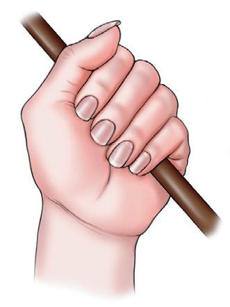
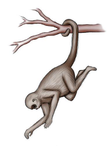
Anatomy and physiology are closely related. Structure and function go together. When you examine the
anatomy of a body part, ask yourself how its structure relates to its function. For example, the structure of the hand is related to its function: its ability to grasp an object (Figure 1-1). The heart pumps blood, and the long, strong, flexible tail of the monkey allows it to hang from the tree. Structure and function go together.
Re-Think
Using any household item, explain what is meant by “structure and function are related.”
Do You Know…
Why This Grave Is Being Robbed, and Why the Grave Robber Is in Big, Big Trouble?
Dissection of the human body during medieval times was not allowed. Thus, the only way that the early anatomists had for obtaining human bodies for dissection was to rob graves. Medieval scientists hired people to rob graves. Punishment for robbing graves was swift and severe. This lad will be in big, big trouble if he is caught, and it looks as if he will be. Surprisingly, grave robbing was common early in this century and in this country. Many a medical student who enrolled in the most prestigious medical schools had to “get” his own cadaver.
MAJOR ORGAN SYSTEMS
THE BODY’S LEVELS OF ORGANIZATION
The body is organized from the very simple to the complex, from the microscopic atom to the complex human organism. Note the progression from simple to complex in Figure 1-2. Tiny atoms form molecules. These in turn form larger molecules. The larger molecules are eventually organized into cells, the basic unit of life. Specialized groups of cells form tissues. Tissues are then arranged into organs such as the heart, stomach, and kidney. Groups of organs, in turn, create organ systems. Each organ system has a function, such as digestion, excretion, or reproduction. All the organ systems together form the human organism. From simple to complex, the body is built from the tiny atom to the human being.
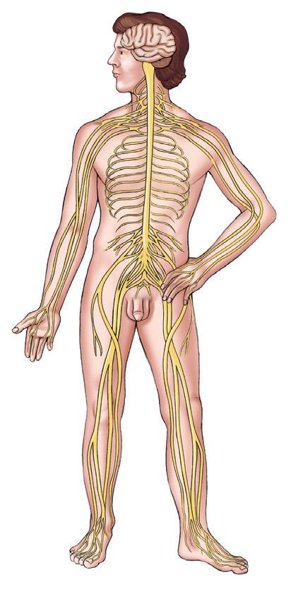
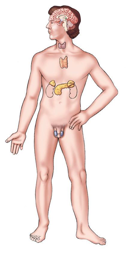
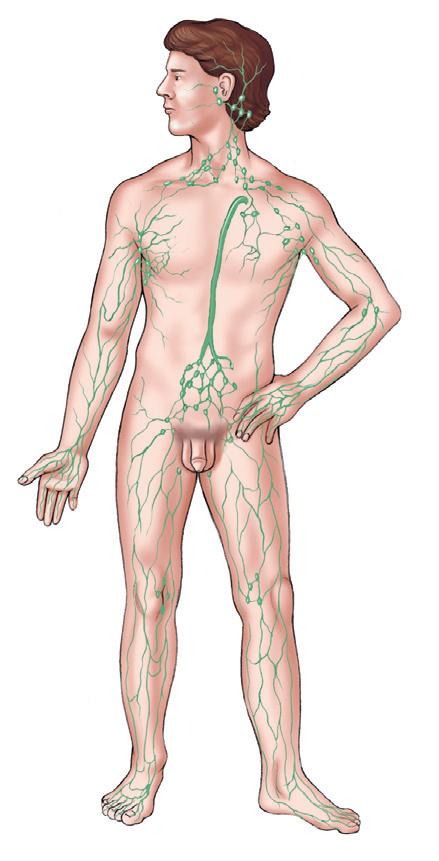
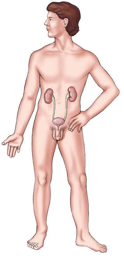
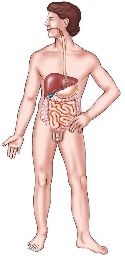
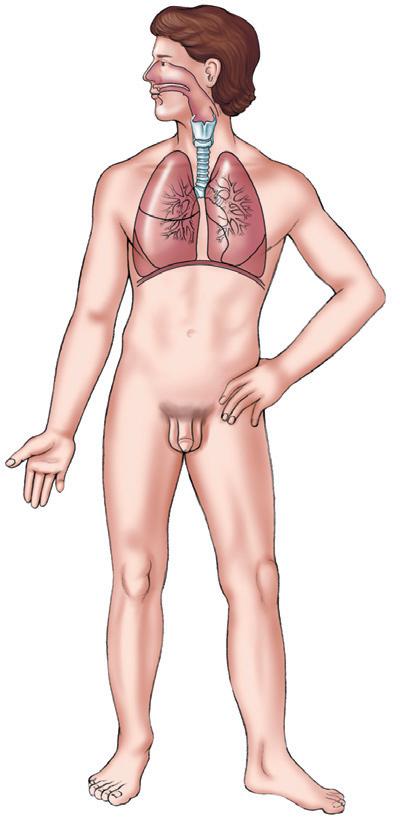
Twelve major organ systems make up the human body. Each performs specific functions that enable the human body to operate as a coordinated whole. Refer to Figure 1-3 and identify the location and distribution of the organs of each system.
• The integumentary (in-teg-yoo-MEN-tar-ee) system consists of the skin and related structures such as hair and nails. The integumentary system forms a covering for the body, helps regulate body temperature, and contains some of the structures necessary for sensation.
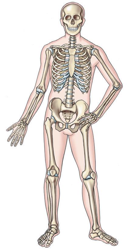
• The skeletal system forms the basic framework of the body. It consists primarily of bones, joints, and cartilage. The skeleton protects and supports body organs and enables us to move around.
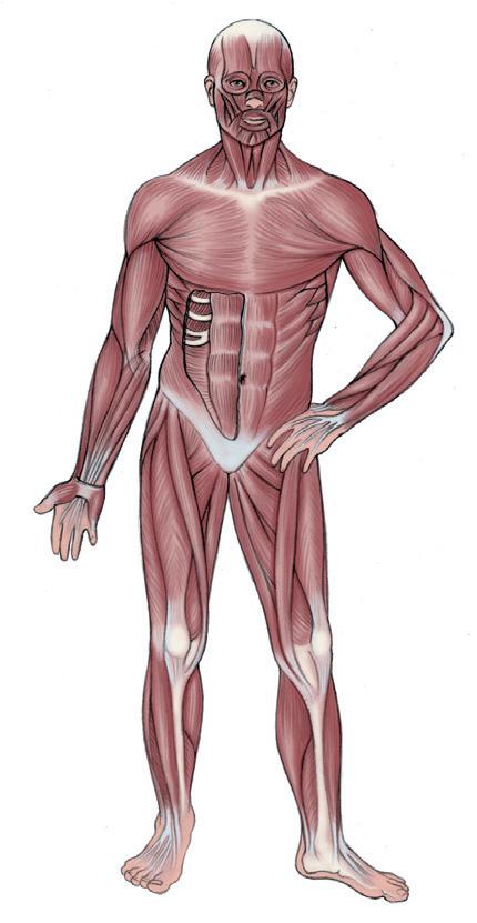
• The muscular system has three types of muscles. Skeletal muscles attach to the bones and are responsible for movement of the skeleton and the maintenance of body posture. Smooth and cardiac muscles are found in various organs and tubes; contraction
FIGURE 1-1 Structure and function are closely related.
• The nervous system is made up of the brain, spinal cord, nerves, and sense organs. Sensory nerves receive information from the environment and bring it to the spinal cord and brain, where it is interpreted. Decisions made by the brain and spinal cord are transmitted along motor nerves to various body structures.
• The endocrine (EN-doh-krin) system contains numerous glands that secrete hormones and chemical substances that regulate body activities such as growth, reproduction, metabolism, and water balance.
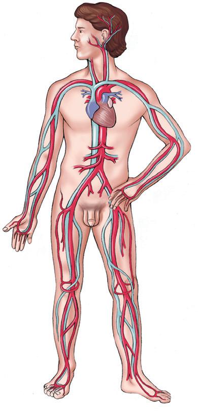
• The circulatory (SER-kyoo-lah-tor-ee) system consists of the blood, heart, and blood vessels. This system pumps (heart) and transports (blood vessels) blood throughout the body. Blood carries nutrients and oxygen to all the body’s cells and also carries the waste away from the cells to the organs of excretion.
• The lymphatic (lim-FAT-ik) system is made up of the lymph nodes, lymphatic vessels, lymph, and other lymphoid organs. Lymph and lymphoid structures play an important role in fluid balance
and in the defense of the body against pathogens and other foreign material.
• The immune system is an elaborate defense system that protects the body not only from pathogens, but also from allergens, such as pollens, bee venom, and some of our own cells that have gone awry (cancer cells). The immune system is widely distributed throughout the body (it is not shown in Figure 1-3).
• The respiratory system contains the lungs and other structures that conduct air to and from the lungs. Oxygen-rich air moves into the lungs; the oxygen is picked up by the blood and distributed throughout the body. Carbon dioxide–rich air moves out of the lungs, thereby ridding the body of waste.
• The digestive system is comprised of organs designed to ingest food and break it down into substances that can be absorbed by the body. Food that is not absorbed is eliminated as waste.
• The urinary system contains the kidneys and other structures that help excrete waste products from the body through the urine. More importantly, the urinary system helps control water, electrolyte, and acid–base balance in the body.
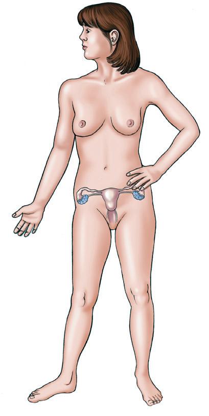
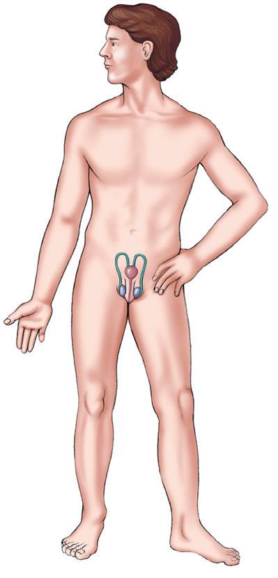
• The reproductive system is made up of organs and structures that enable humans to reproduce. and relaxation of these muscles help the organ systems carry out their functions.
FIGURE 1-2 Levels of organization, from atoms to human organism.
Integumentary system
Nervous system
Skeletal system
Circulatory system
Muscular system
FIGURE 1-3 Major organ systems of the body
Endocrine system
Lymphatic system
Urinary system
Digestive system
Respiratory system
1-3, cont’d
Reproductive system
FIGURE
HOMEOSTASIS: STAYING THE SAME
Homeostasis (ho-me-o-STAY-sis) literally means staying (stasis) the same (homeo). The term refers to the body’s ability to maintain a stable internal environment in response to a changing environment. For example, in a healthy person, body temperature stays around 98.6° F (37° C), even when room temperature increases to 100° F or decreases to 60° F. The amount of water in your cells stays the same whether you drink 2, 3, or 4 liters (L) of water per day. Your blood sugar remains within normal limits whether you have just eaten a turkey dinner or have fasted for 6 hours. Mechanisms that help maintain homeostasis are called homeostatic mechanisms. The body has hundreds of homeostatic mechanisms, including those for temperature control, blood sugar control, water balance, blood pressure regulation, and regulation of plasma sodium levels. Homeostatic imbalance results in disease or dysfunction.
Sum It Up!
Anatomy and physiology describes the structure and function of the body. The body is constructed from simple to complex (atoms to molecules to cells to tissues to organs to organ systems to the human organism). The 12 major organ systems are shown in Figure 1-3. Homeostatic mechanisms enable the body to “stay the same” despite changing internal and external environments.
ANATOMICAL
TERMS:
TALKING ABOUT THE BODY
Special terms describe the location, position, and regions of body parts. Because these terms are used frequently, you should become familiar with them now. People in the medical field are often accused of speaking their own language. Indeed, we do! We always use these terms as if the body were standing in its anatomical position.
ANATOMICAL POSITION
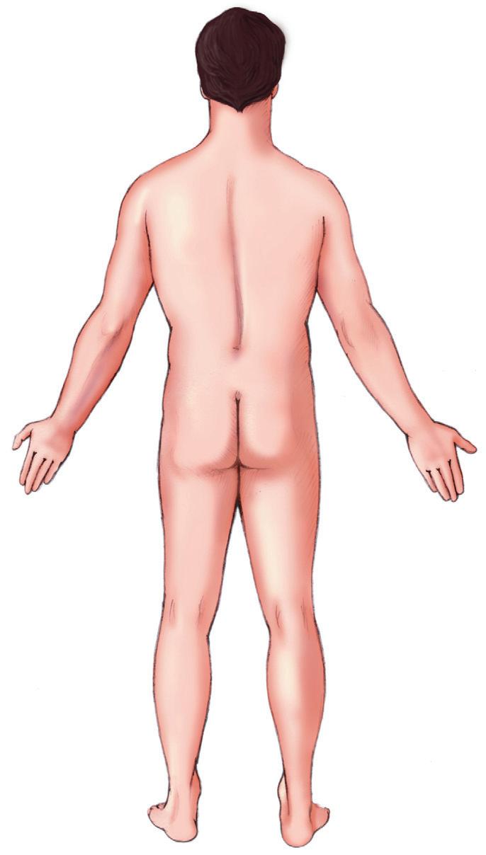
In its anatomical position, the body is standing erect, with the face forward, the arms at the sides, and the toes and palms of the hands directed forward (Figure 1-4).
RELATIVE POSITIONS
Specific terms describe the position of one body part in relation to another body part. These are directional terms. They are like the more familiar directions of north, south, east, and west; however, whereas describing Canada as being located north of the United States would be correct, describing the head as “north of the chest” would sound strange. Therefore, in locating body parts, we use other terminology. The terms come
in pairs. Note that the two terms in each pair are generally opposites. Remember, the references are valid only for the body in its anatomical position.
• Superior and inferior. Superior means that a part is above another part or is closer to the head. For example, the head is superior to the chest. Inferior means that a part is located below another part or is closer to the feet. The chest, for example, is inferior to the head.
• Anterior and posterior. Anterior means toward the front surface (the belly surface). Posterior means toward the back surface. For example, the heart is anterior to the spinal cord, but the heart is posterior to the breastbone. Another word for anterior is ventral, and another word for posterior is dorsal Consider the dorsal fin of a fish. It is the dorsal part of the shark that can be seen moving effortlessly and very quickly toward your surfboard!
• Medial and lateral. Imagine a line drawn through the middle of your body, dividing it into right and left halves. This is the midline. Medial means toward the midline of the body. The nose, for example is medial to the ears. Lateral means away from the midline of the body. For example, the ears are lateral to the nose. In the anatomical position, the hand is closer to the lateral thigh than to the medial thigh.
• Proximal and distal. Proximal means that the structure is nearer the point of attachment, often the
FIGURE 1-4 Anatomical position.
trunk of the body. Because the elbow is closer to the point of attachment than is the wrist, the elbow is described as proximal to the wrist. The wrist is proximal to the fingers, meaning that the wrist is closer to the trunk than are the fingers. Distal means that a part is farther away from the point of attachment than another part. For example, the wrist is distal to the elbow and the fingers are distal to the wrist.
• Superficial and deep. Superficial means that a part is located on or near the surface of the body. The skin is superficial to the muscles. Deep means that the body part is away from the surface of the body. The bones, for example, are deep to the skin.
• Central and peripheral. Central means that the part is located in the center. Peripheral means away from the center. The heart, for example, is located centrally, whereas the blood vessels are located peripherally (away from the center and extending toward the limbs). The brain and spinal cord are called the central nervous system and the nerves are called the peripheral nervous system.
Re-Think
PLANES OF THE BODY
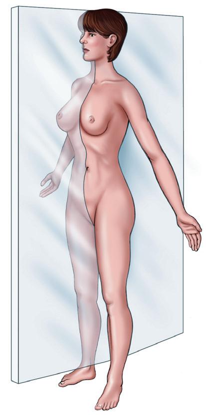
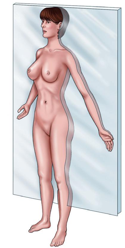
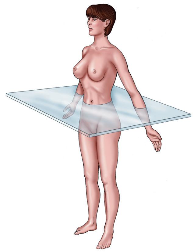
When we refer to the left side of the body, the top half of the body, or the front of the body, we are referring to the planes of the body. Each plane divides the body with an imaginary line in one direction. Figure 1-5 shows the following three important planes:
1. Sagittal plane (see Figure 1-5, A). The sagittal plane divides the body lengthwise into right and left portions. If the cut is made exactly down the midline of the body, the right and left halves of the body are equal. This division is a midsagittal section.
2. Frontal plane (see Figure 1-5, B). The frontal plane divides the body into anterior (ventral) and posterior (dorsal) portions. This plane creates the front part of the body and the back part of the body. The frontal plane is also called the coronal plane. Coronal means “crown,” so the imaginary line for the coronal plane is made across the part of the head where a crown would sit and then downward through the body.
3. Transverse plane (see Figure 1-5, C). The transverse plane divides the body horizontally, creating an upper (superior) and a lower (inferior) body. When the body or an organ is cut horizontally or transversely, it is called a cross section.
Use the terms medial and lateral in describing the parts of the thigh. Do the same with the eye.
FIGURE 1-5 Planes of the body A, Sagittal. B, Frontal (coronal). C, Transverse.
Abdominal
Antecubital
FIGURE 1-6 Regional terms. A, Anterior view B, Posterior view
REGIONAL TERMS
Specific terms describe the different regions or areas of the body. Figure 1-6 illustrates the terms used to identify the regions on the anterior and posterior surfaces of the body.
On the anterior surface, identify the following regions:
Abdominal: anterior trunk just below the ribs
Antecubital: area in front of the elbow
Axillary: armpit
Brachial: arm
Buccal: cheek area; cavity between the gum and cheek
Cephalic: head
Cervical: neck region
Cranial: nearer to the head
Digital: fingers, toes
Femoral: thigh area
Flank: fleshy area along each side between the lower ribs and the top of the hip bones
Inguinal: area where the thigh meets the trunk of the body; often called the groin
Oral: mouth
Orbital: area around the eye
Patellar: front of the knee over the kneecap
Pedal: foot
Plantar: sole of the foot
Pubic: genital area
Sternal: middle of the chest (over the breastbone area)
Umbilical: navel
On the posterior surface, identify the following regions:
Caudal: near to the lower region of the spinal column (near the tailbone)
Deltoid: rounded area of the shoulder closest to the upper arm
Gluteal
Caudal
Lumbar
Flank
Mediastinum
Pleural cavities
Thoracic cavity
Diaphragm
Ventral cavity
Abdomino–pelvic cavity
Abdominal cavity
Pelvic cavity
cavity
Spinal cavity
Dorsal cavity
Gluteal: buttocks
Lumbar: area of the back between the ribs and the hips
Occipital: back of the head
Popliteal: behind, or back of, the knee area
Scapular: shoulder blade area
Sum It Up!
Specific terms describe the relative positions of one body part to the other. The terms are paired as opposites and include superior and inferior, anterior (ventral) and posterior (dorsal), medial and lateral, proximal and distal, superficial and deep, and central and peripheral. The body can be cut into three planes: sagittal (right and left), frontal or coronal (front and back), and transverse (top and bottom) planes. Common terms are used to identify specific areas of the anterior and posterior surface areas.
Re-Think
Of the following terms, which can be seen only on the posterior view of the body: umbilical, antecubital, gluteal, lumbar, sternal, patellar, and popliteal?
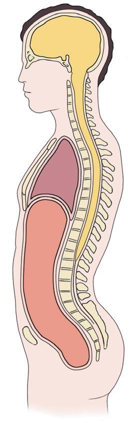
CAVITIES OF THE BODY
DORSAL CAVITY
The organs, called viscera (VISS-er-ah), are located within the cavities of the body. Cavities are large internal spaces. The body contains two major cavities: the dorsal cavity and the ventral cavity (Figure 1-7). The dorsal cavity is located toward the back of the body and has two divisions, the cranial cavity and the spinal (vertebral) cavity
FIGURE 1-7 Major body cavities.
Cranial
Do You Know…
What Your Postsurgical Patient Has Done If He Eviscerated?
His internal organs have protruded through his surgical incision. This word comes from the word viscera (organs). The organs must be kept moist and sterile until they can be returned to their home cavity.
The cranial cavity is located within the skull and contains the brain. The spinal, or vertebral, cavity extends downward from the cranial cavity and is surrounded by bony vertebrae; it contains the spinal cord. The cranial and spinal cavities form one continuous space.
VENTRAL CAVITY
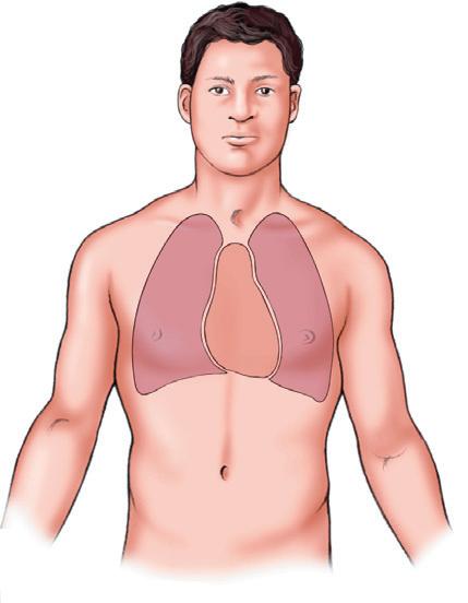
The larger ventral cavity is located toward the front of the body and has two divisions, the thoracic (thohRASS-ik) cavity and the abdominopelvic (ab-DOM-ino-PEL-vik) cavity.
THORACIC CAVITY
The thoracic cavity is located above the diaphragm and is surrounded by the rib cage. The thoracic cavity is divided into two compartments by the mediastinum (MEE-dee-ass-TI-num), a space that contains the heart, esophagus, trachea, thymus gland, and large blood vessels attached to the heart. The pericardial (pair-iKAR-dee-al) cavity (not shown) is located within the mediastinum and contains the heart. The right and left lungs are located on either side of the mediastinum in the pleural cavities. The lungs occupy most of the space within the thoracic cavity.
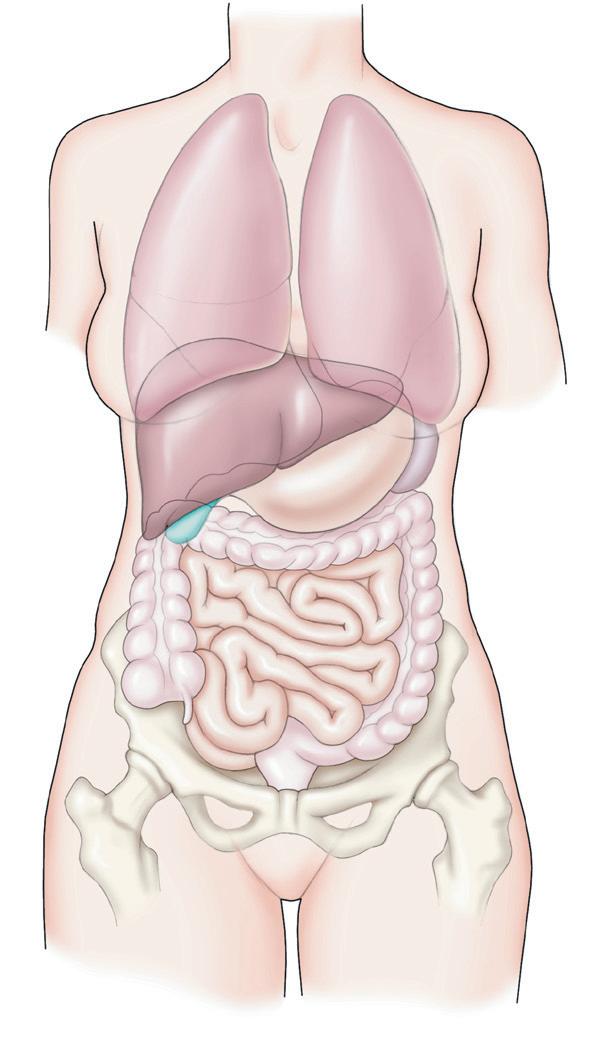
ABDOMINOPELVIC CAVITY
The abdominopelvic cavity is located below the diaphragm. The upper portion of this cavity is the abdominal cavity. It contains the stomach, most of the intestine, liver, gallbladder, pancreas, spleen, and kidneys. The lower portion of the abdominopelvic cavity is called the pelvic cavity. It extends downward from the level of the hips and includes the remainder of the intestines, the rectum, urinary bladder, and internal parts of the reproductive system.
Because the abdominopelvic cavity is so large, it is subdivided into smaller areas for study. Quadrants and regions divide the abdominopelvic cavity. Note the organs located in each quadrant or region, as shown in Figure 1-8.
Division into Quadrants
The abdominopelvic cavity can be divided into four quadrants (see Figure 1-8, A). The quadrants are named for their positions: right upper quadrant (RUQ), left upper quadrant (LUQ), right lower quadrant (RLQ), and left lower quadrant (LLQ).
Quadrant terms are used frequently in the clinical setting. For example, a patient in the emergency room
who has acute pain in the RLQ may be diagnosed with appendicitis. Note that the RLQ appears to be on your left. This is similar to looking in a mirror. Keep this in mind when you are studying the diagrams in the text. Here’s an easy way to remember this: Lower your right arm. It will be lateral to the RUQ and RLQ.
Division into Regions
A second system divides the abdominopelvic cavity into nine separate regions that resemble the squares for tic-tac-toe (see Figure 1-8, B). The three central regions (from top to bottom) include the epigastric, umbilical, and hypogastric regions. The epigastric region is located below the breastbone. Epigastric literally means upon (epi) the stomach (gastric). The umbilical region is the centermost region and surrounds the umbilicus, or navel (belly button). The hypogastric region is located just below the umbilical region. Hypogastric literally means below (hypo) the stomach (gastric). Six regions are located on either side of the central regions. They include the hypochondriac, lumbar, and iliac regions. The right and left hypochondriac regions are located on either side of the epigastric region and overlie the lower ribs. The word hypochondriac literally means below (hypo) the cartilage (chondro) and refers to the composition of the ribs (cartilage). The right and left lumbar regions are located on either side of the umbilical region and are inferior to the hypochondriac regions. The right and left iliac regions, also called the right and left inguinal regions, are located on either side of the hypogastric region. Knowledge of these regions helps you understand terms such as epigastric pain and umbilical hernia. Remember that quadrants and regions refer only to the abdominopelvic cavity and not to the thoracic cavity.
Other Cavities
Four smaller cavities are located in the head. They include the oral cavity, nasal cavities, orbital cavities, and middle ear cavities. (These cavities are described in later chapters.)
Re-Think
Of the following terms, determine which pertain to the ventral cavity: thoracic cavity, brain, vertebral cavity, abdominopelvic cavity, pleural cavity, dorsal cavity, mediastinum, stomach, heart, and spinal cord.
Sum It Up!
The organs are located within body cavities. The two major cavities are the dorsal cavity, located toward the back of the body, and the larger ventral cavity, located in the front of the body. The dorsal cavity is subdivided into the cranial cavity and the spinal cavity. The ventral cavity is divided by the diaphragm into the thoracic cavity (including the mediastinum, pericardial cavity, and pleural cavities) and the abdominopelvic cavity. The abdominopelvic cavity is divided into quadrants and regions.
Right upper quadrant
Left upper quadrant
Right lower quadrant Left lower quadrant
Right hypochondriac region
Right lumbar region
Right iliac region
Epigastric region
Left hypochondriac region
Left lumbar region Umbilical region
Hypogastric region
Left iliac region
A
B
FIGURE 1-8 The abdominopelvic cavity A, Four quadrants. B, Nine regions.
MEDICAL TERMINOLOGY AND DISORDERS
Introduction to Medical Terminology
The medical profession has its own language, called medical terminology. In general, there are four main types or kinds of word parts. By learning these and how they can be put together, you can often “translate” many long and challenging medical words by breaking them up into their word parts.
A word root is the core of the word and provides the basic meaning or “subject” of the word. The other word parts, such as suffixes and prefixes, modify the word root so that it takes on a new meaning. For example, in the word hepatitis, the word root is hepat-, meaning liver.
A suffix is a word part attached to the end of the word root; it modifies the word root. If we add -itis, which is a suffix that means inflammation, to the word root for liver, hepat-, we get hepatitis, which means inflammation of the liver.
A prefix is a word part attached to the beginning of the word root; it modifies the word root. For example, the word nutrition refers to a normal and healthy food intake. By adding the prefix mal- (French for bad), as in malnutrition, the word root has been modified to mean poor or bad nutrition
Combining vowels are word parts used to ease the pronunciation, as in angioplasty (angi/o/plasty). Angi- means blood vessel, and -plasty means repair of. The -o- joining the word parts eases the pronunciation; it does not modify the meaning of the word. When you see angi/o-, you are seeing the word root and its combining vowel.
On the next page you will see many terms with which you are probably familiar, but what you may not be familiar with is what their individual word parts are and how they are put together to make up many of the words you use every day. For instance, take -ectomy, which means excision or removal of. Think of all the terms that have -ectomy at the end—such as lobectomy, vasectomy, hysterectomy, appendectomy, tonsillectomy—and you will see how valuable knowing what this one word part is.
Medical Term Word Parts
Word Part Meaning or Derivation
anatomy ana- up or apart
-tomy incision; to cut
antecubital ante- before; in front of
-cubital
From the Latin word cubitum, meaning “elbow”
biology bio- life
-logy study of diagnosis dia- apart
Description
Anatomy is the branch of science that describes the structure of a body, especially as revealed by dissection. For example, the heart has four chambers.
Antecubital space is the area of the arm anterior to the elbow A sample of blood is commonly drawn from a vein in the antecubital space.
Biology is the study of life and living organisms
Diagnosis is the process of identifying the nature and cause of a disease or injury through an analysis of data such as the patient’s symptoms and laboratory and x-ray studies
-gnos/o- knowing -osis condition or increase pathologist path/o- pertaining to disease A pathologist examines tissue for evidence of disease.
-logist one who specializes in homeostasis home/o- sameness
-stasis stand still
transverse trans- across
-verse From the Latin word vertere, meaning “to turn”
midepigastric mid- middle
-epi- above or upon -gastr/o- stomach -ic pertaining to
prognosis pro- before
-gnos/o- knowing -osis process
quadrant quadr/i- four
Homeostasis refers to the relative constancy of the internal environment of the body despite many challenges to upset the balance.
A transverse plane cuts across the body; an upper and lower body is created.
Related to the middle of the epigastric region of the abdomen. A person often complains of midepigastric discomfort.
Refers to a likely course or outcome of a disease. For example, the prognosis of a person who seeks early treatment of a basal cell carcinoma (skin cancer) is excellent; the prognosis of a person with metastatic cancer is less favorable.
The abdominopelvic cavity is divided into four equal areas called quadrants. -ant performing/promoting
Get Ready for Exams!
Summary Outline
Anatomy is the study of structure; physiology is the study of function. Structure (anatomy) and function (physiology) are related.
I. The Body’s Levels of Organization
A. From simple to complex: Atoms to molecules to cells to tissues to organs to organ systems to human organism
B. Major organ systems (12)
1. Integumentary system
2. Skeletal system
3. Muscular system
4. Nervous system
5. Endocrine system
6. Heart and circulatory system
7. Lymphatic system
8. Immune system
9. Respiratory system
10. Digestive system
11. Urinary system
12. Reproductive system
C. Homeostasis: the body’s ability to maintain a stable internal environment in response to various inter nal and exter nal challenges
II. Anatomical Terms: Talking About the Body
A. Anatomical position: the body standing erect, arms by the side, with palms and toes facing forward
B. Relative positions: superior-inferior, anteriorposterior, medial-lateral, proximal-distal, superficialdeep, central-peripheral
C. Planes (three): sagittal, frontal (coronal), and transverse planes
D. Regional terms: listed in Figure 1-6
E. Cavities of the body
1. Dorsal cavity
a. Cranial cavity: contains the brain
b. Spinal (vertebral) cavity: contains the spinal cord
2. Ventral cavity
a. Thoracic cavity: superior to the diaphragm; contains the pleural cavities (lungs) and mediastinum (pericardial cavity)
b. Abdominopelvic cavity: located inferior to the diaphragm
c. Abdominal cavity: upper part that contains the stomach, most of the intestines, liver, spleen, and kidneys
d. Pelvic cavity: lower part that contains the reproductive organs, urinary bladder, and lower part of the intestines
e. For reference: the abdominopelvic cavity is divided into four quadrants and nine regions
Review Your Knowledge
Matching: Directions of the Body
Directions: Match the following words with their descriptions below. Some words may be used more than once or not at all.
a. posterior
b. distal
c. medial
d. anterior
e. proximal
f. superior
g. deep
1. Toward the midline of the body; opposite of lateral
2. Structure that is nearer to the trunk than another part; opposite of distal
3. Part of the radius (forearm bone) that is closer to the wrist than to the elbow
4. The lungs are located above the diaphragm; their position relative to the diaphragm is described as being above.
5. Toward the front (the belly surface); another word is ventral
Matching: Regional Terms
Directions: Match the following words with their descriptions below.
a. inguinal
b. oral
c. lumbar
d. axillary
e. buccal
f. patellar
g. flank
h. antecubital
i. ster nal
j. scapular
1. Armpit
2. Kneecap area
3. Breastbone area
4. Front part of the elbow area
5. Fleshy area along the side between the ribs and hip bone
6. Pertaining to the mouth
7. Lower back area extending from the chest to the hips
8. Pertains to the space between the cheek and gum
9. Groin region
10. Shoulder blade area
Multiple Choice
1. This part of the humerus (arm bone) is closer to the elbow than to the axillary region.
a. Anterior
b. Superior
c. Distal
d. Proximal
2. Describe the relationship of the mediastinum to the diaphragm.
a. Distal
b. Deep
c. Anterior
d. Superior
3. The umbilical area is located
a. inferior to the inguinal region.
b. superior to the RUQ.
c. inferior to the diaphragm.
d. within the midepigastric region.
4. The ster nal area is
a. superior to the diaphragm.
b. referred to as the breastbone area.
c. superficial to the mediastinum.
d. All of the above are true.
5. Which of the following is not descriptive of the mediastinum?
a. Thoracic cavity
b. Dorsal cavity
c. Ventral cavity
d. Superior to the diaphragm
6. The frontal plane
a. splits the body into right- and left-half sections.
b. is also the coronal plane.
c. splits the body into a top and a bottom section.
d. creates a transverse cross section.
7. Which of the following terms best describes when the blood vessels dilate and the person sweats in order to decrease body temperature?
a. Pathophysiology
b. Evisceration
c. Homeostasis
d. Midsagittal
8. Which of the following is true of these terms: ster nal, umbilical, patellar, and antecubital?
a. All are superior to the inguinal area.
b. All lie within the ventral cavity
c. All can be viewed on the anterior body
d. All lie within the dorsal cavity
Go Figure
1. According to Figure 1-6
a. The brachial, lumbar, and antecubital areas can only be identified on the posterior view of the body
b. The inguinal and flank areas are the same.
c. The gluteal, lumbar, and scapular areas are inferior to the umbilicus.
d. The popliteal and patellar areas are located in the lower extremities.
2. Refer to Figures 1-5 and 1-6. A midsagittal section yields half of a body. Which body regions are preserved in this half-body section?
a. The patellar (right and left), flank (right and left), and brachial (right and left) are preserved.
b. Neither the right nor left patellar areas and neither the right nor left antecubital areas are preserved.
c. All areas displayed in Figure 1-6, B, are preserved.
d. A left or right inguinal, pedal, and axillary area is preserved.
3. According to Figure 1-7
a. The thoracic cavity is a ventral cavity that includes the mediastinum and pleural cavities.
b. The diaphragm separates the two pleural cavities.
c. The dorsal cavity includes the pleural cavities and the mediastinum.
d. All of the above are true.
4. According to Figure 1-8
a. The ventral cavity is divided into quadrants.
b. The dorsal cavity is divided into regions.
c. RUQ, LUQ, RLQ, and LLQ describe only the abdominal cavity.
d. RUQ, LUQ, RLQ, and LLQ are quadrants that define the abdominopelvic cavity
5. According to Figure 1-8
a. The left iliac region is located within the LUQ.
b. The hypogastric region is located within the RUQ.
c. The umbilical region surrounds the navel or belly button.
d. The right lung is located within the RUQ.
Basic Chemistry
Key Terms
acid (p. 23)
adenosine triphosphate (ATP) (p. 25)
atom (p. 16)
base (p. 23) catalyst (p. 22) compound (p. 21)
Objectives
covalent bond (p. 18)
electrolyte (p. 20)
element (p. 15)
energy (p. 24)
enzyme (p. 22)
hydrogen bond (p. 18) ionic bond (p. 18)
1. Define the terms matter, element, and atom, and do the following:
• List the four elements that comprise 96% of body weight.
• Describe the three components of an atom.
• Describe the role of electrons in the formation of chemical bonds.
2. Differentiate among ionic, covalent, and hydrogen bonds.
3. Explain ions, including the differences among electrolytes, cations, and anions.
2
http://evolve.elsevier.com/Herlihy
isotope (p. 17) molecule (p. 20) pH (p. 23)
solution (p. 26) suspension (p. 26)
4. Explain the difference between a molecule and a compound, and list five reasons why water is essential to life.
5. Explain the role of catalysts and enzymes.
6. Differentiate between an acid and a base, and define pH.
7. List the six forms of energy and describe the role of adenosine triphosphate (ATP) in energy transfer.
8. Differentiate among a mixture, solution, suspension, colloidal suspension, and precipitate.
Why a chapter on chemistry? Because our bodies are made of different chemicals. The food we eat, the water we drink, and the air we breathe are all chemical substances. We digest our food, move our bodies, experience emotions, and think great thoughts because of chemical reactions. To understand the body, we must understand some general chemical principles.
MATTER, ELEMENTS, AND ATOMS
MATTER
Chemistry is the study of matter. Matter is anything that occupies space and has weight. Anything that you see as you look around is matter.
Matter exists in three states: solid, liquid, and gas. Solid matter—such as skin, bones, and teeth—has a definite shape and volume. Liquid matter—such as blood, saliva, and digestive juices—takes the shape of the container that holds it. A gas, or gaseous matter— such as the air we breathe—has neither shape nor volume.
Matter can undergo both physical and chemical changes. The logs in a fireplace illustrate the difference
between a physical and a chemical change (Figure 2-1). The logs can undergo a physical change by being chopped into smaller chips of wood with a hatchet. The wood chips are smaller than the log, but they are still wood. The matter (wood) has not essentially changed; only the physical appearance has changed. A chemical change occurs when the wood is burned. When burned, the wood ceases to be wood. The chemical composition of the ashes is essentially different from that of wood.
The body contains many examples of physical and chemical changes. For example, digestion involves physical and chemical changes. Chewing breaks the food into smaller pieces; this is a physical change. Potent chemicals digest or change the food into simpler substances; this is a chemical change.
ELEMENTS
All matter, living or dead, is composed of elements. An element is matter that is composed of atoms that have the same number of positive charges in their nuclei. Even a very small amount of an element such as sodium contains millions and millions of sodium atoms. The same name is used for both the
ATOMS
ATOMIC STRUCTURE
Elements are composed of atoms, the basic units of matter. An atom is the smallest unit of an element with that element’s chemical characteristics. An atom is composed of three subatomic particles: protons, neutrons, and electrons. The arrangement of the subatomic particles resembles the sun and planets (Figure 2-2, A), with the sun in the center and the planets constantly moving around the sun in orbits, or circular paths. The atom is composed of a nucleus (the sun) and shells, or orbits, that surround the nucleus (see Figure 2-2, B).
element and the atom. Although more than 100 elements exist, only about 25 elements are required by living organisms.
Do You Know…
Why Children Should Not Be Allowed to Play in Traffic and Chew on Old Paint?
Aside from the obvious safety issues, old paint and emissions from motor vehicles contain high amounts of lead. Exposure to high levels of lead causes lead poisoning, a serious condition that damages the major organs, including the brain, liver, kidney, and bone marrow. The old name for chronic lead poisoning is plumbism, from the Latin word for lead (plumbum). The chemical symbol for lead is Pb. (By the way, a plumber is called a plumber because ancient water pipes were made of plumbum, or lead.) Why does plumbism have such a great history? Lead was used to make pipes that carried water and was used to make pottery, particularly drinking vessels. This practice killed many of the rich and famous—those wealthy enough to afford leaded wine goblets. Because of the toxic nature of lead, pipes and pottery in the United States are no longer made of lead, gasoline and paint are now leadfree by law, and the disposal of acid lead batteries is regulated. Is the lead problem a done deal? No! Children are still huffing lead fumes from car emissions, playing with toys laced with lead paint, and wearing clothing impregnated with lead. Go figure!
The most abundant elements found in the body are listed in Table 2-1. Four elements—carbon, hydrogen, oxygen, and nitrogen—make up 96% of the body weight. The trace elements are present in tiny amounts, but despite the small amounts required, the trace elements are essential for life. (Not all the trace elements appear in Table 2-1.)
Each of the elements included in Table 2-1 is represented by a symbol, and the first letter of the symbol is always capitalized. For example, the symbol O is for oxygen, N is for nitrogen, Na is for sodium, K is for potassium, and C is for carbon. These symbols are used frequently, so you should memorize the symbols of the major elements.
Where are the subatomic particles located? The protons and the neutrons are located in the nucleus (see Figure 2-2, C). Protons carry a positive (+) electrical charge; neutrons carry no electrical charge. The electrons are located in the shells, or orbits, surrounding the nucleus like planets. Electrons carry a negative ( ) electrical charge. In each atom, the number of protons (+) is equal to the number of electrons ( ). The atom is therefore electrically neutral; it carries no net electrical charge.
All protons are alike, all neutrons are alike, and all electrons are alike. So what makes one atom different from another atom? The difference is primarily in the numbers of protons and electrons in each atom. For example, hydrogen is the simplest and smallest atom.