https://ebookmass.com/product/the-echo-manual-fourthedition/
Instant digital products (PDF, ePub, MOBI) ready for you
Download now and discover formats that fit your needs...
Statistical Mechanics: Fourth Edition. Instructor's Manual R.K. Pathria
https://ebookmass.com/product/statistical-mechanics-fourth-editioninstructors-manual-r-k-pathria/
ebookmass.com
Echo Unbound (Echo Power Trilogy Book 3) Anna Durand
https://ebookmass.com/product/echo-unbound-echo-power-trilogybook-3-anna-durand/
ebookmass.com
Echo Power (Echo Power Trilogy Book 1) Anna Durand

https://ebookmass.com/product/echo-power-echo-power-trilogybook-1-anna-durand/
ebookmass.com
Highland Gladiator Kathryn Le Veque [Veque
https://ebookmass.com/product/highland-gladiator-kathryn-le-vequeveque-3/
ebookmass.com


Microeconomics (Pearson Series in Economics) 9th Edition, (Ebook PDF)
https://ebookmass.com/product/microeconomics-pearson-series-ineconomics-9th-edition-ebook-pdf/ ebookmass.com
Nineteenth-Century Individualism and the Market Economy: Individualist Themes in Emerson, Thoreau, and Sumner 1st Edition Luke Philip Plotica (Auth.)
https://ebookmass.com/product/nineteenth-century-individualism-andthe-market-economy-individualist-themes-in-emerson-thoreau-andsumner-1st-edition-luke-philip-plotica-auth/ ebookmass.com
Python Mini Reference 2022: A Quick Guide to the Modern Python Programming Language for Busy Coders (A Hitchhiker's Guide to the Modern Programming Languages Book 3) Harry Yoon
https://ebookmass.com/product/python-mini-reference-2022-a-quickguide-to-the-modern-python-programming-language-for-busy-coders-ahitchhikers-guide-to-the-modern-programming-languages-book-3-harryyoon/ ebookmass.com
Pucking Revenge : A Fake dating, friends to lovers, hockey romance (The Revenge Games Book 2) Brittanee Nicole
https://ebookmass.com/product/pucking-revenge-a-fake-dating-friendsto-lovers-hockey-romance-the-revenge-games-book-2-brittanee-nicole/ ebookmass.com
A Field Guide for Social Workers: Applying Your Generalist Training – Ebook PDF Version
https://ebookmass.com/product/a-field-guide-for-social-workersapplying-your-generalist-training-ebook-pdf-version/ ebookmass.com




Armenia’s Future, Relations with Turkey, and the Karabagh
Conflict 1st Edition Levon Ter-Petrossian
https://ebookmass.com/product/armenias-future-relations-with-turkeyand-the-karabagh-conflict-1st-edition-levon-ter-petrossian/
ebookmass.com
Departmentof Cardiovascular Medicine
Mayo Clinic
Rochester, Minnesota
Sung-AChang,MD,PhD
Associate Professor
Division of Cardiology
Departmentof Medicine
HeartVascular Stroke Institute Imaging Center
Samsung MedicalCenter
Sungkyunkwan University Schoolof Medicine
Seoul, Republic of Korea
ThaisCoutinho,MD
Chief, Division of Cardiac Prevention and Rehabilitation
Chair, Canadian Women’s HeartHealth Centre
Division of Cardiology
University of Ottawa HeartInstitute Ottawa, Ontario, Canada
Michael W.Cullen,MD
AssistantProfessor of Medicine
Consultant, Departmentof Cardiovascular Medicine
Mayo Clinic Rochester, Minnesota
Raúl E.Espinosa,MD
AssistantProfessor of Medicine
Consultant, Departmentof Cardiovascular Medicine
Mayo Clinic
Rochester, Minnesota
CovadongaFernández-Golfín,MD
Director, Cardiac Imaging Unit
Departmentof Cardiology
University HospitalRamón y Cajal Madrid, Spain
DavidA.Foley,MD
AssistantProfessor of Medicine
Consultant, Departmentof Cardiovascular Medicine
Mayo Clinic
Rochester, Minnesota
William K.Freeman,MD
Professor of Medicine
Consultant, Departmentof Cardiovascular Medicine
Mayo Clinic Scottsdale, Arizona
JeffreyB.Geske,MD
Associate Professor of Medicine
Consultant, Departmentof Cardiovascular Medicine
Mayo Clinic Rochester, Minnesota
ArianaGonzález,MD
Director, Valvular HeartDiseases
Departmentof Cardiology
University HospitalRamón y Cajal Madrid, Spain
DonaldJ.Hagler,MD
Professor of Medicine and Pediatrics
Consultant, Division of Pediatric Cardiology
Departmentof Cardiovascular Medicine
Mayo Clinic Rochester, Minnesota
KyleW.Klarich,MD
Professor of Medicine
Vice Chair, Departmentof Cardiovascular Medicine
Consultant, Departmentof Cardiovascular Medicine
Mayo Clinic
Rochester, Minnesota
IftikharJ.Kullo,MD
Professor of Medicine
Consultant, Departmentof Cardiovascular Medicine
Mayo Clinic Rochester, Minnesota
RobertoM.Lang,MD
President(Emeritus), American Society of Echocardiography
Professor of Medicine, Section of Cardiology
University of Chicago MedicalCenter Chicago, Illinois
GraceLin,MD
Associate Professor of Medicine
Director, HeartFailure Clinic
Consultant, Departmentof Cardiovascular Medicine
Mayo Clinic
Rochester, Minnesota
Lieng-H Ling,MBBS,MD
Associate Professor, Departmentof Medicine
Yong Loo Lin Schoolof Medicine
NationalUniversity of Singapore
Senior Consultant
Departmentof Cardiology
NationalUniversity HeartCentre
Singapore
JosephF.Maalouf,MD
Professor of Medicine
Director, InterventionalEchocardiography
Consultant, Departmentof Cardiovascular Medicine
Mayo Clinic
Rochester, Minnesota
JosephJ.Maleszewski,MD
Professor of Laboratory Medicine & Pathology and Medicine
Departments of Laboratory Medicine & Pathology, Cardiovascular Medicine, and Clinical
Genomics
Mayo Clinic
Rochester, Minnesota
Sunil V.Mankad,MD
Associate Professor of Medicine
Consultant, DepartmentCardiovascular Medicine
Mayo Clinic
Rochester, Minnesota
Robert B.McCully,MD
Professor of Medicine
Director (Emeritus), Stress Echocardiography
Consultant, Departmentof Cardiovascular Medicine
Mayo Clinic
Rochester, Minnesota
HectorI.Michelena,MD
Professor of Medicine
Director, Intraoperative Echocardiography
Consultant, Departmentof Cardiovascular Medicine
Mayo Clinic
Rochester, Minnesota
FletcherA.Miller,MD
Professor of Medicine
Director (Emeritus), Echocardiography Laboratory
Consultant, Departmentof Cardiovascular Medicine
Mayo Clinic
Rochester, Minnesota
William R.Miranda,MD
AssistantProfessor of Medicine
Consultant, Departmentof Cardiovascular Medicine
Mayo Clinic Rochester, Minnesota
VictorMor-Avi,PhD
Research Professor Departmentof Medicine, Section of Cardiology
University of Chicago MedicalCenter
Chicago, Illinois
SharonL.Mulvagh,MD
Professor of Medicine
Dalhousie University
Halifax, Nova Scotia, Canada
Professor (Emeritus), Departmentof Cardiovascular Medicine
Mayo Clinic
Rochester, Minnesota
VuyisileT.Nkomo,MD,MPH
Professor of Medicine
Director, Valvular HeartDiseases Clinic
Consultant, Departmentof Cardiovascular Medicine
Mayo Clinic
Rochester, Minnesota
PatrickO’Leary,MD
Professor of Pediatrics
Division of Pediatric Cardiology
Departmentof Cardiovascular Medicine
Mayo Clinic
Rochester, Minnesota
Sungji Park,MD,PhD
Professor
Director, Imaging Center
HeartVascular Stroke Institute
Samsung MedicalCenter
Sungkyunkwan University Schoolof Medicine
Seoul, Republic of Korea
PatriciaA.Pellikka,MD
President(Emeritus), American Society of Echocardiography
Professor of Medicine
Director, Echocardiography Laboratory
Consultant, Departmentof Cardiovascular Medicine
Mayo Clinic
Rochester, Minnesota
SorinV.Pisralu,MD,PhD
Professor of Medicine
Vice Chair, Division of Cardiovascular Ultrasound
Consultant, Departmentof Cardiovascular medicine
Mayo Clinic
Rochester, Minnesota
DavidPlayford,MBBS,PhD
Professor
University of Notre Dame
Fremantle, Australia
MountHospital, Western Australia
PeterM.Pollak,MD
Director of StructuralIntervention
Consultant, Departmentof Cardiovascular Medicine
Mayo Clinic
Jacksonville, Florida
GuyS.Reeder,MD
Professor of Medicine
Consultant, Departmentof Cardiovascular Medicine
Mayo Clinic
Rochester, Minnesota
Charanjit S.Rihal,MD
William S. and Ann Atherton Professor of Cardiology
Chair (Emeritus), Departmentof Cardiovascular Medicine
Mayo Clinic
Rochester, Minnesota
Hartzell V.Schaff,MD
StuartW. Harrington Professor of Surgery
Consultant, Departmentof Cardiovascular Surgery
Mayo Clinic
Rochester, Minnesota
PeterC.Spittell,MD
AssistantProfessor of Medicine
Consultant, Departmentof Cardiovascular Medicine
Mayo Clinic
Rochester, Minnesota
Geoff Strange,BN,PhD
Professor
University of Notre Dame, Fremantle
Western Australia, Australia
RoyalPrince Alfred Hospital
Sydney, New South Wales, Australia
RakeshM.Suri,MD,DPhil
Professor of Surgery
Cleveland Clinic Foundation and Cleveland Clinic Abu Dhabi
JeremyJ.Thaden,MD
AssistantProfessor of Medicine
Consultant, Departmentof Cardiovascular Medicine
Mayo Clinic
Rochester, Minnesota
YanTopilsky,MD
Associate Professor
Sackler University of medicine TelAviv, Israel
Director of Echo and Non Invasive Cardiology
TelAviv MedicalCenter
HectorR.Villarraga,MD
Associate Professor of Medicine
Consultant, Departmentof Cardiovascular Medicine
Mayo Clinic
Rochester, Minnesota
BrandonM.Wiley,MD,MS
AssistantProfessor of Medicine
Consultant, Departmentof Cardiovascular Medicine
Mayo Clinic Rochester, Minnesota
José
LuisZamorano,MD
Vice President, European Society of Cardiology
Head of Cardiology
University HospitalRamón y Cajal Madrid, Spain
Preface
The first edition of the Echo Manual was originally written in early 1990s asaninternal manual at MayoClinictostandardizetheacquisitionandthe interpretationof echocardiography,whichwas rapidlydeveloping.Whenit was published, we were thrilled by readers’ encouraging response, which has motivated us to update this Manual few times to the current fourth edition. The first edition took 4 years to complete. At that time, all echocardiography images were stored in video tapes. We created a list of educationally valuable illustrative images, which were later retrieved and photographed using 38-roll film per each image. When each roll-film was developed, the best image was selected to be labeled and cropped. The revised images were photographed again to create figures shown in the first edition of the Manual. When echocardiography images were acquired and stored digitally, it became much easier to locate and create images. But, it is remarkable that images created by photography were frequently better than digitized images, some of which are still shown in this edition. The Echo Manual has lived through remarkable growing periods of echocardiography with development of Doppler hemodynamics, color flow imaging, transesophageal imaging, stress echocardiography, contrast (ultrasound enhancing) agent, tissue Doppler, strain imaging, 3D echocardiography and hand-held ultrasound imaging. Echocardiography has matured to have become the most practical and the most widely available imaging and hemodynamic diagnostic tool for the entire field of cardiovascular diseases. Consequently, the utilization of echocardiography is nowin the hands of not only cardiologists, but any physicians who have a need to assess cardiac structure, function, and hemodynamics in their outpatient office, bedside, emergency department, critical care unit, interventional suite, and operating room. The bedside ultrasound imaging
by a hand-held device was recommended to be the fifth pillar to bed-side physical examination in addition to inspection, palpation, percussion, and auscultation (1). There is, however, a large gap between what echocardiography can do and how it is used in clinical practice. Optimal utilization of echocardiography requires dedicated training and it is our sincere hope that this fourth edition of the Echo Manual can help to close the gap for all physicians and sonographers who perform and interpret echocardiography to provide the best care for their patients. Echocardiography has become an amazing tool not only for diagnosis but also guiding many innovative device therapies and procedures. We hope that the Manual also helps interventionalists and cardiac surgeons to use echocardiographyfor obtainingthebest result for their procedures.
As the field of echocardiography has expanded tremendously since the last edition, several new chapters (3D echocardiography, interventional echocardiography, echocardiography for heart failure and LVAD, handcarried ultrasound, and artificial intelligence in echocardiography) were added. Previous chapters were updated with new information, recent references, and new images with corresponding real-time images. We thank all contributing authors for their passion and expertise for echocardiography and for their sacrifice in precious time. We continued to emphasize the interpretation of echocardiography information in clinical context since our ultimate goal of performing echocardiography is to providethebest carefor our patients.
A creation of this fourth edition of the Echo Manual would not have been possible without support and understanding from our families. Most of echocardiography cases and images in this manual came from the extensive clinical materials at Mayo Clinic. We thank Mayo Clinic Echocardiography Laboratory and the Department of Cardiovascular Medicine for providing an amazing environment for practice, education, and research as well as collegiality and friendship. Mark A. Zang, Jeffrey R. Stelley, and Jeffrey W. Gansen of our Echocardiography Laboratory visual section helped creating still and video images used for the Manual. Paul W. Honerman of Illustration and Design revised and created all illustrative figures. Tessa Flies helped me with administrative duties and made sure that I dohave a time tocomplete this Manual intime.We could not thank enough the Wolters Kluwer for their support and patience for
having this fourth edition of the Echo Manual published almost 10 years after thethirdedition.
Finally, we are grateful to echocardiography and numerous pioneers in this field for making our professional life filled with new discoveries, better diagnostic methods, many memorable trips, wonderful meetings, international cousins, mentoring fellows, making friends all around the world, and the most importantly, opportunities to improve the care for our patients.
Jae K. Oh
Onbehalf of all authors
REFERENCE
1. Narula J, Chandrashekhar Y, Braunwald E. Time to add a fifth pillar to bedside physical examination:Inspection, palpation, percussion, auscultation, and insonation. JAMA Cardiology, 2018;3(4):346–350.
List of Contributors
Preface
Abbreviations
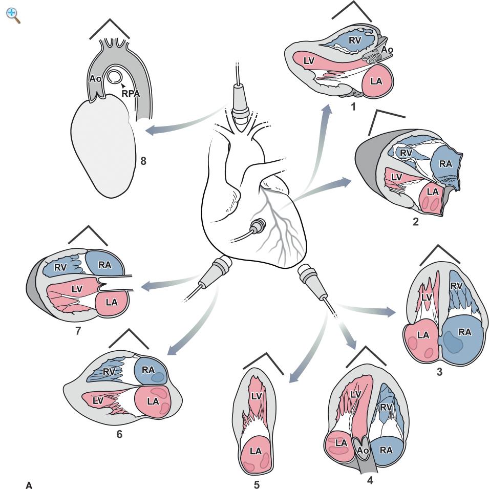
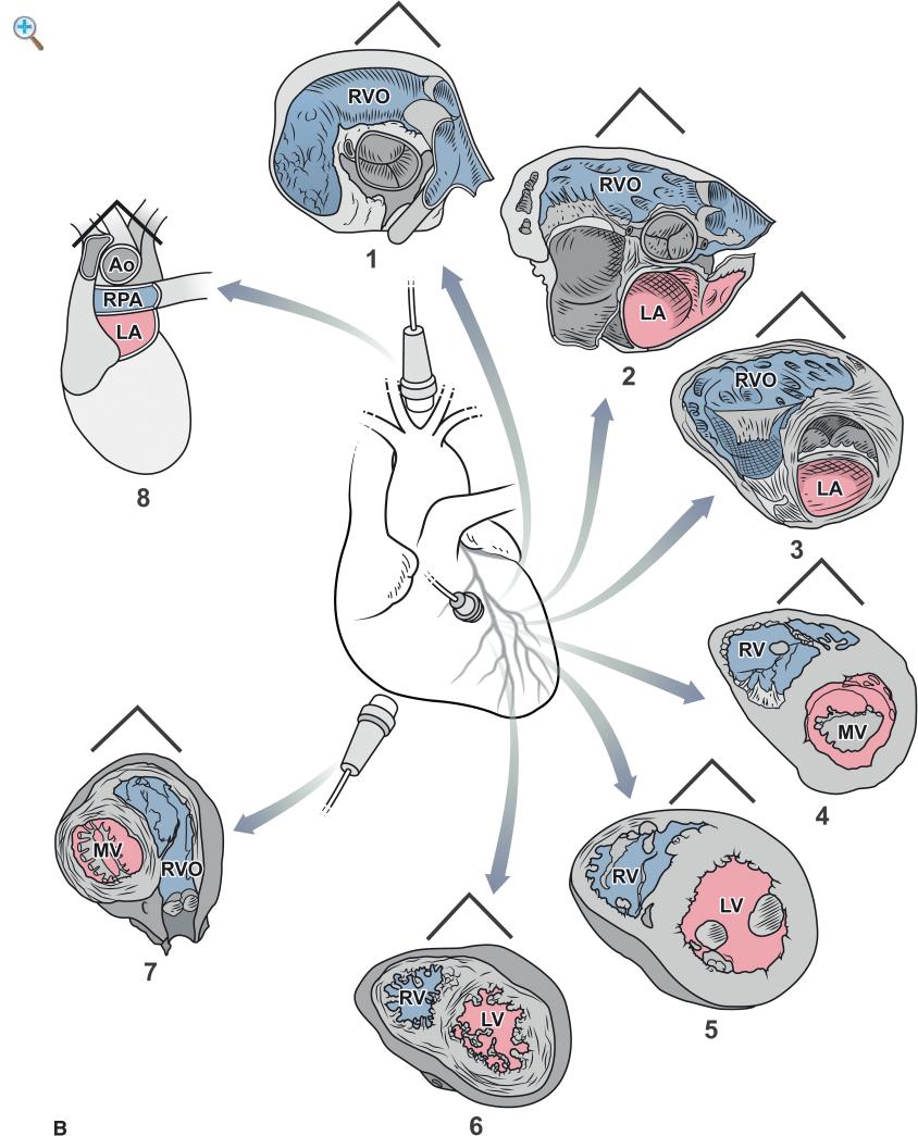
1 Transthoracic M-mode and Two-Dimensional Echocardiography
Jae K Oh and Joseph J. Maleszewski
2 Transthoracic Three-Dimensional Echocardiography
Karima Addetia, Victor Mor-Avi, and Roberto M. Lang
3 Transesophageal Echocardiography
Jeremy J. Thaden, Joseph F. Maalouf, and Jae K. Oh
4 Doppler Echocardiography and Color Flow Imaging: Comprehensive Noninvasive Hemodynamic Assessment
Jae K. Oh and William R. Miranda
5 Tissue Doppler and Strain Imaging
Hector R. Villarraga, Garvan C. Kane, and Jae K. Oh
6 Contrast Echocardiography
Sahar S. Abdelmoneim and Sharon L. Mulvagh
7 Quantification of Left-sided Cardiac Chambers: Mass, Volumes, and Ejection Fraction
Garvan C. Kane
8 Assessment of Diastolic Function
Jae K. Oh
9 Right Heart Assessment and Pulmonary Hypertension
Garvan C. Kane and Sung-A Chang
10 Cardiomyopathies
Jeffrey B. Geske and Jae K. Oh
11 Heart Failure, LVAD, and Transplantation
Yan Topilsky, Grace Lin, and Jae K. Oh
12 Pericardial Diseases
Jae K. Oh, Raúl E. Espinosa, and Lieng-H Ling
13 Native Valvular Heart Disease
Jae K Oh, Sungji Park, Sorin V Pisralu, and Vuyisile T Nkomo
14 Prosthetic Valve Evaluation
Lori A. Blauwet, Fletcher A. Miller, and Jae K. Oh
15 Infective Endocarditis
William K. Freeman
16 Stress Echocardiography
Robert B. McCully, Patricia A. Pellikka, and Jae K. Oh
17 Coronary Artery Disease, Acute Myocardial Infarction, Takotsubo Syndrome
Sunil V. Mankad and Jae K. Oh
18 Cardiac Diseases Due to Systemic Illness, Genetics, or Medication
Garvan C. Kane
19 Cardiac Tumors and Masses
Kyle W. Klarich, Jae K. Oh, and Joseph J. Maleszewski
20 Diseases of the Aorta
Peter C. Spittell
21 Congenital Heart Disease
Patrick O’Leary, Naser Ammash, and Frank Cetta
22 Interventional Echocardiography
Jeremy J. Thaden, Brandon M Wiley, Peter M Pollak, and Charanjit S. Rihal
23 Adult Intraoperative Echocardiography
Hector I. Michelena, Rakesh M. Suri, and Hartzell V. Schaff
24 Intracardiac and Intravascular Ultrasound
Donald J. Hagler, Allison K. Cabalka, and Guy S. Reeder
25 Vascular Tonometry and Imaging for Cardiovascular Risk Assessment
Thais Coutinho and Iftikhar J. Kullo
26 Handheld Cardiac and Point-of-Care Ultrasound
Michael W. Cullen and Brandon M. Wiley
27 Physics of Ultrasound
David A. Foley
28 The Future of Echocardiography
José Luis Zamorano, Ariana González, and Covadonga
Fernández-Golfín
29 Artificial Intelligence and Echocardiography: Current Status and Future Directions
David Playford and Geoff Strange
Appendix
Index
Abbreviations
A latediastolicfillingduetoatrial contraction
a′ latediastolicvelocityof themitral anulus
Aa (sameasa′)
ACTorAT Accelerationtime
Ao aorta
AS aorticstenosis
AVP aorticprostheticvalve
AVR aorticvalvereplacement
CHF congestiveheart failure
CI cardiacindex
CO cardiacoutput
CSA crosssectional area
CW continuouswave
D diastolicforwardflowvelocity
DT decelerationtime
E peakvelocityof earlydiastolicfillingof mitral inflow
e′ peakearlydiastolicvelocityof themitral anulus
Ea mitral anulusearlydiastolicvelocity(samease′)
E/A ratioof EandAvelocities
ECG electrocardiogram (-graphy)
ERO effectiveregurgitant orifice
IVC inferior venacava
IVCT isovolumiccontractiontime
IVRT isovolumicrelaxationtime
LA left atrium (-ial)
LV left ventricle(-icular)
LVEF left ventricular ejectionfraction
LVOT left ventricular outflowtract
MR mitral regurgitation
MS mitral stenosis
MV mitral valve
MVP mitral valveprosthesis
PFO patent foramenovale
PHT pressurehalf-time
PISA proximal isovelocitysurfacearea
PVR pulmonaryvascular resistance
PW posterior wall or pulsedwave
Qp pulmonarystrokevolume
Qs systemicstrokevolume
RA right atrium (-ial)
RV right ventricle(-icular)
S systolicforwardflowvelocity
S′ systolicvelocityof themitral anulus
SV strokevolume
SVC superior venacava
SVR systemicvascular resistance
TAVR transcatheter AVR
TEE transesophageal echocardiography
TR tricuspidregurgitation
TTE transthoracicechocardiography
TVI timevelocityintegral
TVP tricuspidvalveprosthesis
2D two-dimensional
3D three-dimensional
VS ventricular septum


