10 minute read
BONES T
from CerebrumSummer2020
HE STORY BEGINS VERY FAR FROM NEUROSCIENCE. In the early 1990s, as an assistant professor of molecular genetics, I was interested in the molecular basis of bone mineralization. This led me to osteocalcin, a bone-derived hormone found at high concentrations in the skeleton. But there were obstacles to this research. For one, making a knock-out mouse lacking osteocalcin to help determine its functions was anything but easy. More generally, entering a new field made it extremely difficult to find funding, gain acceptance among my peers, and address other real-life issues.
Research by geneticist
Advertisement
Gérard Karsenty at Columbia University
Medical center has revealed that our bones do much more than provide protection and support. A protein called osteocalcin—released as a hormone by the skeleton—has been linked to sugar levels, exercise, and male fertility. More recently, he has shown that osteocalcin triggers a “fight or flight” response to threat.
Eventually, we were able to engineer mice that lacked osteocalcin, and we anticipated that we would find problems with their bones. But their skeletons appeared essentially normal, a result I found intriguing as well as discouraging. Equally intriguing were a number of other issues we found. The mice had unusually fatty abdomens; they had trouble breeding and, unlike typical rodents, never rebelled or tried to bite or escape. The lack of osteocalcin, apparently, had wide-ranging effects on mice’s fat stores, livers, muscles, pancreases, and testes.
Based on such observations, in 1995 I hypothesized that osteocalcin is a hormone that regulates fertility and some aspects of energy metabolism, including fat mass. If so, does this make bone an endocrine organ that directly influences the physiology of other organs, starting with reproductive functions and energy metabolism? At that time, I was not bold enough to follow this hypothesis, and it took me ten years to muster the courage to explore it further.
Evidence eventually suggested that the brain, too, is impacted—and that osteocalcin is a messenger, sent by bone to regulate crucial processes all over the body, including how we respond to danger. The narrative of how these discoveries—much of which controverted accepted scientific dogma—were made over 25 years is anything but linear.
Early Findings
Skeletons do a lot more than just give our bodies their shape. In 2007, we were able to show that through osteocalcin, bones play a crucial role in regulating blood sugar: mice engineered to lack the hormone were essentially diabetic; they were less sensitive to insulin and produced less of it than wildtype mice. When we provided osteocalcin, their insulin sensitivity and blood sugar normalized. When we first presented these findings at a conference, endocrine experts were surprised by the potential implications for the treatment of metabolic diseases, chief among them being type 2 diabetes. Our work also raised provocative questions about the skeleton’s role in fertility. In 2011, we discovered that bones play a crucial role in male reproduction: mice that did not produce osteocalcin had abnormally low levels of testosterone and were sterile, while those producing high levels of osteocalcin had abundant testosterone and bred frequently. (The finding did not appear to be relevant to females.)
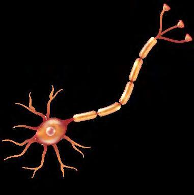
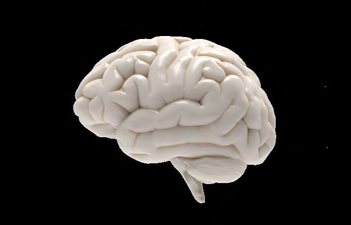
Neuroscience came to the fore in our 2013 paper, published in the journal Cell, when we showed that bone plays a direct role in memory and mood. Mice, whose skeletons did not produce osteocalcin as a result of genetic manipulation, were anxious, depressed, and almost completely unable to master a test of spatial memory. When we infused them with the missing hormone, however, their moods improved and their performance on the memory test nearly normalized. We also found that, in pregnant mice, osteocalcin from the mother’s bones crossed the placenta and helped shape the fetal brain: i.e., bones talk to neurons even before birth.

Human Health Implications
With age, bone mass decreases and memory loss, anxiety, and depression become more common. These may be separate, unfortunate facts about getting old, but they could also be related. Many physicians recommend exercise as a way to prevent agerelated memory loss. Does it help, at least partly, by maintaining bones, which make osteocalcin, which in turn helps preserve memory and mood? In my view, a higher bone mass means a greater capacity for osteocalcin production. A question before us is: Would it ever be possible to protect memory or treat age-related cognitive decline with a skeletal hormone?
My vision of the skeleton as central to energy usage, reproduction, and memory has persuasive evidence in mice, but the extent to which these results translate to people remains an open question. Most hormones have similar functions in mice and humans. Still, osteocalcin is clearly not the only substance that regulates blood sugar, male fertility, and cognition, and its relative importance may be different in rodents and people. In mice, our work has shown that no other substance can compensate for a lack of osteocalcin when it comes to these functions. Is the same true for humans?
We now recognize that the body is far more networked and interconnected than most people think. No organ is an island. The skeleton often seems to play a surprising role. If insulin impacts bone, bone should help regulate insulin. If testosterone has an influence on bone mass, the skeleton should act on the testes. And, most fundamentally, just as the brain talks to the skeleton, bone should help regulate the brain. We continue the search to learn how this bidirectional influence works.
Danger Lurks
While identifying multiple functions for a novel hormone is a good start, it raises a new question: the common link between them. Why does osteocalcin regulate these particular physiological functions? And what others might it regulate?
One common link involves the prototypical human reaction to acute danger: the stress or “fight or flight” response, a suite of physiological processes that were studied and analyzed with great insight by the late Bruce McEwen. These processes that make up the acute stress response include increases in heart rate, blood pressure, energy expenditure, respiration, and adrenal secretion of the hormone cortisol.
Many of the physiological processes regulated by osteocalcin are needed to escape danger. Memory, for instance, enables creatures in the wild to remember where predators are and where there is food. The capacity for exertion (i.e., to run) is also needed to escape danger, as is the ability to mobilize glucose, another function of osteocalcin. Thus, the hypothesis that osteocalcin is a hormone essential in situations of acute danger offers the most convincing argument for links between the physiological functions that it regulates. This, of course, does not exclude additional commonalities among such functions.
As for any physiological process, many questions remain regarding the biology of the acute stress response. For one, how does the activity of the sympathetic nervous system surge so quickly in situations of acute danger? This is a crucial issue, given this system’s key role in controlling bodily functions that are not directed consciously, including heartbeat, breathing, and digesting. In theory, the two arms of the nervous system, the sympathetic nervous system and the parasympathetic nervous system (which conserves energy by slowing body functions like heart rate and digestion), are supposed to work in equilibrium: How is osteocalcin involved in this balancing act?
A key question at this point was whether osteocalcin is actually a stress hormone (i.e., is it implicated in the initiation and/or unfolding of the acute stress response)? To qualify, only two criteria must be met: stress hormone’s circulating levels should rise within minutes in animals facing an acute danger. And such a hormone should regulate, directly or indirectly, the physiological processes, such as those enumerated above, that are recruited to mount an acute stress response.
Concrete Answers
The first observation we made is that in mice, rats, and humans facing danger, circulating osteocalcin levels rise within minutes. How can acute fright quadruple concentration of the hormone so quickly? A chemogenetics approach targeting the fear center of the brain’s bilateral amygdala region provided an answer to this question by showing that, between a stressor and the release of osteocalcin by bone, there was signaling in the brain. A battery of genetic testing, of cell culture assays, and abrogation of neuronal signaling ruled out the possibility that mediators of such signaling are circulating molecules, implicating a neurotransmitter instead.
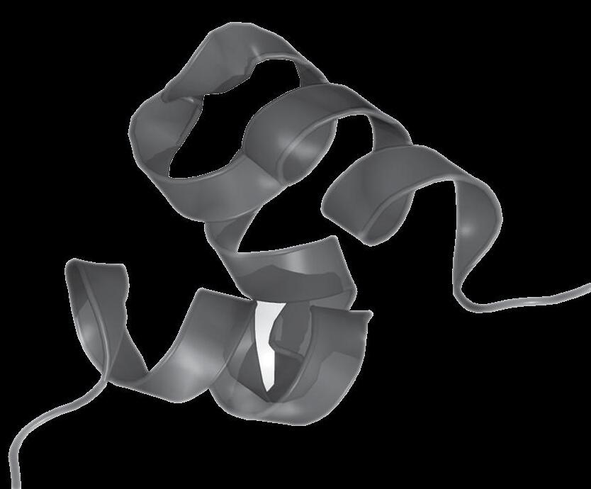
In this regard, osteocalcin’s structure illustrates the cleverness of evolution. The hormone is synthesized in osteoblasts (cells that lead to the formation of bone). It is then modified by the enzyme gamma carboxylase and enters the circulation in the carboxylated form, which is inactive, and the uncarboxylated active form. The inactive form can be blocked by the neurotransmitter glutamate, which is present in both brain and bone.
Here’s how they all come together: Under acute stress conditions, signals in the amygdala (and possibly other fear centers in the brain that we have not yet tested) lead to the release of glutamate. This neurotransmitter goes to the bone, where it enters osteoblasts and blocks the inactive form of osteocalcin, so that only the hormonally active form is released into the circulation.
What allows us to call osteocalcin a true stress hormone is that, furthermore, in its absence (in mice in which osteocalcin has been bred out), typical responses to acute stress are either abolished or severely blunted: These animals show little or no increase in energy expenditure, heart rate, and blood oxygenation. Hence, on genetic grounds, osteocalcin is necessary to trigger the acute stress response.
How, most fundamentally, does osteocalcin work to create an acute stress response? Since it synthesizes the chemical norepinephrine in the brain, we thought that it would do the same in the peripheral nervous system. But it does not. Instead, studies show that osteocalcin sends its signals through a receptor found on neurons, and these signals then travel from the central nervous system to peripheral organs. The net result: The surge in circulating levels of osteocalcin inhibit the recycling of acetylcholine molecules and the electrical activity of certain neurons, reducing activity of the parasympathetic nervous system (the system that slows down responses such as breathing and heartbeat), and leaving the sympathetic nervous system unopposed and ready to initiate the acute stress response. We cannot rule out the possibility that osteocalcin signaling has other effects as well, such as contributing directly to the increase in energy expenditure in target organs.
Acute Stress Response in the Absence of Adrenal Glands
If osteocalcin is necessary to trigger the acute stress response, the next question is whether it is sufficient to trigger this response. There is ample evidence that, at the time, the acute stress response develops a surge of circulating glucocorticoid hormones— the role of which has not been fully understood. So, in view of the results we had obtained, the elephant in the room was to determine whether adrenal glands are actually necessary to mount an acute stress response.
In a totally unambiguous manner, mice or rats that have had their adrenal glands removed—and thus cannot synthesize glucocorticoid and mineralocorticoid hormones or epinephrine—develop an absolutely normal acute stress response when exposed to stressors. The same is true in patients with adrenal insufficiency. In other words, the acute stress response does not seem to require the adrenal glands to develop. An added and precious benefit of asking if adrenal glands are necessary to mount an acute stress response was to illuminate the role of osteocalcin in the initiation of the acute stress response.
How could we explain that the acute stress response does not need glucocorticoid hormones? The answer came from general endocrinology. It has been known for decades that glucocorticoid hormones (corticosterone in rodents, cortisol in humans) are powerful inhibitors of osteocalcin expression in osteoblasts. As a result of that, mice and rats whose adrenal glands have been removed have elevated circulating osteocalcin levels at baseline, and these levels surge higher under stress than in sham-operated animals. This raises the hypothesis that adrenalectomized animals develop a normal acute stress response simply because they are hyper-osteocalcinemic. We reasoned that if this hypothesis was true, then reducing circulating osteocalcin levels and preventing them from surging in mice that had been adrenalectomized, should prevent the development of an acute stress response when exposed to stressors.

We found that mice with osteocalcin that lack only one allele were not hyperosteocalcinemic and that their circulating osteocalcin levels did not increase when exposed to stressors. When these mice with osteocalcin were adrenalectomized, they were now unable to develop an acute stress response when exposed to stressors, indicating genetically that osteocalcin is not only necessary, it is also sufficient to mount an acute stress response, at least in the case of adrenal insufficiency. From an evolutionary standpoint, this study also illustrated, one more time, that with the appearance of bone, the way many physiological functions are achieved or regulated had changed.
When it comes to the acute stress response per se, the data cited above raise many questions. Is osteocalcin needed for the development of all stress manifestations, or only for the ones we studied in the mouse? Does osteocalcin act only through regulation of parasympathetic tone, or does it also signal directly in organs whose activity is recruited under stress? What is the molecular identity of the glutamatergic neurons found in bone? Where are their cell bodies? Where do they connect in the brain? Can we use osteocalcin signaling in the parasympathetic nervous system to identify novel functions of the hormone? Finally, why do circulating glucocorticoid levels go up during the acute stress response? Although we have learned a great deal about this mysterious but elusive hormone along this long and winding road, so much more needs to be explored. l
> From Our Podcast with Gérard Karsenty

“Do our bones influence our minds?” “There is no question, absolutely. There are always naysayers who said I prefer the world as it was 50 years ago, and I prefer bone as it was before it became an endocrine organ. But, no, the evidence is overwhelming, and they're overwhelming because they come from so many labs, in so many countries. So, there is no way to stop data right now.”
“Is osteocalcin being studied by other geneticists or neuroscientists? It's interesting in the sense that it's kind of the merging of two, almost completely different, fields. It is a great example of how science works cross-functionally.”
“About 25 to 30 labs in the world are working on osteocalcin in mice and in humans. Neuroscience is part of our project as you see with this article, but we are also delving into new areas of research that are all centered around the notion that the bone may have been invented by evolution, in part, as a survival tool when animals left the sea to go to land. And what we are trying to write now is a new chapter of the physiology of danger through osteocalcin. But the function of bones, the classical function of bones, fits this definition because while you are sitting and listening to me, if the ceiling falls on your head, you will be unhappy, but you will not die, because bone protects your brain.”
