4 minute read
DiSCo MRI SFN


MRI scans provide a detailed picture of the brain, enabling doctors to both diagnose disease and monitor the effectiveness of therapies. We spoke to Dr Karin Shmueli about her work in developing a new MRI method that will provide a deeper picture of tissue electromagnetic properties and may help diagnose Alzheimer’s disease at an earlier stage than currently possible.
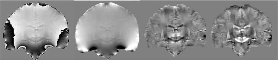
A technique developed in the ‘70s, magnetic resonance imaging (MRI) has become an important diagnostic tool across many areas of medicine, including neurodegenerative disease. An MRI scanner is typically used for around 30 minutes, during which different images are collected. MRI brain images illustrating the stages in quantitative susceptibility mapping: raw phase images (left) are “Those images are acquired in different ways unwrapped, then background fields are removed so the final susceptibility map (right) can be calculated. to emphasise different types of tissue. One In functional MRI (fMRI), information on at an earlier stage than is currently possible,” says image might highlight fluid, and another patterns and networks of brain neural activity is Dr Shmueli. “We are now working on optimising might show lesions in white matter in the obtained by assessing variations in the magnitude these techniques in healthy volunteers.” brain,” explains Dr Karin Shmueli, Principal signal over time. Measuring changes in the The next step would be to apply these Investigator of the DiSCo MRI SFN project. phase signals over time should allow extraction techniques to diagnose different diseases, These images provide a very rich picture of of functional susceptibility and conductivity including not just Alzheimer’s, but also the condition of soft tissue in the brain, while changes. However, this is a demanding task and potentially several other conditions. Dr Shmueli’s functional MRI scans are also an extremely sophisticated image processing will be required. team and collaborators are looking at brain valuable diagnostic and neuroscientific tool. “A lot of things change over time; we’re breathing, susceptibility changes in individuals with sickle“With functional MRI scans we can look at the our hearts are beating and blood is flowing. We cell anaemia, and are also studying arterioway the brain works, and how brain activity want to remove those physiological fluctuations venous malformations in the brain. “With varies over time,” says Dr Shmueli. from the phase signals, to detect the fluctuations arterio-venous malformations you get shunting MRI maps of the magnetic susceptibility and electrical conductivity provide new information about the chemical makeup veins, which would normally contain a lot of deoxygenated blood,” she outlines. “We have been able to detect that non-invasively for the first of tissue and how it changes in a variety of neurodegenerative diseases. time, using quantitative susceptibility mapping.”
DiSCo MRI SFN project
An image is acquired in MRI through what is called a pulse sequence (literally a sequence of pulses of radiofrequency energy and magnetic field gradients), which can be varied to highlight particular features of the brain. Now Dr Shmueli and her colleagues in the project are designing a rapid pulse sequence lasting between 5-10 minutes which will provide images containing several different pieces of information. “We can get some of this information using current scans, while some of it is totally new information that hasn’t been collected before,” she outlines. The signals acquired in MRI are numerically complex in nature, with both a magnitude and a phase; the latter is often discarded, but Dr Shmueli makes use of both. “We use these signal components to give us different pieces of information. We can use the phase to give us both a susceptibility map and a conductivity map,” she explains. These maps of the magnetic susceptibility and electrical conductivity have been found to provide new and useful information about the chemical makeup of tissue and how it changes in a variety of neurodegenerative diseases. in susceptibility and conductivity that reflect neural activity,” says Dr Shmueli.
The aim is to remove the physiological noise, leaving only small signals from which researchers can gain deeper insights into brain activity patterns. “The brain has a mechanism called functional hyperaemia, which means that lots of oxygenated blood is sent to active regions of tissue. This blood oxygenation is measured indirectly in standard fMRI and we aim to quantify it much more directly by looking at blood susceptibility changes over time,” continues Dr Shmueli.
This research holds wider relevance to the diagnosis of neurodegenerative disease. The priority at this stage in the project is to improve the pulse sequence, before testing it on healthy volunteers and eventually a group of patients with early stage Alzheimer’s disease. “That will be in the last year of the project, once we’ve developed the acquisition and processing techniques,” explains Dr Shmueli. Her team aims to reduce the amount of time these patients have to spend inside a scanner, while still gathering detailed information. “We want to assess the structural, compositional, and functional changes that happen in the brain of oxygenated arterial blood straight into the
DiSCo MRI SFN Developing Integrated Susceptibility and Conductivity MRI for Next Generation Structural and Functional Neuroimaging
Dr Karin Shmueli Department of Medical Physics & Biomedical Engineering, University College London Malet Place Engineering Building, Rm 2.02 T: +44 (0)207 679 0256 E: k.shmueli@ucl.ac.uk W: https://www.ucl.ac.uk/ medical-physics-biomedicalengineering/research/ research-groups/magneticresonance-imaging-group
DrKarin Shmueli is an Associate Professor in Magnetic Resonance Imaging in the UCL Department of Medical Physics and Biomedical Engineering. Karin pioneered MRI susceptibility mapping (QSM) techniques as a Postdoctoral Visiting Fellow at the USA National Institutes of Health. She leads a group of researchers optimising QSM techniques for several applications from neurodegenerative diseases to cancer.