5 minute read
Complications in phaco for small eyes
Small eyes can lead to a range of problems in surgery. Soosan Jacob MS, FRCS, DNB reports
Small eyes can be classified as microphthalmos [short anterior chamber (AC) depth and short axial length], relative anterior microphthalmos (short AC depth and normal axial length) and axial hyperopia (normal AC depth with short axial length).
Microphthalmic eyes may be short but otherwise normal (nanophthalmos) or may have associated abnormalities like coloboma, glaucoma, corneal opacity or other complex malformations.
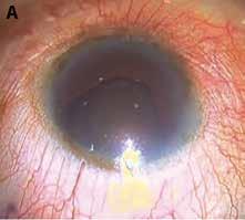
Nanophthalmos is bilateral with affected patients having small eye, microcornea, small anterior segment, normal or increased lens thickness, convex iris, shallow and crowded AC, high hypermetropic errors (+8 to +20DS) and axial length <20.5mm. Blindness may occur if left untreated and even after successful cataract surgery, retinal problems such as macular hypoplasia can limit vision.
Problems encountered during cataract surgery are secondary to small size, peripheral anterior synechiae, chronic angle closure glaucoma (CACG), poorly dilating pupil and thickened choroid and sclera. Spontaneous as well as postoperative uveal effusions and exudative retinal detachments may occur.
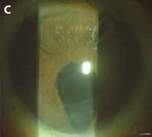
Microphthalmic eyes with choroidal coloboma may require retinal laser preoperatively and those with iris coloboma may require iridoplasty together with cataract surgery. Additional surgery for glaucoma or corneal opacity may be required in complex cases. Postoperative vision may be limited by associated ocular comorbidity.
In relative anterior microphthalmos, the normal sized lens causes crowded anterior segment, shallow AC and CACG. It is not associated with scleral abnormalities or uveal effusions.
Cataract surgery in small eyes has the benefit of decreasing anterior chamber crowding. Preoperatively, pupillary Problems encountered during cataract surgery are secondary to small size, peripheral anterior synechiae, chronic angle closure glaucoma (CACG), poorly dilating pupil and thickened choroid and sclera
dilatation can precipitate angle closure and prophylactic YAG peripheral iridectomy may be necessary. Fundus evaluation to look for uveal effusions and B-scan to measure thickness of choroid and sclera are important.
The eye should be softened with oral acetazolamide and/or glycerol, IV mannitol and ocular pressure with Pinkie ball/ Honan balloon before surgery. Well-constructed tunnels prevent iris prolapse. Pupillary dilatation techniques such as mydriatics, viscomydriasis, synechiolysis, pupil stretch, mini-sphincterotomies or pupil expanders are used if indicated. Lack of manoeuvring space within the AC may be a challenge.
Rhexis with high molecular weight cohesive viscoelastics and microinstruments passed through a partially opened incision is generally successful. Bimanual phaco may be preferable in very small eyes.
Arshinoff’s soft shell technique maintains space and protects endothelium. Soft nuclei may be partially hydroprolapsed and emulsified in parts. However, hard nuclei are often encountered because both patient and surgeon tend to delay surgery. Debulking by shaving away epinucleus followed by divide and conquer or crater and chop techniques are helpful. Manoeuvring space can be maintained by increasing bottle height or by using pressurised air infusion. Loose or defective zonules cause vitreous hydration and further AC shallowing. Limited dry anterior vitrectomy with highspeed 25-gauge vitrector helps deepen AC. However, pars plana dimensions may be different from normal eyes and sclerotomies should be placed carefully. High-powered IOLs are difficult to inject and the incision may need to be enlarged. Prophylactic sclerotomies or sclerectomies may be placed in microphthalmic eyes for decompression of vortex veins and allow fluid drainage without causing uveal effusions. Sudden changes in intraocular pressure should be avoided.
Phacoemulsification can generally be carried out using normal techniques but with extra care in eyes with axial hyperopia. In these eyes, surgery may be done for cataractous lens or alternatively as refractive lens exchange for correction of the refractive error; a good choice in older hyperopic patients who have started to lose accommodation. Surgery is generally easy as the nucleus is soft and can easily be hydroprolapsed out of the capsular bag and aspirated.
Patients should be counselled regarding increased risk of surgery and poor visual prognosis secondary to any associated retinal abnormalities or amblyopia. Risk of posterior capsular rent (PCR) and endothelial damage are higher in these eyes because of positive vitreous pressure
and lack of surgical space. Uveal effusion, suprachoroidal haemorrhage, aqueous misdirection syndrome and prolonged uveitis are other complications. In case of a PCR, secondary IOL fixation may be done using a glued IOL (using small diameter optic and with trimmed haptics for tucking) or other preferred technique.
IOL power calculation is difficult in these eyes with a higher chance of errors as small errors in axial length measurements and estimation of ELP can get magnified. Ultrasound biometric machines are calibrated for normal eyes with fixed anatomical proportions and this may affect accuracy in microphthalmic eyes where the normal sized lens takes up a larger volume. Both immersion and optical biometry should be used and repeated measurements taken. The Holladay 2, Kane, EVO 2.0, Barrett Universal II, Hill-RBF, Haigis and Hoffer Q formulae are better relied on. It is advisable to calculate using different formulae before deciding final IOL power. Intra-operative aberrometry (ORA Inc, Wavetec Vision) can also be useful.
For lower degrees of hyperopia, standard choices may be made but higher degrees require customised special highpowered IOLs.
An alternative option is to piggyback an IOL in the same sitting or at a second sitting after checking residual refractive error and available space in the sulcus. The power may be calculated by using the Gills nomogram: {(1.5 x Spherical equivalent) +1}. Placing two IOLs in the bag should be avoided as this can lead to interlenticular fibrosis, decrease in vision and late hyperopic shift.
It should, however, be remembered that many of these small eyes may not have enough space or may not tolerate two IOLs with resultant crowding, uveitisglaucoma-hyphema syndrome etc. Also, interlenticular membranes may still develop even with one IOL in the bag and other in the sulcus and even if made of different materials. Hyperopic eyes may have a large angle kappa and multifocal IOLs should be avoided in such eyes. IOL implantation may be deferred in very severe microcornea where the IOL optic may cause crowding.
A: Microcornea

C; Iris coloboma
FEMTOSECOND LASER ASSISTED CATARACT SURGERY (FLACS) FLACS can be useful in shallow anterior chambers where manoeuvring space is less. Longer tunnels should be programmed

B: Choroidal coloboma

D; Repaired iris coloboma
and positioned carefully to prevent iris prolapse. A femtosecond-created rhexis decreases the chances of a runaway and is a major advantage. Pre-treatment of a dense nucleus with femtosecond can break the nucleus into smaller fragments, thus making removal easier and thereby decrease possible endothelial damage.
Dr Soosan Jacob is Director and Chief of Dr Agarwal’s Refractive and Cornea Foundation at Dr Agarwal’s Eye Hospital, Chennai, India and can be reached at dr_soosanj@hotmail.com
EuroTimes is your magazine!
Do you have ideas for any stories that might be of interest to our readers?

Contact EuroTimes Executive Editor Colin Kerr at colin@eurotimes.org


