22 minute read
Medicine and Technology


Healthcare and the treatment of diseases are currently undergoing significant changes. Innovative measurement technology, mobile sensors, high-precision analytical devices and 3D printing techniques are transforming the healthcare system. Medicine can be customised and patients have more say in their treatment. Researchers at the FHNW HLS are therefore developing practical solutions for the digital age. With modern pharmaceutical technology they are driving the development of new drugs and applications.
De-coding complex diseases
Despite decades of research, many diseases cannot be treated successfully as science still does not fully understand what causes them. This is particularly true for illnesses in which multiple factors interact, such as chronic liver disease or Alzheimer’s disease, and which are often diagnosed at advanced stages. Researchers at the FHNW HLS are using elaborate cell culture systems to model these diseases and gain a better understanding of the mechanisms behind them. This ground-breaking work also contributes to a reduction in animal experiments.
Some diseases have a single cause, such as the bacterium Vibrio cholerae for cholera. Many others develop via the interaction of several factors, for example genetic predisposition, an unhealthy lifestyle and environmental effects. Since identifying a clear cause is often impossible, such illnesses are called complex diseases and are often difficult to diagnose and treat. Cell biologist Laura Suter-Dick from the Institute for Chemistry and Bioanalytics at the FHNW HLS has dedicated herself to researching these diseases on a cellular level: “Understanding the molecular mechanisms that underlie a complex disease is essential for successful treatment. Various factors can interact and trigger the development of the disease – or they may trigger cellular processes which then cause the disease.”
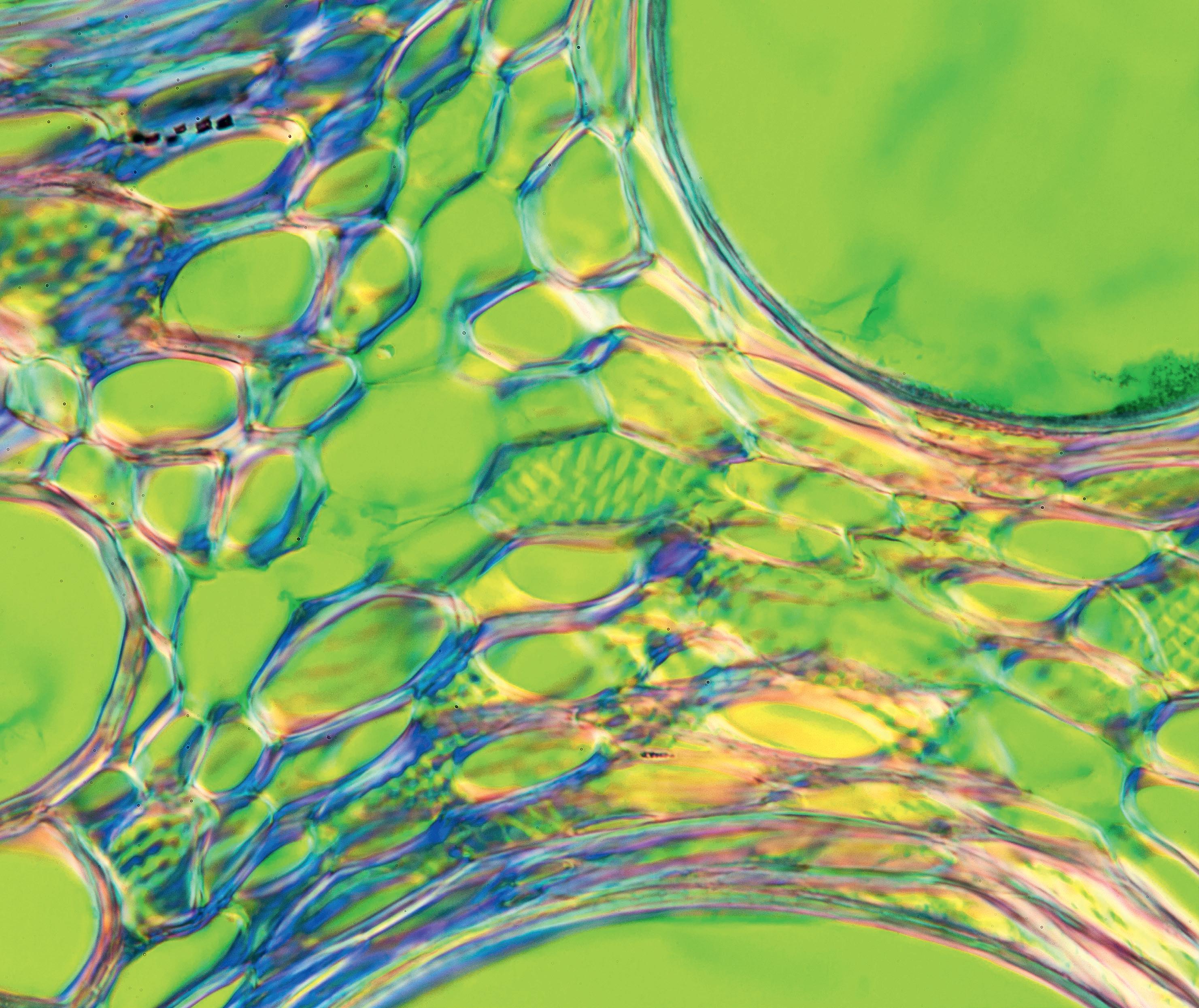
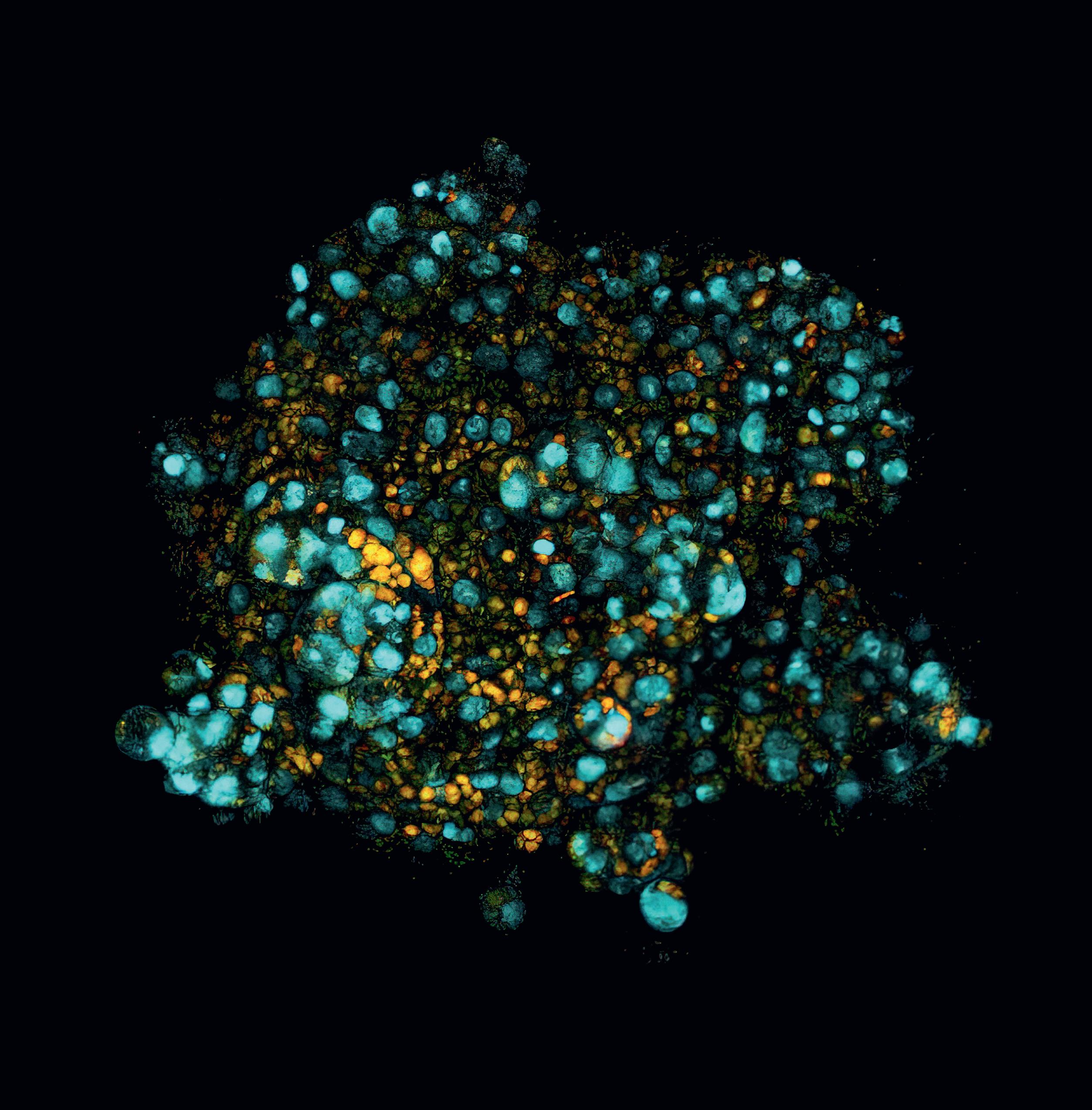
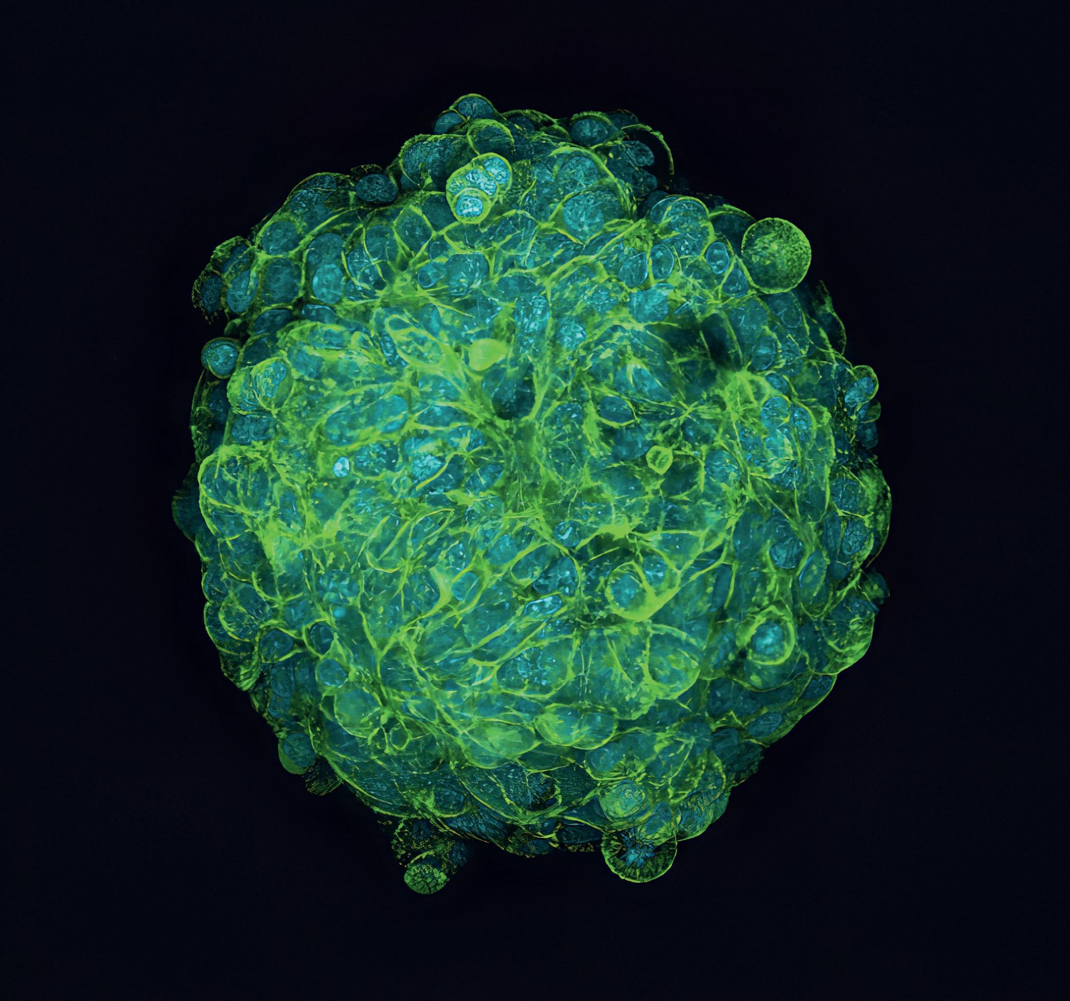
To study such processes, researchers like SuterDick work with miniature replicas of organs called 3D models or organoids in which diseases can be simulated. These models contain organ-specific cell
types that closely mimic a real organ in their function and layout. In her cell biology and in vitro toxicology laboratory at the FHNW HLS, Suter-Dick is developing sophisticated 3D cell culture systems which she uses, for example, to simulate chronical“In our models, we can ly diseased liver tissue or Alzheimer‘s disease. The researcher and her team want to use this technology watch human cells develop to find markers that could enable early diagnosis and treatment options, as well as to look for ways the characteristics of liver to prevent the development of the disease altogethfibrosis or Alzheimer’s er. “The earlier you detect a disease, the more effectively you can treat it,” says Suter-Dick. disease.” Laura Suter-Dick
From cell to model
The scientist bases her research on the 3R principle – “replace”, “reduce” and “refine” –, a guideline that aims to work responsibly with laboratory animals and to reduce and replace animal experiments. “There is often no need for an animal model,” explains Suter-Dick. “If we recreate the physiological architecture of a tissue in the lab, we reproduce its physiological function.” Hence the researcher builds her cell systems under specific conditions to
resemble the tissue they are designed to imitate. Since real organs need a range of cell types running different processes in order to function, the models also do not consist of just a single cell type. Imitation of the disease under investigation begins during the cell growth phase: if the researcher wants to simulate a disease, the cells are exposed to the causes from the start.
The example of liver disease
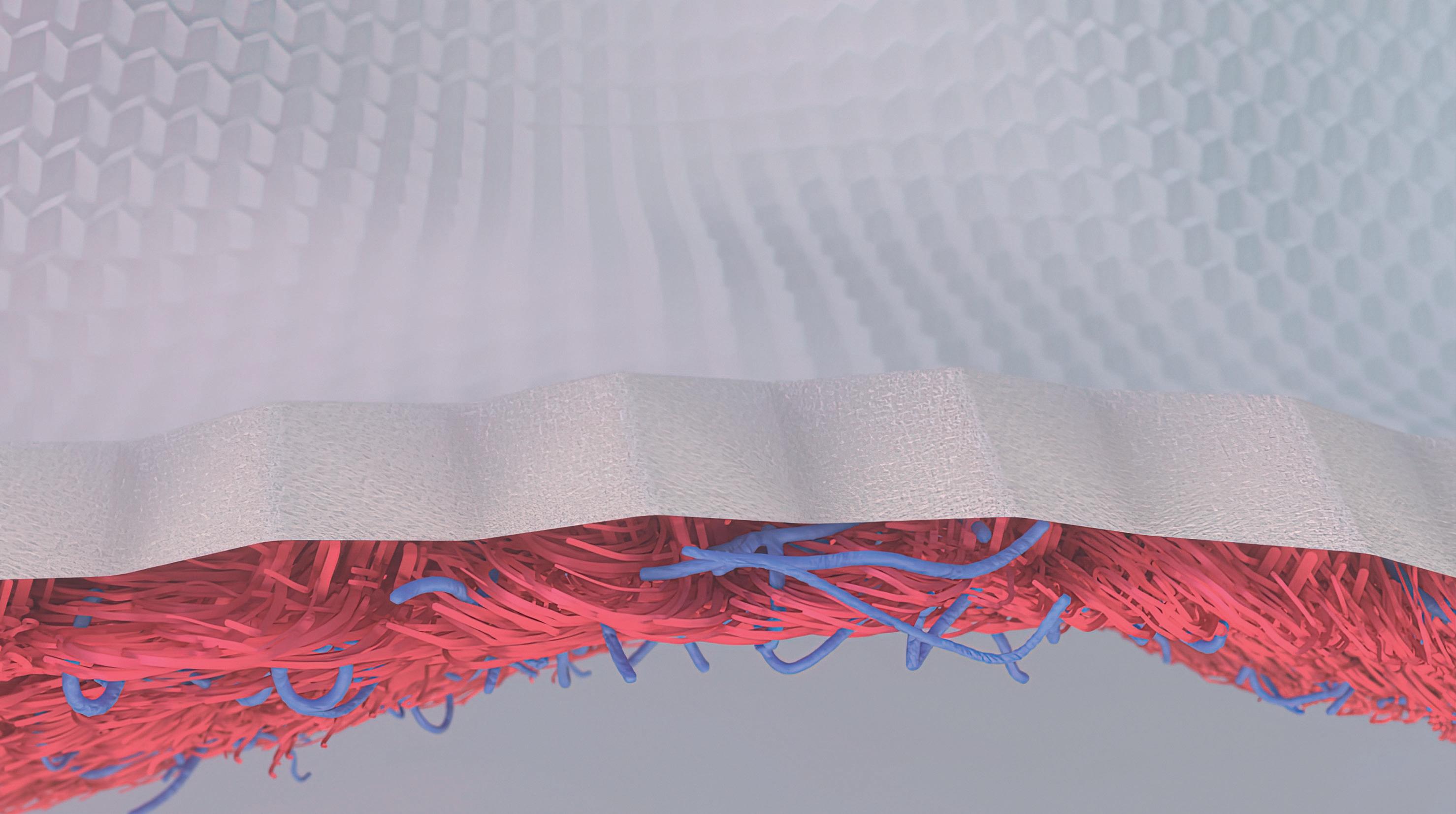
A disease model with which Suter-Dick‘s group has many years of experience is liver fibrosis, a chronic condition that causes pathologically activated cells to deposit collagen fibres. Liver tissue is gradually replaced by connective tissue and the organ becomes scarred. There are many causes, including chronic inflammation from viral infections such as hepatitis C, but also medications or toxins that stress the liver over a long period. For example, prolonged high alcohol consumption can cause a fatty liver, which leads to fibrosis. However, fatty liver can also develop for other reasons and is then referred to as Non-Alcoholic Fatty Liver Disease (NAFLD), which is the most common liver disease in western countries. It can be caused by an undiagnosed metabolic disease or an unhealthy lifestyle, is most common in people who are overweight or obese, and even children are increasingly suffering from it. Fatty liver can lead to liver fibrosis and from there to cirrhosis: the liver stops working and has to be replaced by a transplant. Liver cirrhosis also often leads to liver cancer.
Liver fibrosis in the culture dish
Suter-Dick uses three types of human cells for her liver cell cultures: hepatocytes (the main cells in the liver), Kupffer cells, which are responsible for local immune response, and stellate cells. The latter produce the collagen fibres in a scarring liver and thus play a key role in fibrosis and cirrhosis. Using her culture dish liver models, Suter-Dick is investigating how liver fibrosis develops and which molecular signals the cells display. “After extended treatment with bile acids and other stimuli, our models showed clear signs of fibrosis,” the scientist reports. “The cells in the model also released specific liver damage markers called microRNAs. This is significant, as elevated microRNAs, especially
microRNA-122, can also be detected in blood in animal studies and in humans with liver damage.” Hence, microRNAs are good candidates for biomarkers that could be relevant not only in vitro, but also in the clinic, opening the way to better diagnosis and therefore enabling earlier treatment.
Using the new single-cell sequencing technology in her models, the researcher also measured which sections of the genetic material are read and transcribed in individual cells. This enabled her to compare the metabolic processes which are particularly active in healthy and fibrotic liver cell
cultures. She also observed how cell populations change over the course of the disease. This work should help identify specific approaches for the treatment of liver fibrosis.
Hunting down Alzheimer‘s
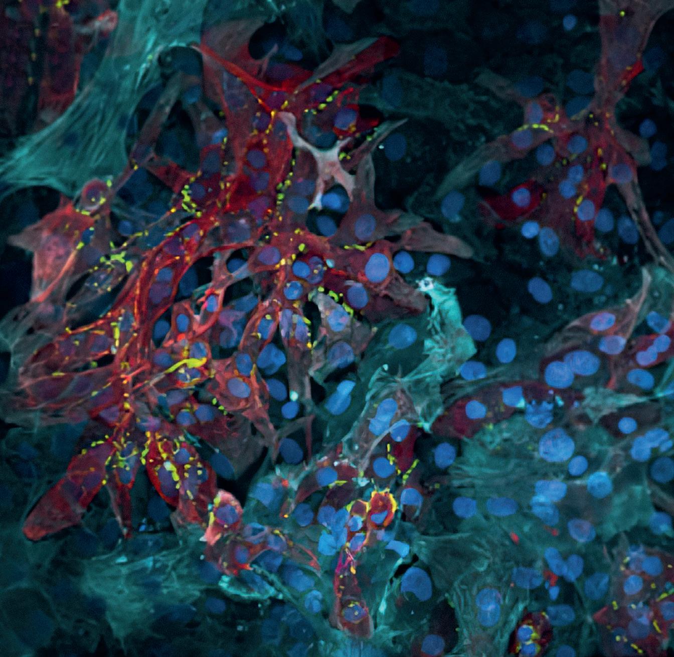
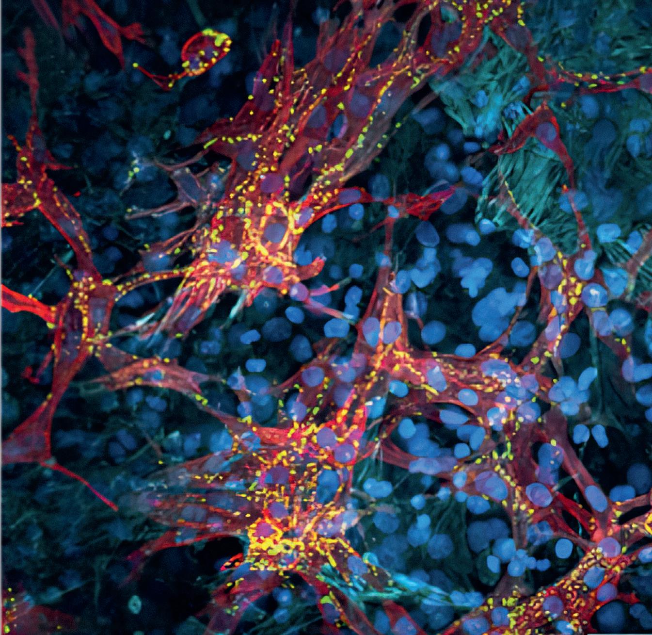
Such new techniques are also urgently needed for Alzheimer‘s disease. This neurodegenerative dementia condition leads to neuron cell death in the brain, but the mechanisms that cause it are still unclear. However, a hereditary form of Alzheimer‘s could be linked to genetic mutations. Suter-Dick’s team genetically modified neuron precursor cells to show characteristic features of Alzheimer‘s and were then able to differentiate these cells in cultures. This resulted in mature neurons and astrocytes, two key cell types in the brain. In this process, “healthy” progenitor cells developed into healthy neurons and “diseased” progenitors developed into diseased neurons. “Of course, Alzheimer‘s disease does not show up in cell culture with memory impairment and other cognitive problems,” Suter-Dick explains, “but we see the same biochemical changes that occur in the brains of Alzheimer‘s sufferers: the formation of beta-amyloid plaques and the alteration and accumulation of tau protein.” Disease development in the model takes about six to ten weeks, relatively long for a cell culture process. But this slowness corresponds closely to the chronic progress of the disease in humans.
In the culture dish, diseased and healthy neurons can now be compared. This new approach will be used in a follow-up project to further investigate the basic mechanisms of Alzheimer‘s disease and related therapeutic approaches. These 3D cell culture systems not only help to reduce the use of animal experiments; they make it possible to model disease processes on human cells and better investigate the development and treatment of those diseases.
Methods and infrastructure
– General cell culture methods – 3D cell culture models and microfluidic systems (MPS: microphysiological systems) – Enzymatic, biochemical and immunodetection methods (to determine protein expression) – Flow cytometry and cell sorting (FACS) – Imaging, incl. confocal microscopy – Molecular biological methods, incl. single cell sequencing
Support
– Swiss Center for Applied Human
Toxicology Foundation – SCAHT – Innosuisse – F. Hoffmann-La Roche AG – BRIDGE Programme, Swiss National
Science Foundation and Innosuisse
Collaboration
– InSphero AG – CSEM Swiss Center for Electronics and Microtechnology Inc. – Haute École Arc, HES-SO
Sensor socks instead of plaster casts
For an orthopaedic splint to fit the patient well, a precise model is critical. In future, the painstaking work of making a plaster cast model will be replaced by measuring textiles. Researchers at the FHNW HLS have developed a prototype sock that measures foot shapes in 3D with millimetric precision, thanks to tiny sensors in the sock which measure magnetic field values. The application only needs a weak magnetic field above the treatment table and the precise shape of the foot in the sock can be acquired in a few seconds.
Orthopaedic splints correct joint position, relieve pain and provide stability, but a high level of skill and manual dexterity is required for their manufacture. The orthopaedic technician has to place the patient‘s foot in exactly the right position; only by successfully recording this position digitally can a precisely fitting splint be produced. To do this, the technician must hold the foot in the desired position with one hand while with the other hand wrapping it in a plaster bandage. Once the plaster is dry, it is cut open and the inner shape of the cast can be recorded with an optical 3D scanner. This provides a computer model, on the basis of which a patient-specific corrective splint – the orthosis – is made. Researchers at the FHNW HLS have shortened this complex procedure to a simple push of a button: they are developing an electronic networked sock that can record foot position and shape in a matter of seconds. Patients would only need to put on the sock. With the foot correctly
positioned, a specialist can trigger the measurement via pedal switch or voice command. Nothing gets wet or dirty, the specialist has both hands free and the outcome is a high-precision model for the orthosis. “Our measuring textiles “Senior orthotist Thomas Ruepp had the idea of an intelligent sock for orthosis production in the provide the same data as a early 1990s; together with my FHNW colleague Ralf Schumacher we then patented the integration plaster cast at the push of sensors into textiles,” says Joris Pascal of the Inof a button. And because stitute for Medical Engineering and Medical Informatics at the FHNW HLS. But it is only now becomthey are more accurate, ing reality thanks to a successful interdisciplinary they can be used to make project between FHNW electrical engineers and medical technology start-up Bellwald Tec to develbetter orthoses.” op a prototype sock. It enables foot measurement to an accuracy of two to three millimetres within a Joris Pascal few seconds, thanks to over a hundred small magnetic sensors incorporated into the sock. With the sock on the foot in a suitable magnetic field, the sensors deliver measured values to an electronic control unit which accurately calculates each sensor’s position. Software uses the resulting point
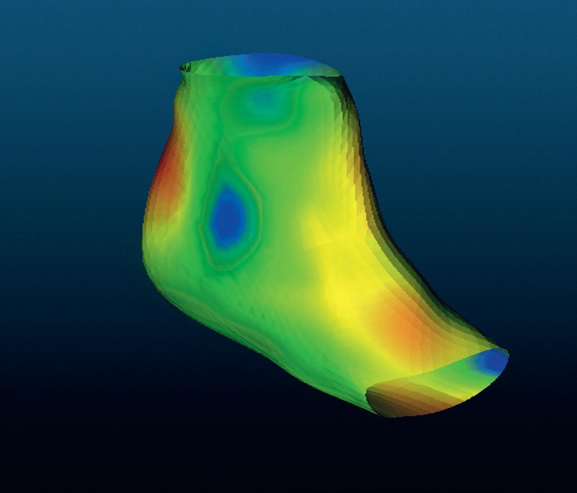
cloud to reconstruct the foot shape as a digital 3D model which, as in conventional orthosis production, is then sent to a 3D printer to create a splint. It can also be used as a direct template for a computer-designed splint. “We wanted a solution that seamlessly follows orthotists’ usual workflow,” says Pascal. He and his team hope that the intelligent textile will not only make the splint production process easier for everyone involved, but also lead to greater therapeutic success. “A splint that fits well is also worn more regularly,” says the researcher.
The sensor sock is more accurate than a plaster model since, as plaster dries, even small foot movements can change its shape. Moreover, the accuracy of the plaster cast can be affected by cutting, opening and removal, and thus two or three different splints often have to be made in order to eliminate pressure points. However, since health insurance companies pay a lump sum for orthoses, some sufferers do not get a second version for cost reasons and rarely wear their uncomfortable splint. The result can be a permanent deformity, possibly leading to problems walking and an inability to work. On the other hand, if a child‘s pointed foot or other joint deformity is corrected early enough, he or she can walk better as an adult. Good orthoses therefore improve quality of life as well as giving economic advantages.
For their prototype, the researchers developed sophisticated electronics and a special communication protocol, with all the sensor chips on a single flexible circuit board and transmitting simultaneously. This avoids a tangle of dozens of individual wires and means that the circuit board can be wound in a spiral and woven into the sock. The weak magnetic field, whose magnitude and direction the sensors measure, is generated with small current-carrying coils located a few centimetres under the foot. In a medical practice, the coils could be positioned under the treatment table, for example.
In practice, the smart sock still has to become more robust so that it can regularly be put on, taken off and washed. This is the team‘s follow-up project. “With the foot, we have chosen a body part with a relatively complex shape,” says Pascal. “When the sock is ready for practical use, we could adapt the system for other orthoses and applications, for example, corsets or a smart cap that scans head shapes.”
Methods and infrastructure
– Computer Aided Design (CAD) – Sensor and electronics development – Hardware-related software development – Algorithm development for magnetic tracking – 3D mapping and modelling of magnetic fields – Modelling of anatomical shapes – Electronics laboratory – Calibrated Helmholtz coil – 3D scanner, digitiser – 3D printer
Support
– Bellwald Tec GmbH
Collaboration
– Bellwald Tec GmbH – Basler Orthopaedics René Ruepp AG
A folding heart model
A heart model would be an ideal solution for some drug tests, provided that it could mimic the heartbeat and deliver meaningful results. A research team including the FHNW HLS has now developed an artificial miniature heart that comes very close to this. The basis is a square-shaped paper structure with fishbone-like folding elements that allow it to be compressed and stretched. When coated with heart cells, this origami ‘scaffold’ contracts and expands in response to tension stimuli – just like a real heart muscle.
To test a drug’s efficacy or side effects in the lab, researchers need suitable models, for example of the heart – the organ in the human body with the highest stamina muscle. Heart muscle cells can already be grown in small clusters known as organoids, on which pharmacological substances are tested. However, these are very simple models compared to an anatomic heart, which is a hollow muscle with four chambers that continually contract and expand. To better represent this complex organ, a research group from the FHNW HLS and the University of Basel has developed a miniature heart model that can beat.
In the KOKORO Nano Argovia project funded by the Swiss Nanoscience Institute, the team applies thin layers of living heart cells onto cellulose paper using bioprinting. The pre-folded paper can be compressed and stretched in two dimensions, a little like an accordion. If the printed cells grow and develop properly, the “origami” heart tissue can contract and expand repeatedly, like a heart muscle.
3D printing with cells
Bioprinting – the technology for manufacturing this heart model – is similar to 3D printing with polymers, but the result is biological tissue. Experts at the University of Basel provided the cell cultures: newborn mouse heart cells, which are not yet fully mature and can still proliferate. In order for them to be printed like polymers, they must first be embedded in hydrogel – a gelatine-like substance made of water and a strong mesh of hydrophilic fibres that swell up in it. For the heart model hydrogel, the researchers used collagen fibres also found in gelatine. “Our hydrogel has about the same consistency as jelly,” says Maurizio Gullo from the Institute for Medical Engineering and Medical Informatics at the FHNW HLS. “Even when we add cells, the gel can still be pressed through a thin nozzle, giving us a fine filament that we use to print a layer of cells line by line and layer by layer on our paper scaffold.” Using this technique, Gullo’s team first printed a thin layer of heart muscle cells, then a second layer with vascular cells. They supply the muscle cells with nutrients and remove harmful
metabolic products. Each heart model contains several million cells and the surface measures one square centimetre; it is thus slightly larger than the cross-section of a mouse heart.
Fine-tuning
The foldable paper for the heart model was made by industry partner Omya from biocompatible cellulose fibres, to make it as cell-compatible as possible. It looks like typical square origami paper and was pre-folded by machine at the FHNW HLS into fine structures only a few hundred micrometres in size. The researchers coated the folded paper structures with hydrogel for structural stability and printed them with the hydrogel cell layers. “With the paper as a structural support, we were able to use a slightly softer hydrogel for printing,” explains project partner Joachim Köser from the Institute for Chemistry and Bioanalytics at the FHNW HLS. “For the cells to develop well, the gel has to be as soft as an embryonic mouse heart. If it is too stiff, they don’t get enough nutrients, waste products don’t disperse fast enough, and they suffocate.” Köser and his team have therefore developed available
hydrogels into a new product that meets the requirements of the heart model. In addition to gel stiffness, they also analysed the optimal density and length of the hydrogel fibres. The final criterion was price. “If 3D print-based heart tissue can ever be used to treat heart patients in the future, it should be affordable,” says Köser.
Active and aligned
In this project the research team has shown that the origami-based heart model principle works: thanks to the protective hydrogel coating, the paper scaffold does not dissolve, and it is compatible with the cells. The two different cell layers – muscle and vascular – are also compatible. The researchers succeeded in finding a hydrogel and nutrient solution in which both cell types thrive: “We dyed the cells in viability tests and analysed the different factors they released,” Gullo explains. “This allowed us to confirm that the cells are surviving and are sufficiently active.” However, this is not enough, he adds: “It is important that the cells feel comfortable, since after application, they still have to mature: they have to differentiate and become functional.”
The vascular cells, for example, form a network while the muscle cells develop the protein structures they need for contraction: actin and myosin fibres. In this context, the paper scaffold has a third purpose in addition to its folding and support functions: its cellulose fibres provide the cells with a structure. “By aligning the muscle cells along the paper fibres, we enable the muscle fibres to contract directionally later,” explains Köser. “We have strengthened this effect by structuring the hydrogels in such a way that the cells can align themselves with them. To this end, we gave the hydrogels linear structures.”
Maurizio Gullo
synchronously and directionally in response to stimulation.” Other researchers can now use the model to study the effect of drugs on the heart, not just at the cellular level but also in functional terms. For example, they will learn whether the model heartbeat changes when a drug is added.
Looking forward, the research team hopes for a second possible application: with a different base structure than paper, the heart model could be developed into a patch that replaces scarred tissue with the patient’s own cells after a heart attack. In this case, too, the newly attached heart muscle cells would follow the contraction of the neighbouring cells, helping the still healthy tissue to beat and thus reducing the risk of a second heart attack.
Successful interaction
In order for the muscles to function like real muscle fibres, they have to synchronise with each other during maturation. Only then can they all contract together in response to an impulse. Moreover, they have to exert enough force while doing it. To test this, the researchers at the University of Basel stimulated the cells with electrical impulses and measured the voltage needed to trigger a contraction. The smaller this value, the stronger the muscle cells. “In a real heart muscle, the nervous system triggers the heartbeat; this is not possible in the model,” explains Gullo. “Nevertheless, in our model the impulse is transmitted in the same way as in the heart: it is transferred from one muscle cell to the next in a kind of domino effect, and one row of cells contracts after the other.” Gullo and Köser are pleased with the outcome: “Our heart model really ‘beats’. The cells contract
Methods and infrastructure
– Cell culture laboratory – Chemistry laboratory – Bioprinting (RegenHU 3D Discovery) – Hydrogel chemistry – Rheometry – Soft lithography (impression) – Mechanical paper folding
Support
– Swiss Nanoscience Institute (Nano Argovia project KOKORO A14.07)
Collaboration
– Omya AG – University of Basel
Well packaged for the large intestine
Tablets or capsules usually release their active ingredients in the stomach or small intestine. If the effect of the drug is needed in the large intestine, this is clearly a disadvantage – the drug is absorbed too early into the bloodstream or acts in the wrong place. Researchers at the FHNW HLS have therefore developed a method of embedding active substances in a framework of dietary fibres. This framework can reach the large intestine intact, at which point large intestine bacteria break down the fibres, releasing the active ingredient exactly where it is needed.
Chronic inflammatory bowel diseases such as Crohn‘s disease and ulcerative colitis are common - around 20,000 people are affected in Switzerland alone. Despite their suffering, therapy options are limited. Standard treatments include anti-inflammatory drugs such as cortisone or the salicylic acid derivative mesalazine, as well as liquid diets. These are designed to give patients the necessary nutritional intake despite disease-related pain, diarrhoea and loss of appetite, but even so, the disease can often only be partially or temporarily halted.
Biochemist Georg Lipps from the Institute for Chemistry and Bioanalytics at the FHNW HLS is convinced that if drugs could act at the inflammation site itself, treatment could be more successful. In colitis, and to some extent in Crohn‘s disease, the target site is the large intestine. However, before a drug can get there, it has to pass through the five-metre-long small intestine. It is here that many medicines dissolve so that the intestinal mucosa can absorb the active ingredients. But of course, this also affects medicines that are actually supposed to reach the large intestine. On their long journey through the small intestine, they are removed too early.
Bacteria as helpers
A team led by Lipps and Georgios Imanidis from the Institute for Pharma Technology has discovered a packaging material that ‘smuggles’ active substances past the small intestine’s digestive enzymes and into the colon. They had this brilliant idea while browsing through scientific journals, where they read that colon bacteria break down the dietary fibre xyloglucan. The human body cannot digest these plant cell wall fibres, but our gut bacteria feed on them. By breaking down the fibres in the large intestine, they release metabolic products that we can use. The small intestine on the other hand has almost no bacteria. Lipps assumed that a xyloglucan-based tablet could therefore reach the large intestine intact, only decomposing once there. “Up to now, a pH-based approach has been used,” the biochemist explains. “Medicines are given a coating that is insoluble in acid, but which dissolves at neutral to mildly alkaline pH. Tablets should thus

disintegrate in the lower small intestine, but the pH of the gastrointestinal tract varies both between people and within the same person, depending on food and physiological conditions. Therefore with this method, where the tablet actually dissolves varies considerably, and the administration of the drug is neither reproducible nor efficient.”
To test whether an active substance embedded in xyloglucan reaches the large intestine, the researchers led by Imanidis produced two different tablets at the FHNW HLS Process Technology Centre: one with the active substance mesalazine and a control with caffeine. To do this, they pre-processed the polysaccharide xyloglucan fibres, mixed them with the active or control substance and then pressed the tablets. “Pressing creates a coherent polysaccharide scaffold which encloses the active substance,” explains Imanidis. “Only when bacteria break down the scaffold is the mesalazine released. ” Imanidis‘ team also sealed the tablets with a polymethacrylate film that protects them from stomach acid. “When the tablet enters the neutral environment of the small intestine after going through the stomach, this first protective layer gradually dissolves,” says Imanidis. “The key is that once it is gone, the xyloglucan matrix takes over the protection role so that no active ingredient is released before the end of the small intestine. When the tablet enters the large intestine, the bacteria go to work.”
After producing the tablets, the researchers tested active ingredient release in a three-stage experiment funded by the BRIDGE programme of the Swiss National Science Foundation and Innosuisse. First, they simulated conditions in the large intestine by exposing the tablets to an enzyme that breaks down xyloglucan. Next, instead of the enzyme, they used bacteria that control the digestion, such as Bacteroides ovatus. Finally, they followed the passage of the tablets through the digestive tract in pigs by analysing concentrations of mesalazine and caffeine in continuous blood and stool samples. “The active ingredient mesalazine is well absorbed in the small intestine but hardly at all in the large intestine,” explains Lipps. “If the tablet only releases mesalazine in the large intestine, the amount of active substance in the blood circulation will therefore be low, and this is exactly what we observed. The caffeine control on the other hand can also reach the systemic blood circulation from the colon. Since only caffeine was detectable in the blood, the protective dietary fibre shell functions exactly as we had hoped and only dissolves in the colon. Stool samples confirm this result.”
In addition to therapeutic use in intestinal disease, xyloglucan tablets are suitable for other applications. For example, they could transport specific nutrients into the large intestine that have a positive effect on intestinal flora and thus also prevent inflammation.
Methods and infrastructure
– Fluid bed granulation – Tabletting by eccentric press – Drum coating – Detection of xyloglucanase activity (viscometer, UV/ VIS spectroscopy) – Cultivation and analysis of the microbiome (anaerobic box) – In vitro dissolution test
USP II/release – Pre-clinical in vivo test – Chemical analysis of plasma and faeces samples by LC-MS/MS
Support
– FreeNovation, Novartis – BRIDGE Programme, Swiss National
Science Foundation and Innosuisse
Collaboration
– Vetsuisse Faculty University of Zurich