
152 minute read
INSECTS AS VECTORS OF APPLE PROLIFERATION PHYTOPLASMA Apple proliferation spread over long distance Biology, ecology and vectoring capability of AP vectors Cacopsylla picta and Cacopsylla melanoneura Fieberiella florii Insect vector monitoring Visual inspection Beating tray Sticky traps Sweep-net Control strategies against the vectors Development of sustainable control strategies
TABLE OF CONTENTS
1
2
3
Preface Introduction
PLANT HOSTS OF APPLE PROLIFERATION PHYTOPLASMA
Geographic distribution and impact of AP in European apple growing regions
Germany
Northern Italy - South Tyrol, Trentino, Piedmont and Valle d’Aosta
AP in other European regions and the Middle East Symptoms
Specific symptoms
Non-specific symptoms
Co-occurrence of symptoms
Symptom assessment to determine the degree of infestation
Legal regulations Host-pathogen interactions
Molecular aspects of symptom development in the apple tree Insect vector independent 'Ca. P. mali' transmission Latent infected plant material – an infectious time bomb? The phenomenon of recovery Resistant plant material – the ideal solution? 'Ca. P. mali' host plants Interactions between the endophytic microbial community and phytoplasmas
THE CAUSATIVE AGENT OF APPLE PROLIFERATION
Taxonomy, phylogeny and molecular characterization of 'Ca. P. mali' Molecular diagnosis
INSECTS AS VECTORS OF APPLE PROLIFERATION PHYTOPLASMA
Apple proliferation spread over long distance Biology, ecology and vectoring capability of AP vectors
Cacopsylla picta and Cacopsylla melanoneura
Fieberiella florii Insect vector monitoring
Visual inspection
Beating tray
Sticky traps
Sweep-net Control strategies against the vectors Development of sustainable control strategies 5 6
9 10 10 11 14 15 15 15 17 18 18 19 20 23 24 26 27 28 29
33 34 36
39 40 41 41 44 45 46 46 46 47 48 49
4
Biological control: microbial symbionts of the insect vectors and their potential role in phytoplasma transmission
Biotechnological control: intraspecific and interspecific communication Unraveling environmental factors on apple proliferation using statistical models Fauna of apple agroecosystems and potential new vectors
CONCLUSIONS
AUTHORS REFERENCES
49 50 52 54
59
63 71
PREFACE
In the last years the cooperation between the Fondazione Edmund Mach (Trentino) and the Laimburg Research Center (South Tyrol) was intensified to bundle the expertise of both research institutions. The cooperation in the field of apple proliferation (AP) disease became exemplary of how good scientific collaborations between the partner institutions is supposed to be. Both provinces - Trentino and South Tyrol - have large areas of apple cultivation, which were affected by recurrent outbreaks of apple proliferation disease. It was therefore evident that relevant discoveries can only be achieved in close collaboration and exchange. We discussed, planned, worked together, argued, but altogether and most important we grew as a team. The complexity of the topic is reflected by the different scientific and technical backgrounds of the researchers involved in the projects. This work was also possible thanks to the external contribution of prestigious national and international researchers known for their studies on apple proliferation. It should thus not be concealed that we are proud of what we achieved together in the last years. These achievements are reflected by numerous scientific publications, presentations and by this book that we edit as a joint work. The book is aimed at scientists in the field, local farmers, students and anyone who is interested in apple proliferation. We provide an overview of the current state of apple proliferation related research (with a focus on Trentino and South Tyrol) and provide an extended list of references for further reading. The book is available in three languages, Italian, German and English to make its content accessible to national and international readers. Our collaboration is ongoing and we are curious to see which interesting discoveries the future will bring.
Enjoy reading!
Gianfranco Anfora Center Agriculture Food Environment - University of Trento/Fondazione Edmund Mach and Katrin Janik Laimburg Research Centre
INTRODUCTION
Figure 1 Schematic overview of interactions involved in phytoplasmoses Phytoplasmas are responsible for several plant diseases worldwide with a large economic impact (Weintraub and Beanland 2006). One of the most economically important phytoplasmal diseases is Apple Proliferation (AP, often also referred to as “apple witches’ brooms”), caused by the cell wall-less bacterium 'Candidatus Phytoplasma mali' ('Ca. P. mali'), which reduces fruit size, weight and quality in affected apple trees. Yield reduction caused by AP in Italy has led to an economic loss of about 100 million Euro in 2001 (Strauss 2009). The phloem-limited phytoplasmas are transmitted by phloem-sucking insects but can also be spread by humans through grafting and infected plant material. The aetiological agent of AP is mainly transmitted by the two psyllids Cacopsylla picta (Förster 1848) (synonym C. costalis) and Cacopsylla melanoneura (Förster 1848) (C. picta: Frisinghelli et al. 2000; Jarausch et al. 2003; Carraro et al. 2008; C. melanoneura: Tedeschi and Alma 2004). In addition, transmission by the leafhopper Fieberiella florii (Stål 1864) (Hemiptera: Auchenorrhyncha: Cicadomorpha) has been demonstrated (F. florii: Krczal et al. 1988; Tedeschi and Alma 2006; General: Alma et al. 2015). As there are no curative treatments for the disease, a combination of different preventive strategies, such as vector control by means of insecticide treatments, eradication of infected trees and use of certified non-infected planting material, are currently the only measures to prevent AP spread. During the last decade, this management approach allowed an effective
Microbiology Molecular Biology Genetics
Bacterium
Insect
Entomology Molecular Biology Genetics Environmental and abiotic factors
Meteorology / Climatology Topography Agronomic Factors Agronomic Measurements Plant
Plant Pathology Plant Physiology Molecular Biology Genetics Agronomy
limitation of disease incidence in Northern Italy. Nevertheless, in some years an alarming recrudescence of witches’ brooms emerges. The reasons of new epidemics are largely unknown; in addition, the reasons for the spatial AP outbreaks clustering in certain regions are unknown as well. Therefore, there is an increasing need to deepen our knowledge about the disease, the biological system and to develop and implement innovative and environmentally sustainable disease and pest management programmes. Successful AP spread involves bacterial replication in host plants and in insect vectors followed by dispersal of the bacteria by the latter ones. Tackling AP thus involves a systemic approach interconnecting multidisciplinary fields such as bacteriology, plant physiology, entomology and environmental sciences on the molecular, macroscopic and geographical level (Fig. 1). Improving cooperational, interdisciplinary research is thus crucial and a prerequisite to curtail AP outbreaks in the future. With this book, we give an overview about the current situation and the different interdisciplinary fields relevant in AP research.
1
PLANT HOSTS OF APPLE PROLIFERATION PHYTOPLASMA
Dana Barthel, Pier Luigi Bianchedi, Andrea Campisano, Laura Tiziana Covelli, Gastone Dallago, Claudio Ioriatti, Wolfgang Jarausch, Thomas Letschka, Cecilia Mittelberger, Mirko Moser, Sabine Öttl, Josef Österreicher, Wolfgang Schweigkofler, Rosemarie Tedeschi, Michael Unterthurner, Katrin Janik
Figure 2 Germany and its federal states. Federal states shown in blue are affected by apple proliferation
Germany Apple proliferation (AP) has been described in Southern Germany already in the 1950s (Kunze 1989). For a long time it was thought that the northern range of AP distribution crosses Germany from Bonn in the West to Thuringia in the East (Kunze 1989). In 1995 'Ca. P. mali' was detected in F. florii in Southern Germany (Bliefernicht and Krczal 1995) and in 1998, Seemülller et al. published a first survey of AP in Germany based on PCR detection of the pathogen. These authors extended the northern range of AP in Germany to Ibbenbüren in North Rhine-Westphalia not far from the Dutch border (Fig. 2), though they were not able to confirm AP infections in the important apple growing region of “Altes Land” near Hamburg. In the past years, however, AP has spread also into this region and has become an increasing concern (Weber and Zahn 2013). In 2004, AP was first detected in Saxony (Eastern Germany) and a survey carried out from 2008 to 2010 showed a disease incidence of up to 36 % in 15 – 20 year old orchards (Herzog et al. 2012). Historically, the major distribution of AP – accompanied with the highest economic losses – is in the southwestern regions of Germany, mainly in Rhineland-Palatinate and Baden-Württemberg (Kunze 1989; Seemüller et al. 1998; Jarausch 2007; Jarausch et al. 2007; 2011a). A survey conducted in 2005 and 2006 revealed infection rates of up to 57 % infected trees per orchard (Jarausch 2007). In some abandoned orchards infection rates higher than 75 % were reported (Jarausch et al. 2011a). The results of these surveys indicated that AP is widespread in Germany including the Northern, Eastern and Southern regions. The data of Seemüller et al. (1998) demonstrated that AP is not only present
H “Altes Land” AMBURG Lower Saxony
IBBENBÜREN
North Rhine-Westphalia
BONN
Thuringia
Rhineland
Palatinate BERLIN
Saxony
BadenWürttemberg
in commercial orchards but also widely distributed in low-intensity or scattered orchards and infected trees from these partially abandoned orchards are presumed to be an important reservoir of infection. AP is considered one of the most important diseases of apple in South-West Germany, although extensive yield-loss data are missing.
Northern Italy - South Tyrol, Trentino, Piedmont and Valle d’Aosta In the late 1950s and early 1960s first cases of AP were reported in South Tyrol (Fig. 3) (Österreicher and Thomann 2003, 2015a). Since that time symptomatic trees on vigorously growing rootstocks appeared regularly but sporadically. In 1998 first cases of affected trees on M9 rootstocks in Eisacktal/Valle Isarco were reported and an increased number of symptomatic trees was documented. Within the following two years the disease was reported in all apple growing districts of South Tyrol, however the areas Burggrafenamt/Burgraviato and Vinschgau/Val Venosta were predominantly affected and in other districts most incidences were found on hill-sites. In some orchards, about 60 % of the trees on vigorous rootstocks were affected, while orchards with apples on dwarfing M9 rootstocks were not that affected (about 5 %), and in the subsequent years the number of symptomatic trees declined (Österreicher and Thomann 2015a). This first severe outbreak of AP coincided with high densities of C. melanoneura; in 1994, high densities of this insect were found in Eisacktal/Valle Isarco and in the end of the ‘90s in Vinschgau/Val Venosta. In 2001 on hill-sites up to 70 C. melanoneura individuals were found per apple tree branch; approximately four times less than in orchards located in valleys (Österreicher and Thomann 2003). From that year on measures against C. melanoneura were implemented in South
Val Venosta Burgraviato Valle
Val d’Adige BOLZANO Isarco
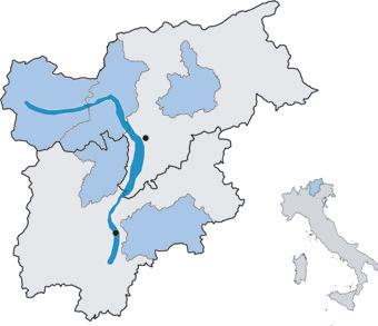
Val di Non Figure 3 Autonomous region of TrentinoSouth Tyrol (Italy). Districts and areas shown in blue are affected by apple proliferation
TRENTO
Valsugana
Tyrol and the density of this psyllid was reduced dramatically, with the result that in 2005 this insect was only found on every second tree on average (Österreicher and Thomann 2015a). In 2004, disease manifestation increased in several orchards and the following year AP became a South Tyrol wide concern again. In 2006, symptomatic trees were found in about 75 % of the monitored orchards (Fig. 4). However, the problem was not equally distributed: while Eisacktal/Valle Isarco was hardly affected at all, in Vinschgau/Val Venosta, Burggrafenamt/Burgraviato and Etschtal/Val d’Adige numbers of new reported cases increased (Österreicher and Thomann 2003, 2015a). In 2004 first individuals of C. picta were reported in South Tyrol and this vector was found in the following years all over the main apple growing regions in South Tyrol, but not in Eisacktal/Valle Isarco (Wolf and Zelger 2006). The highest densities of this insect were found in Vinschgau/Val Venosta, Burggrafenamt/Burgraviato and Etschtal/Val d’Adige; thus, additionally to the treatments against C. melanoneura, in 2006 vector management was extended also against C. picta and in the following years vector densities decreased (Österreicher and Thomann 2015a; Mittelberger et al. 2016). After a few years of relative relief, disease manifestation increased again starting in Vinschgau/Val Venosta and Burggrafenamt/Burgraviato and peaked in 2013. Interestingly, other regions like Eisacktal/ Valle Isarco remained not or much less affected (Österreicher and Unterthurner 2014). In the two peak years 2006 and 2013 AP led to a total economic damage of about 50 million € in South Tyrol (Österreicher and Thomann 2015b). In 2014 till 2018 the situation improved and less than 1 % of symptomatic trees in the orchards were counted (Fig. 4). Due to an enforced control strategy, the densities of C. picta and C. melanoneura decreased from 2012 to 2014 in all monitored regions in South Tyrol (Mittelberger et al. 2016). Fischnaller and co authors (2017) confirmed this trend also for the years 2014 till 2016. In Trentino (Fig 3), the first report of AP infected apple trees dates
Figure 4 AP infestation in the districts of South Tyrol with apple cultivation
% of symptomatic trees in apple orchards
3.5
3.0
2.5
2.0
1.5
1.0
0.5
0.0
2005 2006 2007 2008 2009 2010 2011 2012 2013 Burggrafenamt / Burgraviato Vinschgau / Val Venosta Etschtal / Val d'Adige Überetsch / Oltradige Unterland / Bassa Atesina Leifers / Laives Eisacktal / Valle Isarco
2014 2015 2016 2017 2018
back to the early 1950s (Refatti and Ciferri 1954), but the disease appeared rather sporadically. An outbreak was reported in the early 1990s, causing considerable economic damage (Vindimian et al. 2002), primarily in Val di Non (Vindimian and Delaiti 1996). In order to quantify the disease spread and to understand the predisposing factors, growers in Val di Non carried out a survey of infected trees in 1999 and 2000 (Springhetti et al. 2002). 4.9 and 6.8 million trees were controlled, i.e. 65 % and 91 % of apple trees asset in Val di Non, respectively. The average infection rate increased from 0.8 to 1.7 %. In general, the infection rate was higher at higher altitudes and in older orchards on more vigorous rootstocks (Springhetti et al. 2002). Nevertheless, about 5-10 % of infected trees in two-years-old orchards and up to 20 % in some three-years-old orchards were found. Since 2001, the Phytosanitary Office of the Province of Trento performed an official monitoring activity. The survey was extended to the whole apple growing area of the province and a potential effect of differential agronomic measures, cultivar asset or different altitudes was analyzed (Vindimian 2002). Until 2005, the average percentage of infected trees ranged from 2.5 to 2.9 %. During this period, the highest percentage of infected trees was reported for hill–sites of the district Val di Non whose apple growing area account for nearly 60 % of the total apple growing area of the Province of Trento. In this district, the average disease incidence reached up to 5.5 %, but in some older orchards planted on vigorous rootstocks up to 70 % of the trees showed an infection. The infection rate rapidly decreased when uprooting of infected trees became mandatory and chemical control measures against the insect vectors were implemented in 2006. The adoption of recommended control actions was fostered by granting growers with a subsidy for uprooting orchards older than 20 years or orchards with more than 20 % of infected trees. The average percentage of infected trees constantly decreased during the subsidized uprooting program from 2006 to 2010, when the lowest rate of 0.27 % was reached. The infection rate started to upsurge in 2012, significantly in Val d’Adige and Valsugana, regions that account for 30 % of the total apple growing area of the province. In these two districts, the average infection rate rose to 6 % in 2014, pushing the average infection rate of the Province of Trento to 2 %. In North-Western Italy (Fig. 5), AP has been recorded in two apple growing regions, Piedmont and Valle d’Aosta where at the end of the 1990s and in the first years of the 2000s a severe epidemic occurred. In Piedmont, the territories of Alto Canavese (Province of Turin) are the historic hotspots of the disease. In Alto Canavese typical cultivars are grown in organic and in integrated managed orchards. The apple production is mainly for the local market. Some records of AP infestations arose also from localities of the Province of Turin and from the Province of Cuneo, areas characterized by more intensive apple cultivation. However, AP outbreaks were rather sporadic and
Figure 5 Autonomous region of Aosta Valley and the region of Piedmont. Districts shown in blue are affected by apple proliferation
Alto Canavese
Provincia di Torino AOSTA
Aosta Valley TORINO
Piemonte
Province of Turin Alto Canavese Provincia di Cuneo
TURIN CUNEO
Piedmont
Province of Cuneo
CUNEO
not severe (Minucci et al. 1996; Alma et al. 2000; Pinna et al. 2003; Spagnolo et al. 2005). On the other hand, in Valle d’Aosta AP is widespread and represents a serious threat since the 1990s, especially in older orchards. In this region disease incidence reached 100 % in some orchards (Tedeschi et al. 2002; 2003). After a Ministry decree issued in 2006 obligating control measures against AP (Ministero delle Politiche e Agricole Forestali 2006), a sanitation programme was implemented. Trees should be regularly inspected for the presence of typical symptoms and infected trees should be removed. In case that more than 25 % infection was observed, the whole orchard had to be uprooted. As a consequence, the spread of AP declined, but surveys are constantly being carried out and treatments against the vectors are prescribed in both regions.
AP in other European regions and the Middle East AP is widespread in apple growing regions in Europe and has been recorded in Austria, Belgium, Bosnia and Herzegovina, Bulgaria, Croatia, Czech Republic, Finland, France, Germany, Hungary, Italy, Norway, Poland, Romania, Serbia, Slovenia, Spain, Switzerland, the Netherlands and Turkey (Tedeschi et al. 2013). To provide up-todate information about the distribution of different phytoplasmal diseases, including AP, a survey about the disease distribution and the prevalence of their confirmed and putative insect vectors throughout Europe has been performed (Bertaccini 2014). Based on these results, a database consisting of maps and detailed tables from 28 European and Middle East countries was compiled (COST FA0807 2013).
Symptoms
Current measures against AP are consequent treatments against the phytoplasma transmitting insect vectors and the removal of infected plant material. Removal of infected plant material requires reliable recognition of infected trees. Thus, the ability to recognize trees with specific AP symptoms is an indispensable skill of any fruit grower in affected regions. An AP infection induces a broad range of symptoms in wild and commercial Malus species (Bovey 1963; Blattny et al. 1963; Rui 1950; Refatti and Ciferri 1954; Morvan and Castelain 1975; Kartte and Seemüller 1988) but symptom expression can vary enormously (Schmid 1975).
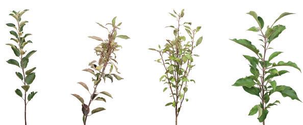
Specific symptoms A symptom is classified as AP specific when unambiguously related to an AP infection. AP specific symptoms are the formation of witches’ brooms (an abnormal bush like cluster of dwarfed weak shoots) and enlarged and dentate stipules with the latter symptom being more difficult to determine on the tree (Seemüller 1990; Jarausch 2007; Seemüller et al. 2011a) (Fig. 6 and 7). Size and shape of stipules depend on the developmental stage of the affected branch and the respective cultivar (Mattedi et al. 2008b; 2008f). Thus, a careful comparison of stipules from symptomatic and asymptomatic trees of the same cultivar is necessary for adequate disease evaluation. Although the presence of specific symptoms indicates an AP infection, the absence of enlarged stipules or witches’ brooms is not a proof that the respective tree is 'Ca. P. mali' free.
Non-specific symptoms Some infected apple trees may show symptoms that – when appearing alone - cannot clearly be linked to an AP infection, consequently these symptoms are considered non-AP-specific. The contemporaneous expression of two or more non-AP-specific symptoms may though, reliably indicate an AP infection (Thomann and Tumler 2000;
Figure 6 A healthy shoot (A) compared to shoots showing apple proliferation characteristic witches’ brooms (B-D)
A) B) C)
D)
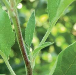
Figure 7 Enlarged and dentate stipules - a specific symptom of apple proliferation

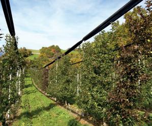
Mattedi et al. 2008b; 2008f). Early leaf reddening is easily visible and one of the most evident non-specific symptoms of AP disease (Bovey 1963) (Fig. 8). However, mechanical tree damages, fungal infections or certain physiological conditions can also induce foliar reddening (Schmid 1975; Mattedi et al. 2008b; 2008f). AP-induced premature bronzing is characterized by a certain colour shade and papery leaf texture (Mattedi et al. 2008f). Moreover, early leaf reddening as an indication for AP infection is considered to vary between cultivars and over the years due to climatic conditions (Mattedi et al. 2008f). In a study with the cultivar ‘Golden Delicious‘ it could be revealed that early leaf reddening affecting the complete canopy was correlated to an AP-infection in 86 % of the observed cases (Öttl et al. 2008). Instead of early leaf reddening, certain apple varieties (e.g. ‘Gala‘) may show pre-harvest chlorosis, which is a hint for an AP infection (if it is not caused by a nutrient deficiency). Infected trees often show an earlier bud break in springtime (Schmid 1975; Mattedi et al. 2008f), which can only be observed in a short time interval, making this symptom rather difficult to recognize. AP induced stunted branches and rosette formation of apical leaves can emerge during summer as well as new shoots from auxiliary buds of the old wood (Zawadzka 1976). These symptoms are often coupled with an increased susceptibility to powdery mildew (Bovey 1963; Zawadzka 1976; Maszkiewicz et al. 1979). In some cases, the interpretation of new budding may be difficult since it is affected by abiotic factors, such as pruning or damaging of the bark. Late blossoming (late flowering) is often defined as a non-specific AP symptom (Kartte and Seemüller 1988); however, Mattedi et al. (2008b; 2008f) described this symptom as not characteristic for an infection. The autors point out that the severity of this symptom depends on the apple variety and that new planted trees are particularly affected because of previous hormonal treatments in the nursery. The non-specific symptom of small, taste- and colourless fruits with a long pedunculus (Blattny et al. 1963; Zawadzka 1976; Seidl 1980; Schmidt et al. 2009; Seemüller et al. 2011a) (Fig. 9) is the economically important
symptom of AP since these fruits are not marketable and result in a notable reduction of earnings (Herzog et al. 2012; Österreicher and Thomann 2015b; Seemüller et al. 2011a). Further non-specific symptoms described for AP disease which are not directly evident are root malformation (Kunze 1989) or root branching (Guerriero et al. 2012b), formation of abnormally formed suckers, growth suppression, reddish winter wood and hooked apical buds (Mattedi et al. 2008f). For some Malus taxa also leaf malformation and leaf roll, degreening of veins and even vegetation dieback were reported (Kartte and Seemüller 1988).
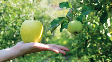
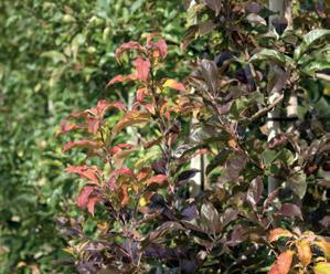
Figure 8 Early leaf reddening - an unspecific symptom of apple proliferation
Figure 9 Small fruits - an unspecific symptom of apple proliferation. Left: Apple from a healthy tree. Right: Apple from an AP infected tree
Co-occurrence of symptoms Specific symptoms or certain symptom combinations are often distinct for AP (Thomann and Tumler 2000), but to date, symptom expression is rather irregular and no systematic pattern can be observed (Carraro et al. 2004). It is still unclear if differential symptom development is related to different pathogen strains, sensitivity of certain apple cultivars, phytoplasmal colonization behavior of the aerial parts of the tree, environmental factors or certain plant-physiological conditions (Seemüller et al. 1984b; Seemüller 2002; Carraro et al. 2004; Seemüller and Schneider 2007; Herzog et al. 2010; Baric
et al. 2011b). Thus, comprehensive studies addressing the development of certain symptom patterns would be helpful to optimize the monitoring strategies for the field. The presence or absence of symptom is also associated to the presence or absence of the phytoplasma in the aerial parts of the trees. Remission of symptoms, a phenomenon known as recovery, was described to occur either transiently or permanently in infected trees (Seemüller et al. 1984b; Osler et al. 2000; Carraro et al. 2004). Recovery is characterized by a cessation of symptoms, but with bacteria persisting in the roots (see also chapter “The phenomenon of recovery”). The bacteria residing in the roots of recovered trees can spontaneously recolonize the canopy and induce symptoms again (Carraro et al. 2004). Treatment with resistance inducers and plant growth regulators showed only limited and transient effects on symptom expression, infection rates and growth rates of apple trees infected with 'Ca. P. mali' (Schmidt et al. 2015). Healing of infected apple trees, i.e. the eradication of the pathogen has never been reported.
Symptom assessment to determine the degree of infestation The best period for visual symptom assessment is during harvest since most symptoms develop in autumn and compared to spring, the number of trees with specific symptoms is nearly doubled (Jarausch 2007). However, an iterative AP symptom assessment allover the vegetation period is recommended because several symptoms, such as early bud break and premature leaf reddening are only displayed in certain time periods. Nevertheless, visual symptom assessment does not guarantee that the entirety of infected plants in an orchard can be identified, since symptomless trees can be infected as well (see chapters “Latent infected planting material – an infectious time bomb?” and “The phenomenon of recovery”). This is mainly because the physiological state of the tree, the phase of infection, climatic conditions and other factors can affect symptom expression.
Legal regulations Since 2006 the Italian Ministry of Agriculture prescribes that AP infected plant material must be eliminated to reduce the risk of a further AP spread (Ministero delle Politiche e Agricole Forestali 2006). The decrees currently in force in both provinces (Province of Bolzano and Trento) define witches’ brooms as a specific symptom for AP (Provincia Autonoma di Trento, 2003; Autonome Provinz Bozen / Provincia Autonoma di Bolzano 2016). Enlarged and dentate stipules, small fruits, early bud break and premature foliar reddening are considered AP symptoms when at least two of these symptoms occur in combination. Apple trees exhibiting specific symptoms or a certain symptom combination must be uprooted by law.
Host-pathogen interactions
Like all phytoplasma, 'Ca. P. mali' is a plant pathogen that resides in the phloem. 'Ca. P. mali' is present in the apple tree throughout the whole year (Baric et al. 2011b), but the concentration of phytoplasma in the aerial parts of the tree varies drastically in the course of the year (Schaper and Seemüller 1984; Seemüller et al. 1984a; 1984b; Loi et al. 2002; Pedrazzoli et al. 2008; Baric et al. 2011b). 'Ca. P. mali' recolonizes the aerial parts of the tree starting in late spring/early summer, while colonization peaks in late summer and lasts until December. With ongoing phloem degradation, the phytoplasma concentration in the canopy is further reduced during dormancy (Schaper and Seemüller 1984; Pedrazzoli et al. 2008). Therefore, the infectivity (i.e. in this case the ability to infect the insect vector) of an apple tree might vary throughout the year. In the root system 'Ca. P. mali' is present throughout the whole year (Schaper and Seemüller 1982; Seemüller et al. 1984a; Baric et al. 2011b). The constant phloem renewal in the root system permits the survival of the phytoplasmas during winter (Schaper and Seemüller 1982). Phytoplasmas interact with their plant host on different levels. They move through sieve plate pores, interfere with physiological and biochemical processes of plants and block the phloem transport by obstructing the sieve tubes (Kartte and Seemüller 1991; Lepka et al. 1999). 'Ca. P. mali' lacks many genes considered to be essential for cell metabolism and thus relies on the uptake of nutrients from the plant. It can be speculated that phytoplasma assimilate a wide range of nutrients and organic compounds from the host cells, probably with detrimental effects on the host metabolism. Phytoplasmas can influence plant metabolism directly through a set of membrane proteins and indirectly through effector proteins (see chapter “Molecular aspects of symptom development in the apple tree”). In vitro studies demonstrated that the immunodominant membrane protein Imp from 'Ca. P. mali' interacts with plant proteins like actin (Boonrod et al. 2012). This resembles the situation of another phytoplasma, 'Ca. P. asteris', whose immunodominant antigenic membrane protein Amp binds actin and is hypothesized to play a role in phytoplasmal mobility in its host (Galetto et al. 2011). Furthermore, Amp of 'Ca. P. asteris' interacts with subunits of the ATP synthase of the insect, suggesting that ATP synthase plays a role as receptor for cell entry in mid gut and salivary glands of the insects (Galetto et al. 2011). These findings indicate an important role of Amp in determining insect vector specificity (Suzuki et al. 2006; Galetto et al. 2008; 2011; Rashidi et al. 2015). Interestingly, 'Ca. P. asteris' can modulate its gene expression according to the stage of infection and to the host species (Oshima et al. 2011). Infection with 'Ca. P. mali' can also lead to the production of different defence proteins, an increase of phenolic compounds and it seems to alter hydrogen peroxide (H2O2) production in host plants (see chapter “Molecular aspects of symptom development in the
apple tree”). The availability of the AP genome sequence (Kube et al. 2008) provides the basis to investigate phytoplasma-host relationships and bacterial virulence factors. Sequence analyses revealed that the 'Ca. P. mali' genome carries a high number of membrane-associated AAA+- ATPases and proteases (e.g. FtsH encoded by hflb/also known as ftsh genes) that may degrade proteins for nutrient uptake or dampen the host’s defence reactions (Kube et al. 2008) or act as virulence factors (see chapter “Molecular aspects of symptom development in the apple tree”). As a consequence of these complex interactions, the content of chlorophyll, leaf biomass and the amount of soluble proteins considerably decreases, whereas the content of sugars, starch, amino acids and total saccharides significantly increases (Bertamini et al. 2002). Changes in contents of pigments, chlorophyll-protein complex and photosynthetic activities were observed (Bertamini et al. 2003), as well as changes in volatile organic compounds (Rid et al. 2016). An increased level of saccharides has been often interconnected with AP induced symptom development in the apple tree, but many questions remain, e.g.: is the phytoplasma able to induce all metabolic modifications such as hormonal imbalance, sugar reallocation, starch accumulation, etc. in the host actively or are they a subsequent result of structural changes that can be observed in the colonized phloem? Moreover, disturbances of the photosystem can be observed when stress at phloem level is taking place (Lemoine et al. 2013). Although phytoplasmas directly interact through effectors and membrane proteins with its host, the number of phenotypic changes and their intensity could be an indirect consequence of changes in phloem physiology, tissue occlusion, callose deposition, etc. as described in many studies (Musetti et al. 2010; 2011a; 2013a; 2013b; Guerriero et al. 2012a; Patui et al. 2013; Zimmermann et al. 2015). Thus, first interactions during infection might be very specific and localized but it cannot be excluded that later consequences are rather “chain reactions” that lead to physical changes in the phloem tissues and “simply” cause un-concerted physiological imbalances in the host. Which pathways, genes and proteins are directly affected by the phytoplasma is still poorly understood and unspecific metabolic downstream changes in diseased plants might further hamper analyses aiming to detangle what is cause and what is consequence.
Molecular aspects of symptom development in the apple tree For most apple cultivars, an infection with AP does not lead to death of the infected tree. Furthermore, a high number of 'Ca. P. mali' in the canopy has been described to be a pre-requisite for severe AP symptom development (Schaper and Seemüller 1984; Seemüller et al. 1984a; 1984b; Bisognin et al. 2008b). Phytoplasma concentration in the roots, on the other hand, does not seem to impact colonisation intensity of the canopy and AP symptom expression (Baric et al. 2011b) (see chapter “Host-pathogen interactions”). However, the
exact molecular mechanisms underlying symptom development are not known in detail, yet. In a study using tobacco and periwinkle as model plants, an AP infection induced impaired carbohydrate translocation to sink tissue (Lepka et al. 1999). This impairment of carbohydrate transport has been correlated to the development of growth inhibition (stunting) and the converse accumulation of carbohydrates in the source tissue as the reason for leaf degreening (chlorosis) (Lepka et al. 1999). In line with the hypothesis that an alteration of the carbohydrate metabolism is a reason for stunting and chlorosis, it could be shown that key genes of the Calvin cycle and of chloroplast photosynthesis pathways are altered in AP infected plant material. These alterations could be involved in AP symptom development (Aldaghi et al. 2012; Luge et al. 2014). A study on the early onset of chlorophyll degradation in leaves of AP infected apple trees revealed that the pathway underlying AP-induced chlorophyll degradation is the same pathway involved in seasonal leaf senescence. The authors of this study also showed that AP infected plants contain less chlorophyll, degrade it earlier but slowlier and contain less catabolites when chlorophyll breakdown is completed (Mittelberger et al. 2017b). Giorno et al. (2013) describe a decrease of glucose, fructose and sorbitol contents in AP infected apple plantlet tissues, whereas sucrose and starch were increased. The authors of this study also found PR-6, PR-8 and Mal d 1 genes being upregulated. The encoded gene products are thus likely involved in the induction of the immune response in the AP-infected plantlets. Giorno and co-authors (2013) also hypothesized that the increased amount of soluble sugars might act as a signalling strategy in the plant which affects gene expression. The fact that symptom development might result from an effect of 'Ca. P. mali' on plant hormonal regulation has been suggested by many authors and shown in different studies (Luge et al. 2014; Zimmermann et al. 2015; Tan et al. 2016; Janik et al. 2017). Aldaghi et al. (2012) claim that profound disturbances in the balance of growth regulators are the cause of a broad array of symptoms in affected trees. In their study, they cluster the AP affected apple genes into three ontology groups: i) genes involved in photosynthesis pathways are de-regulated and might thus be relevant for AP symptom development, e.g. leaf chlorosis and carbohydrate metabolism mediated symptoms; ii) genes that affect senescence and auxin accumulation and might thus be responsible for the inhibition of apical dominance (stunting) and have an effect on flavonoid biosynthesis (leaf reddening); iii) genes that regulate plant defence, in particular leading to the reduction of H2O2 and thus increase the susceptibility and multiplication of the bacterium in the host (Aldaghi et al. 2012). In line with this, Musetti et al. (2004) found that H2O2 is accumulated in recovered, symptomless AP infected trees. The authors thus concluded that a H2O2 accumulation counteracts AP symptom development. Even though many authors interpret findings on the metabolomic level as the reason for certain symptoms, direct proofs and molec-
ular links of this interconnection remain scarce. The spatio-temporal programme of disease development in the tree is not easy to dissect. Especially since the natural infection is difficult to model, most studies rely on analyses performed with model plants (e.g. Arabidopsis spp. or Nicotiana spp.) or with apple seedlings but rarely involve fully grown apple trees. However, even if the apple tree is used to study disease progress, the seasonal and physiological state (e.g. the immunological defence abilities) plays a crucial role for the respective analyses. Bacterial factors that allow the pathogen to establish and maintain an infection, so called virulence factors or effector proteins, are key players during disease development of different phytoplasmoses (Hoshi et al. 2009; MacLean et al. 2011; Sugio et al. 2011a; 2011b; Maejima et al. 2014). Often knowledge about Aster yellow witches’-broom (AYWB) phytoplasma or other phytoplasma species is used as a proxy to explain findings of AP symptom development. Just recently the AP effector ATP_00189, which shares sequence and functional homology to the AYWB effector SAP11, has been identified (Siewert et al. 2014; Janik et al. 2017). Similar to AYWB-phytoplasma SAP11, ATP_00189 of 'Ca. P. mali' binds teosinte/cycloidea/pcf (TCP) class II transcription factors of its host plant (in case of 'Ca. P. mali' TCPs from Malus × domestica) (Sugio et al. 2011a; Janik et al. 2017). These transcription factors are regulating hormone expression during different growth, defence and developmental plant processes (Cubas et al. 1999; Lopez et al. 2015; Ikeda and Ohme-Takagi 2014). Furthermore, a study with Nicotiana benthamiana expressing the SAP-like ATP_00189 of 'Ca. P. mali' revealed that this effector changes volatile expression, leads to defects in glandular trichome development and suppressed jasmonic acid responses (Tan et al. 2016). It is hypothesized that phytoplasmal AAA+ ATPases play a role in 'Ca. P. mali' virulence (Seemüller et al. 2011b; 2013). In particular the AAA+ ATPase AP460 might be part of a phytoplasmal secretion system acting as an AP virulence factor (Seemüller et al. 2018b). However, the exact function and the role of these proteins during infection remain elusive.
Since phytoplasma are phloem-restricted inside the plant, they can only be transmitted from plant to plant by phloem-sucking insects or by intact phloem-phloem connections. Transmission by insect vectors is considered the most important and most relevant way of AP spread. However, transmission of 'Ca. P. mali' can be obtained also by grafting, by natural root bridges or by dodder (Cuscuta sp.) (see also chapter “'Ca. P. mali' host plants”). Transmission from infected apple to the experimental host periwinkle (Catharanthus roseus) or from periwinkle to tobacco was achieved by different dodder species (Carraro et al. 1988; Seemüller et al. 2011a; Luge et al. 2014). Dodder is a parasitic plant that forms phloem connections between different plants and hence can transmit phytoplasma between different host plants. Several tree species have been shown to be interconnected through their root systems (Tarroux et al. 2014). This phenomenon, called anastomosis, has been reported in more than 150 species (Bormann 1966). Root anastomoses assume vital functions for the tree community by improving nutrient’s absorption, root system longevity, tree stability and by mitigating competition between old and young trees (Drénou 2003). Formation of natural root bridges seems to be very common also with apple trees (Vindimian et al. 2002; Ciccotti et al. 2008). Epidemiological studies suggested a possible role of root bridges in the spread of AP (Bliefernicht and Krczal 1995), especially in medium-aged and old apple orchards (Vindimian et al. 2002; Baric et al. 2008). Applications of the systemic, phloem transported herbicide glyphosate to apple tree stubs and the subsequent translocation of this herbicide to adjacent trees via root connections were used to proof the presence and frequency of root contacts between trees within an apple orchard (Ciccotti et al. 2007; Baric et al. 2008). Root bridges occur not only between viable apple trees, but also among just planted young plants and vital residuals of old roots left in the soil after uprooting the previous orchard. These leftover roots have been found to be viable up to 5-6 years from the uprooting of the actual tree and they still could be tested 'Ca. P. mali' positive (Mattedi et al. 2008a). Evidence of 'Ca. P. mali' transmission through root bridges has been demonstrated in an experimental trial where infected seedlings were potted together with uninfected seedlings (Ciccotti et al. 2008). Root bridge formation occurred from the first year on and increased in frequency in the following years. The natural transmission of 'Ca. P. mali' by root bridges was proven by specific PCR and immunofluorescence studies (Ciccotti et al. 2008). Experimental data suggest that root connections seem to play a role for the spread of 'Ca. P. mali' in older orchards and between trees on vigorous rootstocks (Baric et al. 2008). However, root-to-root transmission of 'Ca. P. mali' could also be observed in trees growing on the less-vigorous rootstock M9, commonly used in
commercial orchards (Lesnik et al. 2008). 'Ca. P. mali' can be transmitted experimentally from tree to tree by grafting. Scion grafting is usually a more efficient method to transmit 'Ca. P. mali' than grafting small tissue pieces (Seemüller et al. 2011a). However, the success of graft-transmission by scions is dependent on the season since colonisation of the aerial parts of the tree is subjected to seasonal fluctuation of 'Ca. P. mali' presence (Seemüller et al. 1984b; Pedrazzoli et al. 2008; Baric et al. 2011b; Schmidt et al. 2015) (see also chapter “Host-Pathogen interactions”). Pedrazzoli et al. (2008) demonstrated that transmission rates by chip budding were very low between March and May but highest between June and August. Consequently, experimental inoculations, e.g. for AP resistance screening, are done in the latter period of the year. Vice versa, grafting of scions removed during the dormancy period lowers the risk of accidental transmission from latent infected material during the production of planting material. Roots are constantly colonised by the phytoplasma (Seemüller et al. 1984b) and are therefore a good donor tissue for 'Ca. P. mali' transmission by root scion grafting in other periods of the year. Grafting material from trees of unknown infectious status to susceptible indicator plants (indexing) has become a common method to determine if the donor plant is infected with phytoplasma or viruses (European and Mediterranean Plant Protection Organization 1999, 2017). In vitro culture was successfully employed to maintain 'Ca. P. mali' in micropropagated Malus cultivars (Jarausch et al. 1996). Using in vitro grafting, phytoplasma could be transmitted to healthy in vitro plants with very high efficiency (Jarausch et al. 1999). This in vitro approach has then been applied to screen Malus sieboldii and M. sieboldii × M. × domestica hybrids for AP resistance (Bisognin et al. 2008a). On the other hand, grafted rootstocks derived from nurseries that unwittingly used infected scions constitute a risk of introducing infected material to an orchard (see chapter “Latent infected planting material – an infectious time bomb?”). However, 'Ca. P. mali' is not transmitted by seeds (Seidl and Komarkova 1974).
The latency period referred to AP is defined as the time span from the infection to the development of visible symptoms in the plant. Until now it is not exactly defined wheather the latency period ends with the development of specific or unspecific symptoms. Bovey (1963) reported an average latency period of 1.8 years after artificial infection by chip budding or bud insertion. Infection by scion grafting might result in faster symptom development, due to higher phytoplasma transmission rates/size of the plant of the rootstock. Scion grafting from symptomatic field-grown trees on healthy M9 rootstocks in February resulted in symptom development in 67.4 % of the plants in July and 84.8 % in October of the same year (Schmidt
et al. 2015). In a six-year study of a new planted orchard, Unterthurner and Baric (2011) demonstrated that AP symptoms are expressed in most cases within one-and-a-half to two years after infection. However, the authors of this study also reported a prolonged latency period of four years in one of the infected trees. In one study, conducted in north-eastern Italy it could be determined that 10 % of randomly chosen non-symptomatic trees are actually AP infected (Mattedi et al. 2008c). In the years 2003 and 2006 a similar study was performed in South Tyrol; 2.3 % and 10.5 %, respectively of non-symptomatic trees from two apple orchards were infected (Baric et al. 2007). In the years 2015 till 2017 one out of 1000 non-symptomatic trees from two South Tyrolean apple orchards were AP positive (unpublished data). Attention must be drawn on the fact that only healthy, 'Ca. P. mali' free and certified planting material is used for production and propagation. Infected planting material might facilitate the spread of the disease to areas so far free of AP. Therefore, 'Ca. P. mali' is listed as a quarantine organism in many countries and must be absent in planting material. The import of Malus material from known AP host countries is regulated throughout the world (https://gd.eppo.int/ taxon/PHYPMA/categorization). It is known that – despite all efforts – AP is sometimes detected in mother stock and nursery material (W. Jarausch, personal communication). Due to the latency period, visual inspections are not sufficient to recognize the entirety of infected but asymptomatic plant material. This applies to mother stocks which are pruned severely every year for scion production as well as for the young plants in the nursery. Therefore, random PCR screening of mother plants is applied as a new strategy to ensure healthy planting material (at least in Germany) (W. Jarausch, personal cummunication). The risk of transmission by grafting using a latent infected cultivar scion is considered lower, especially if the budwood has been taken in the dormant season (see chapter “Insect vector independent 'Ca. P. mali' transmission - root bridges and graft transmission”). There are reports of symptomatic trees already in the first year of plantation, e.g. Mattedi et al. (2008a) detected 1.1 % infected trees in a newly planted experimental orchard. From 2001 to 2004, PCR tests of material ready for planting revealed latent infections from about 1 ‰ plants in Germany (unpublished data) and 3 ‰ plants in Italy (unpublished data). Mattedi et al. (2008c) found two out of 300 trees infected before plantation in the Province of Trento (Italy). In the roots of latent trees, 'Ca. P. mali' is present throughout the whole year in concentrations comparable to those in symptomatic trees (Baric et al. 2011b). In contrast, 'Ca. P. mali' is colonizing just sporadically and in very low concentrations the aerial parts of latent trees (Seemüller et al. 1984a; 1984b; Loi et al. 2002; Pedrazzoli et al. 2008; Baric et al. 2011b). Since insect vectors feed on leaves and green parts of the plant, latently infected or recovered, i.e. symptomless trees (see also the chapter “The phenomenon of recovery”)
might thus represent an inoculum source for vector dependent 'Ca. P. mali' transmission. Still it remains unclear, how many phytoplasma need to be taken up by an insect vector to establish an infection in the latter one. Therefore, the question, if 'Ca. P. mali' host plants with a very low titer in aerial parts have an infective potential at all, remains unanswered.
The phenomenon of recovery
Field grown apple trees infected with 'Ca. P. mali' can show a spontaneous remission of symptoms, a phenomenon described as recovery (Osler et al. 2000). It was found that within 10 years, 71 % of AP symptomatic trees of the cultivar ‘Florina’ recovered; this corresponds to a mean annual rate of symptom remission of 29 % (Osler et al. 2000). Recovered trees are not free of bacteria and did thus not literally recover from the infection, but only from the typical symptoms. Despite the disappearance of symptoms, the bacteria can still be detected in the roots, but not in the canopy (Carraro et al. 2004). However, the apple tree remains infected and is able to transmit the phytoplasma via root grafting but not via bud grafting (Carraro et al. 2004). After a non-symptomatic period, the tree can change its status from asymptomatic to symptomatic and phytoplasma can be detected in the aerial parts of the tree again (Osler et al. 2000; Carraro et al. 2004; Seemüller et al. 1984b; 2010b). Interestingly, so far this phenomenon could only be observed in the field but could not be induced under experimental conditions, yet (Carraro et al. 2004; Schmidt et al. 2015). Unravelling the mechanisms underlying recovery may provide important insights to better understand the AP disease process. The molecular mechanism of recovery involves the activation of several branches of the plant immune response. An increase of H2O2 in affected tissues is characteristic for recovered plants, possibly directly or indirectly counteracting the bacterium (Musetti et al. 2004). Furthermore, recovered trees show increased levels of Ca2+ concentrations which might be connected with the observed increased callose deposition and protein accumulation in the leaf phloem (Musetti et al. 2010). The increased callose expression has been hypothesized to lead to (reversible) phloem plugging and thus blocks an effective distribution of the bacteria or hampers bacterial effector protein translocation leading to a loss of symptoms (Musetti et al. 2011b; Guerriero et al. 2012a). However, no correlation between phloem mass flow limitation and phytoplasma titre was found in Arabidobsis infected with 'Ca. P. asteris', which suggests that sieve element proteins are involved in defence mechanisms other than mechanical limitation of the pathogen (Pagliari et al. 2017). Musetti and co-workers (2013b) hypothesize that during infection a cascade of hormonal activations leads to either symptom-development or recovery: salicylic acid (SA) is immediately increased upon infection and antagonizes jasmonic acid (JA)-dependent defences, leading to a symptomatic infection
and accumulation of H2O2 which in turn leads to increased levels of SA. The authors suggest that this SA induction might counteract symptom development and thus leads to recovery (Musetti et al. 2004; 2013b). The idea of such SA induction is supported by the fact that recovered trees are less prone to symptom induction by re-infection than trees that have not been previously recovered, suggesting a type of induced resistance (Osler et al. 2000). Patui et al. (2013) showed that recovered trees accumulated JA via an induction of the oxylipin pathway and demonstrated that SA levels are declined in recovered trees, underlining the reciprocal antagonism between JA and SA pathways. These authors further suggest that the observed peroxidase and oxidase activity in combination with a decreased reactive oxygen species (ROS) scavenging activity could lead to an H2O2 accumulation during recovery. However, despite the lack of clarity if H2O2 accumulation is cause or consequence, the simultaneous activation of JA and SA during recovery is a possible scenario which might indeed enhance defence responses (van Wees et al. 2000).
Resistant plant material – the ideal solution?
Phytoplasma diseases are difficult to control due to the biphasic phytoplasmal life cycle in the plant and in the insect vector and because of the different ways of transmission. Since direct treatments are lacking, resistant plant material would be a great benefit. Search for natural genetic resistance to AP within the taxa Malus has been carried out extensively (Kartte and Seemüller 1991; Seemüller et al. 1992). Among Malus × domestica, the following cultivars are mentioned as (relatively) tolerant to AP infection: ‘Lord Lambourne’ (Friedrich 1993), ‘Clivia’, ‘Herma’ (Friedrich and Rode 1996), ‘Roja de Benejama’ (Invasive Species Compendium 2017), ‘Antonovka’, ‘Cortland’, ‘Spartan’, ‘Yellow Transparent’, ‘Wealthy’ (Németh 1986; Thakur and Handa 1999), ‘Melrose’ (Richter 2003), ‘Goldstar’, ‘Rubinola’, ‘Lotos’ and ‘Rosana’ (Pflanzenschutzdienst Baden-Württemberg 2003). However, tolerance evaluation of these cultivars was rather based on empirical field observations assessing symptom occurrence than on a screening through targeted infection trials. In extensive studies done by Seemüller and co-workers (1992) a systematic and controlled survey was performed using different genotypes comprising hundreds of established and experimental rootstocks of Malus × domestica as well as wild and ornamental Malus genotypes. These were tested by graft-mediated infection in long-term field trials leading to the observation that resistance (i.e. lower symptom expression and/or lower bacterial titers in the infected plants) was characteristic only to some experimental apomictic rootstocks derived from one specific accession of M. sieboldii (Kartte and Seemüller 1991; Seemüller et al. 1992; 2008; 2018a; Bisognin et al. 2008b; Seemüller and Harries 2010).
Nevertheless, these promising resistant M. sieboldii rootstocks are not directly suitable for commercial apple growing, given that scions grafted on these varieties mostly develop vigorous and less productive trees than on the standard rootstock M9. To maintain the resistance characteristics and improve the agronomic value of the rootstock resistant M. sieboldii genotypes were crossed within the SMAP project with the standard rootstock M9 (Bisognin et al. 2009; Seemüller et al. 2010a; Seemüller and Harries 2010; Jarausch et al. 2010; 2011b). The development of simple sequence repeat (SSR) markers and the selection of true recombinant clones were a challenging aspect due to the high level of apomixis of M. sieboldii. Apomixis is the production of genetic identical offspring despite pollination of the mother plant with a genetically diverse cultivar (Koltunow 1993), a characteristic that highly reduces the generation of recombinant genotypes. The breeding program was further hampered by the fact that M. sieboldii introduced hypersensitivity to latent apple viruses into some of the progeny and that the M. sieboldii derived clones had to be micropropagated to achieve efficient clonal production (Bisognin et al. 2008b; Liebenberg et al. 2010). Nevertheless, a screening for resistance using the in vitro graft technique (Jarausch et al. 1999) followed by controlled infections allowed the selection of several resistant genotypes. Data from eight years field trials confirmed that the resistance could be inherited to the breeding progeny and that mainly among the offspring of progeny 4608 x M9, resistant genotypes were identified showing pomological properties similar to M9 (Seemüller et al. 2018a). This progeny is also tolerant to latent apple viruses. The most promising rootstocks are now entering the phase of agronomic field studies in Germany and Italy (Seemüller et al. 2018a).
The insect vector’s feeding behaviour is relevant for spreading the phytoplasma. A polyphagous vector likely may inoculate a larger variety of plant species compared to a monophagous vector (Weintraub and Beanland 2006). C. picta is described as monophagous on apple (Malus spp. and M. × domestica) (Ossianilsson 1992; Weintraub and Beanland 2006; Alma et al. 2015). Instead, C. melanoneura is described as widely oligophagous on hawthorn (Crataegus spp.) and on apple (Ossianilsson 1992; Tedeschi et al. 2008). F. florii is polyphagous, feeding on several plants, mainly Rosaceae (Swenson 1974; Tedeschi and Alma 2006). Using PCR detection, the pathogen could be observed in naturally infected trees of several wild Malus species and of domestic apple (Seemüller et al. 2011a). Controversial reports exist regarding the natural occourrence of 'Ca. P. mali' in hawthorn. Tedeschi and Alma (2007) and Tedeschi et al. (2009) found this plant naturally infected, though, Mayer et al. (2009) did not find naturally infected hawthorn in another geographical context. There are also several reports
of 'Ca. P. mali' detection (by PCR) in wild or cultivated plants: hazel (Corylus avellana) (Marcone et al. 1996), pear (Pyrus communis), Nashi pear (Pyrus pyrifolia), Japanese plum (Prunus salicina) (Lee et al. 1995), hornbeam (Carpinus betulus), bindweed (Convolvulus arvensis) (Seemüller 2002), sweet cherry (Prunus avium) and oak (Quercus robur and Quercus rubra) (Seemüller et al. 2011a). In graft inoculation experiments 58 ornamental and wild Malus species and subspecies as well as 40 hybrids of different Malus species, which were used as rootstocks, could be infected with 'Ca. P. mali' (Kartte and Seemüller 1988, 1991). However, hawthorn could not be successfully infected experimentally (Mayer et al. 2009). As described in the chapter “Insect vector independent 'Ca. P. mali' transmission – root bridges and graft transmission”, 'Ca. P. mali' can also be experimentally transmitted to other plant species by dodder or by grafting. With these methods 'Ca. P. mali' could be transmitted to periwinkle (Catharanthus roseus) (Marwitz et al. 1974; Carraro et al. 1988), celery (Apium graveolens) and tomato (Solanum lycopersicum) (Seemüller et al. 2011a), as well as to the different tobacco species Nicotiana occidentalis, N. tabacum, N. clevelandii, N. quadrivalvis (Seemüller et al. 2011a; Luge et al. 2014) and N. benthamiana (Boonrod et al. 2012). The potential role of the above-mentioned 'Ca. P. mali' host plants for AP spread remains unclear, since successful transmission in the field furthermore requires an insect vector that feeds on the phloem of the respective plant host and is adapted to enable 'Ca. P. mali' transmission. So far, there is no evidence of an involvement of wild plants other than hawthorn as reservoirs for the pathogen in the epidemic cycle.
The plant microbiome is the entirety of microorganisms living in or on a plant. It plays a fundamental role in plant health and productiveness (reviewed in Turner et al. 2013). Non-pathogenic bacteria residing inside the plant tissues are called endophytic bacteria and the entity of these plant colonizing bacteria makes up the endosphere. This endosphere is densely populated by non-pathogenic microbial endophytes inhabiting all possible niches within the plant (Hardoim et al. 2015). Endophytic colonization of the xylem vessels has been frequently reported (Germaine et al. 2004; Compant et al. 2005; Lòpez-Fernàndez et al. 2015), and even the phloem, the most microbe-restricted part of the plant, is –albeit to a lower extent- colonized by endophytes (Bulgari et al. 2011; Pažoutová et al. 2012; Hilf et al. 2013). Infection with phytoplasmas affects the composition of endophytic communities and it is conceivable that microbial endophytes may in turn influence the infectious process or trigger plant recovery (Kamińska et al. 2010; Grisan et al. 2011; Bulgari et al. 2011; 2014). Some studies exploring how the plant microbiome alleviates phy-
toplasma disease focussed on mycorrhizal symbioses (Lingua et al. 2002; Garcia-Chapa et al. 2004). An effect of the widespread and well-characterized biocontrol fungal endophyte Epicoccum nigrum on 'Ca. P. mali' infection has been documented in the model plant Catharanthus roseus (Musetti et al. 2011a). This study showed that co-inoculation of E. nigrum in 'Ca. P. mali' infected C. roseus leads to a reduction of AP symptoms. However, the underlying mechanisms of the E. nigrum-phytoplasma interaction are still unclear. It can only be speculated whether the positive effects induced by E. nigrum are directly exerted by the endophyte (e.g. by producing antimicrobials active against the phytoplasma) or indirectly mediated by an altered plant immunity. Bulgari et al. (2012; 2014) reported that 'Ca. P. mali' influences bacterial endophyte communities present in the roots of apple trees. The authors observed that in AP diseased plants these endophyte communities were less diverse and differentially assembled compared to those present in uninfected plants. The 16S rDNA of Pseudomonadales and Sphingomonadales was detected in roots of healthy, but not in diseased plants. DNA from Burkholderiales was also more frequently isolated from healthy roots than from diseased ones. Certain bacterial taxa such as Xanthomonadales, Actinomycetales, Legionellales and Acidimicrobiales appear to preferentially colonize infected plants. On the other hand, Lysinibacillus colonies were isolated only from healthy plants (Bulgari et al. 2012). Several microorganisms associated with either non-infected or AP diseased plants comprise strains that are known to exert a potential biocontrol or protective role against certain plant pathogens (Duffy and Défago 1999; Ait Barka et al. 2000; Schouten et al. 2004; Kavino et al. 2007; Compant et al. 2008; Choudhary and Johri 2009; Verhagen et al. 2010; Trivedi et al. 2011). It is known that many endophytes produce secondary metabolites and other active compounds and thus have antibacterial and antifungal properties against pathogens. Some endophytes can elicit plant-defence mechanisms and therefore act as resistance inducers (reviewed in Romanazzi et al. 2009 and Compant et al. 2013). Several studies have shown that systemic acquired resistance (SAR) is involved in the recovery phenomenon (Osler et al. 2000; Musetti et al. 2005; 2007) (see also chapter “The phenomenon of recovery”). Therefore, understanding the role of endophytes during the induction of recovery of AP-infected plants is interesting since this might provide a possibility to identify a sustainable control measure against AP. Recent studies have been shown preliminary results on a promising activity of a set of symbiotic microorganisms isolated from host plants and insect vectors of phytoplasmas. These include one Xanthomonadaceae and several Bacillus isolates (Naor et al. 2015). In particular, the Dyella-like bacterium (DLB; Iasur-Kruh et al. 2017b) isolated from the planthopper Hyalesthes obsoletus, the main vector of 'Ca. P. solani' (aetiological agent of Bois noir disease of grapevine,
Quaglino et al. 2013), was able to inhibit growth of the cultivable Mollicute Spiroplasma melliferum (a model for phytoplasma inhibition studies, Naor et al. 2011). Furthermore, DLB reduced symptom severity and led to an increased recovery rate of infected plants (Iasur-Kruh et al. 2017a; Naor et al. 2017). According to these recent findings, non-fastidious microorganisms that share both host organisms (plant and insect) with the respective pathogen may hold the key to a novel, up-to now hardly explored approach to discover new microbial biocontrol tools also against AP.

2
THE CAUSATIVE AGENT OF APPLE PROLIFERATION
Katrin Janik, Sabine Öttl, Federico Pedrazzoli, Omar Rota-Stabelli, Rosemarie Tedeschi, Thomas Letschka
AP phytoplasma are taxonomically defined as 'Candidatus Phytoplasma mali' (Seemüller and Schneider 2004). The provisional 'Candidatus' status is used because these microorganisms are unculturable and Koch’s postulates cannot be fulfilled, 'Ca. P. mali' belongs to the family of Acholeplasmataceae (order Acholeplasmatales) of Mollicutes, a large class of bacteria containing various pathogens including Spiroplasma (Entomoplasmales) (Oshima et al. 2013; Siewert et al. 2014). Like all other members of the class Mollicutes, 'Ca. P. mali' is characterized by the absence of a cell wall and by an obligate parasitic life cycle in the phloem of the plant which hampers its in vitro culturing. The genus 'Candidatus Phytoplasma' experienced a large genetic radiation that generated at least 40 different species affecting a variety of host plants; the timing of this radiation is still unknown as well as the age of diversification of Phytoplasma from other Acholeplasmatacae (Kube et al. 2012). Although there is a certain specificity between phytoplasmas and their hosts, phytoplasmas of the same species can occasionally infect different plants, rendering the epidemiology extremely complex (Lee et al. 2000). 'Ca. P. mali' is closely related to 'Candidatus Phytoplasma pyri', the causal agent of pear decline; these two are in turn closer related to 'Candidatus Phytoplasma prunorum' (causing European Stone Fruit Yellows) and more distant to 'Candidatus Phytoplasma spartii' (Seemüller and Schneider 2004). These four phytoplasmas belong to the 16SrX phytoplasma group (Lee et al. 1998; Marcone et al. 2004). 'Ca. P. mali' is characterized by a small genome arranged in a single, linear chromosome. The genome differs in several aspects from that of other phytoplasma species (Kube et al. 2008) and is genetically highly dynamic with a low GC-content (Jarausch et al. 2000; Bai et al. 2006; Sugio and Hogenhout 2012). Phytoplasma classification systems were based on 16S rRNA sequence diversity. Primers that specifically amplify phytoplasmal 16S rRNA genes have been widely described and are used for diagnosis (Deng and Hiruki 1991; Ahrens and Seemüller 1992; Lee et al. 1993; Namba et al. 1993; Schneider et al. 1993; Gundersen and Lee 1996). A commonly used phytoplasma classification system involves the analysis of the RFLP pattern of a 16S rRNA amplicon (Lee et al. 1998): based on this classification, 'Ca. P. mali' belongs to the 16SrX-A subgroup as described above. Discriminating among genetic variants of the 'Ca. P. mali' species is a key prerequisite to study AP outbreaks in different European regions. A classification of 'Ca. P. mali' based on the analysis of more (also non-ribosomal) genes and with higher discriminating power has been provided. This molecular typing allows a clearer differentiation between different strains within a phytoplasm species (Smart et al. 1996; Schneider et al. 1997; Jarausch et al. 2000; Danet et al. 2011; Baric et al. 2011a; Martini and Lee 2013; Seemüller et al. 2013;
Šeruga Musić and Skorić 2013; Valiunas et al. 2013). In particular, Multi-locus sequence typing (MLST), a method based on the analysis of variations at multiple genome sites, allows the analysis of intra- and inter-species relationships (Danet et al. 2011; Casati et al. 2011; Janik et al. 2015). In this method, several different genetic areas of the phytoplasma genome (loci) are analyzed and a strain-specific typing code is generated. The ability to detect higher genetic diversity allows studying the geographical distributions of different phytoplasma strains, identifying mixed infections, and evaluating virulence and associations between certain strains and insect vectors. The advantage of MLST studies lies in the possibility of a fine-tuned typing by analyzing and comparing sequences of different loci. A drawback, however, is that even though evolutionary processes can be analyzed, many common MLST analysis programmes (e.g. eBURST) do not consider differential likelinesses of certain mutational events in different gene groups (e.g. considering conserved/unconserved functions). Seemüller and colleagues (Seemüller et al. 2010b; 2011b) for example used the hflB and imp genes to suggest an association between certain strains and virulence. A role of hflB and imp in virulence is assumed and the authors showed a correlation between certain sequence variants of the hflB gene and strain virulence. However, the biological role of the encoded proteins during phytoplasmal infection is not yet fully understood (see chapter “Molecular aspects of symptom development in the apple tree”). While these two molecular markers turned out to be highly polymorphic, stable SNPs have been found in the nitroreductase gene which led to the commonly used subtype definition of “AT-1”, “AT-2” and “AP” strains (Jarausch et al. 2000). Another typing approach based on the 16S rRNA and the rpl22 gene (encoding L22) revealed an insect (vector)-strain correlation in South Tyrol (Northern Italy) (Baric et al. 2011a). C. melanoneura harbored 'Ca. P. mali' strain AT-1 and C. picta harbored AT-2 (Baric et al. 2010a; 2011a). Results further indicated a spatio-temporal distribution pattern: the AT-1 strain was prevalent before 2005 in this region, while the AT-2 strain was detected in 2006 for the first time, i.e. two years after the first discovery of C. picta (Baric et al. 2011a). Shortly after the appearance of C. picta, a strong outbreak of AP appeared peaking in the year 2006 (see chapter “Northern Italy - South Tyrol, Trentino, Piedmont and Valle d’Aosta”). Transmission of 'Ca. P. mali' by C. picta was shown by different independent studies in Italy and Germany (Frisinghelli et al. 2000; Seemüller et al. 2004; Jarausch et al. 2004a; Carraro et al. 2008; Oppedisano et al. 2019b). Interestingly, transmission trials of 'Ca. P. mali' with C. melanoneura succeeded only in North-Western Italy and Trentino (Tedeschi et al. 2003; Tedeschi and Alma 2004; Mattedi et al. 2008d; Oppedisano et al. 2019b) but failed in Germany (Seemüller et al. 2004; Mayer et al. 2009). These findings led to the hypothesis that certain 'Ca. P. mali' subtypes might be specifically associated with certain C. melanoneura populations in different regions (Mayer
et al. 2009). Thus, genetic typing data may help to explain local and periodically occurring outbreaks of AP and its association to certain insects. A future approach should be to combine data from phytoplasma typing, strain-vector associations, and insect population genetics and behaviour to develop models and allow a prediction of AP spread. This indeed requires a profound knowledge about parameters affecting the highly complex biological system of AP dissemination. Applying knowledge from molecular typing and sequence analyses could further reveal factors involved in virulence, vector association and eventually allow a kind of molecular source tracking to trace the source of infection. Any typing approach is limited to the kind of loci analyzed and thus strongly depends on the relevant problem and question addressed in the respective study. Thus, the analysis of different loci often makes it difficult to compare and interpret data from different authors and studies. In future it is thus recommended to use a uniform typing method to study the spatio-temporal distribution and spread of 'Ca. P. mali'.
Molecular diagnosis
In the last decades, different assay types have been used for phytoplasma diagnostics. These diagnostic tests range from biological assays in which suspicious infected material is grafted to woody indicator plants or serological assays using specific antibodies against 'Ca. P. mali', e.g. enzyme-linked immunosorbent assays (ELISA) or immunofluorescence detection. However, these techniques are often work-intense, have a low sensitivity or are prone to generate false-negative results. For a reliable and less cumbersome detection of 'Ca. P. mali' in plants and in insects, different molecular tools have been established. All of them are based on the detection of AP specific DNA. PCR amplification of AP specific DNA regions is the most sensitive and reliable diagnosis tool. Most authors follow procedures developed by Kirkpatrick et al. (1987), Ahrens and Seemüller (1992) and Maixner et al. (1995) for DNA extraction and phytoplasma DNA enrichment using phloem tissue of apple trees. The method of Doyle and Doyle (1990) is widely used for DNA extraction from plants and insects as well (Firrao et al. 1994; Tedeschi et al. 2002; Carraro et al. 2008). Nested PCR, a highly sensitive DNA amplification involving two separated PCR runs, has been employed for the detection of 'Ca. P. mali' in plants and psyllids using universal primers (P1/P7 + F2n/R2) and 16SrX group specific primers (P1/P7 + fO1/rO1) (Lee et al. 1995; Lorenz et al. 1995). Nested PCR is advisable if low concentration or an uneven distribution of the pathogen in the host is suspected. Due to the genetic similarity within AP group phytoplasma, specific identification often requires further steps, such as amplicon digestion with different restriction enzymes and subsequent RFLP analysis or sequencing (Kison et al. 1994; Lee et al. 1995; Lorenz et al. 1995; Razin and Tully 1995; Gundersen and Lee 1996; Smart et al. 1996; Jarausch
et al. 2000). An immunocapture PCR (IC-PCR) protocol has been proposed by Heinrich et al. (2001) for a sensitive, reliable and reproducible large-scale detection of 'Ca. P. mali'. Protocols for differentiation of AP strains were published by Jarausch et al. (2000), Casati et al. (2010) and Baric et al. (2011a). Different quantitative real-time PCR protocols have been developed for AP in plants and insects based on SYBR Green (Jarausch et al. 2004b; Galetto et al. 2005; Torres et al. 2005), TaqManTM (Baric and Dalla Via 2004; Aldaghi et al. 2007; 2008) and EvaGreen® technologies (Monti et al. 2013). The described PCR methods require specific lab equipment and must be performed by experienced personnel. In the last years, loop-mediated isothermal amplification (LAMP) has become an interesting, fast, cheap and sensitive on-site diagnostic method for phytoplasma detection (Notomi 2000; Dickinson 2015). Several reports present promising results that LAMP might be a reliable diagnostic method for AP in the future (Neumüller et al. 2014; De Jonghe et al. 2017).
3
Tiziana Oppedisano, Gino Angeli, Mario Baldessari, Dana Barthel, Gastone Dallago, Stefanie Fischnaller, Claudio Ioriatti, Valerio Mazzoni, Cecilia Mittelberger, Sabine Öttl, Bernd Panassiti, Federico Pedrazzoli, Omar Rota-Stabelli, Hannes Schuler, Rosemarie Tedeschi, Tobias Weil, Gianfranco Anfora
Apple proliferation spread over long distance
After the first report on apple proliferation from the 1950s in the Province of Trento (Rui 1950), it was Bovey (1963) who described the epidemiology and was the first to link the disease to certain vector insects that allow a progressive - but not rapid - spread in the area (Bovey 1971; Amici et al. 1972). The investigations immediately focused on some groups of Hemiptera, in particular on leafhoppers and planthoppers (collectively known as Auchenorrhyncha) which were already demonstrated to be vectors of several other phytoplasmas. For this reason, Auchenorrhyncha species were strongly suspected of being potential vectors of the causative agent of AP (Kunze 1976). Field collections in apple orchards were conducted and molecular analyses performed to find the responsible disease vectors (Refatti et al. 1986; Carraro et al. 1988; Kunze 1989). The first transmission experiments conducted with 'Ca. P. mali' indicated that two hoppers, the leafhopper Artianus interstitialis (Germar 1821) and the xylem feeding froghopper (or spittlebug) Philaenus spumarius (Linnaeus 1758) were able to acquire and transfer the causative agent from infected celery to apple seedlings (Hegab and El-Zohairy 1986). However, these findings could not be confirmed by other researchers (Frisinghelli et al. 2000). So far, although various phloem feeding hemipterans showed to be occasional carriers of 'Ca. P. mali', only three species have been proven and are acknowledged to be AP vectors. They are the two psyllids Cacopsylla picta (Foerster 1848) (syn. C. costalis) and Cacopsylla melanoneura (Foerster 1848) (Hemiptera: Sternorryncha: Psyllidae), and the leafhopper Fieberiella florii (Stål 1864) (Hemiptera: Auchenorrhyncha: Cicadellidae) (Frisinghelli et al. 2000; Tedeschi et al. 2002; Jarausch et al. 2003; Tedeschi and Alma 2006; Carraro et al. 2008; Alma et al. 2015; Oppedisano et al. 2019b). The vector ability of these species has been proven by laboratory transmission trials and, similar to other phytoplasma vectors, they were able to transmit the etiological agent 'Ca. P. mali' in a persistent-propagative manner (Weintraub and Beanland 2006). Propagative means that the pathogen can multiply in insects; on the other hand, persistent means that the insect remains inoculative for life (Fletcher et al. 1998). Phytoplasmas can be ingested during phloem sap feeding, but do not necessarily multiply in the respective insect. Therefore, is the detection of a phytoplasma in a phloem-feeding insect not per se proving that the respective insect is also able to transmit the pathogen. The replicative process in the insect body requires the passage of phytoplasmas from the gut epithelium to the salivary glands – a highly concerted process of bacterial adaptation to the vector insect (Hogenhout et al. 2008; Alma et al. 2015). Thus, the capacity of an insect vector to transmit the ingested phytoplasma can be proved only by transmission trials. Here, infected insects are released to feed on healthy host plants which are subsequently analyzed to evaluate infection status (Bosco and Tedeschi 2013).
Cacopsylla picta and Cacopsylla melanoneura The genus Cacopsylla is the largest genus of the family Psyllidae including more than 400 recognized species (Ouvrard 2017). The genus comprises all important vectors of fruit tree phytoplasmas (Hodkinson 1974). Cacopsylla picta (Fig. 10) is distributed across Europe and Asian Minor whereas C. melanoneura (Fig 10) is widespread across the Palearctic region. Both species are univoltine (i.e. they have one generation per year) and overwinter in adult stage (Lauterer 1999; Mattedi et al. 2008d; Jarausch and Jarausch 2010; Jarausch et al. 2011a; 2014; Tedeschi et al. 2012) on shelter plants, mainly conifers (Čermák and Lauterer 2008; Pizzinat et al. 2011). At the end of the winter adults migrate back (remigrants) to their host plants where copulation, oviposition and nymphal development take place. Cacopsylla melanoneura is oligophagous on plants of the genera Malus spp., Crataegus spp., and occasionally Pyrus spp. while C. picta is strictly monophagous on Malus spp. In Italy, C. melanoneura migration from overwintering sites to the orchards has been recorded between the end of January and mid-March while C. picta migrate from end of March to April. The newly emerged adults progressively leave the host plants until June (C. melanoneura) and July (C. picta) (Mattedi et al. 2008d; Tedeschi et al. 2012). Larval development takes four to five weeks; the newly hatched imagines (emigrants) remain in the orchards for about two weeks before migrating to their overwintering sites. AP psyllids young adults are light green, with a mesothorax yellowish banded. Later their color is dirty yellow or orange-colored with more or less extensive dark brown or black markings (Ossianilsson 1992). During hibernation the body coloration changes to black-brown (Lauterer 1999). Forewings are colorless, veins in old specimens are dark brown or black, pterostigma is fuscous. In C. picta, the overall length of males is 2.86-3.24 mm,
Figure 10 Example of an adult female of C. picta (Emigrant) and of C. melanoneura (Remigrant)
C. picta
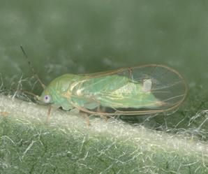
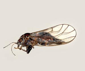
C. melanoneura
of females 3.14-3.43 mm and, a female may lay approximately 160 eggs (Ossianilsson 1992). In C. melanoneura, overall length of males is 2.52-3.10 mm, of females is 2.95-3.30 mm (Ossianilsson 1992) and each female may lays about 200 eggs. Morphological discrimination of adult C. melanoneura and C. picta from other Cacopsylla species is difficult (Tedeschi et al. 2009) and even more complicated at nymphal stages. PCR-based approaches have been developed that allow discrimination of different Cacopsylla species and thus complement classical morphological species determination (Tedeschi and Nardi 2010; Oettl and Schlink 2015). The spatial distribution, natural infection rate and transmission capacity of C. picta and C. melanoneura are heterogeneous among different geographic regions. In North-Eastern Italy, Germany and other East European countries both C. picta and C. melanoneura occur sympatrically, i.e. the insects can occur together in the same geographical area, but C. picta has a major vector role (Frisinghelli et al. 2000; Jarausch et al. 2003; 2004a; 2007; Mattedi et al. 2008d; Oppedisano et al. 2019b). Carraro et al. (2008) proved that C. picta adults are already highly infective when moving from shelter plants to apple trees and Jarausch et al. (2011a) showed that the vectors remain infective during their entire presence in apple orchards. Tedeschi et al. (2003) evidenced the importance of the overwintered adults of C. melanoneura in vectoring 'Ca. P. mali', due to the longer period spent in the apple orchards and a higher proportion of 'Ca. P. mali'-infected specimens compared to newly emerged adults. In Germany, Jarausch et al. (2007; 2011a) found transmission rates of 8 to 45 % and 0 % in overwintering adults of C. picta and C. melanoneura, respectively. A six years-study carried out in Trentino (Northeastern Italy) confirmed the higher transmission efficiency of C. picta with 4.1 % infected test plants compared to C. melanoneura with 0.36 % (Mattedi et al. 2008e). After a sudden outbreak of the disease observed in some apple-growing areas of Trentino-South Tyrol in 2011, acquisition and transmission trials were again carried out to evaluate the new vectoring status of the two main vectors and, among all life stages, the transmission efficiency reached 1.5 % in C. melanoneura and 10.2 % in C. picta (Oppedisano et al. 2019b). Other studies have also shown, that psyllids transmit phytoplasma as both adults and nymphs (Tedeschi and Alma 2004; Jarausch et al. 2004a; Jarausch et al. 2011a; Oppedisano et al. 2019b). For C. melanoneura infection rates were found to be less than 1 % in Germany, Northern Switzerland and Eastern France (Mayer et al. 2009). This is in contrast to C. melanoneura in North-Western Italy where Tedeschi et al. (2003) reported that 45 % of C. melanoneura individuals collected from a 100 % AP-infected orchard were tested positive for 'Ca. P. mali'. For C. picta, in contrast, the natural infection rate was found to be high across several studies (Jarausch et al. 2003; 2004a; Mattedi et al. 2007; 2008e; Carraro et al. 2008; Baric et al. 2010b). A characterization of C. picta in North-Eastern Italy, showed an average infection of 'Ca. P. mali' of 45 % in the overwin-
tered and 14 % in newly emerged C. picta adults (Carraro et al. 2008). Another important factor in the vector capacity of C. melanoneura is the role of host plants. C. melanoneura collected from different host plants subsequently showed differential host plant preferences on hawthorn Crataegus monogyna (Jacquin 1775) and apple and might be genetically separated (Malagnini et al. 2013). This is in line with German C. melanoneura populations that prefer hawthorn as their primary host that might explain the scarce importance of C. melanoneura as a vector of AP in Germany (Mayer et al. 2009). Different transmission efficiencies due to different adaptations of psyllid populations and certain phytoplasma strains cannot be excluded (Tedeschi and Nardi 2010). In this respect, Baric et al. (2011a) showed a genetic correlation between certain 'Ca. P. mali' strains and C. picta or C. melanoneura, respectively. Even after all the experiments conducted in the past years the actual impact of C. melanoneura in the AP spread remains not fully clear. The phytoplasma concentration in an insect vector depends on the phytoplasma concentration in the source plants (Tedeschi et al. 2012), the duration of the acquisition period and the ability of the phytoplasma to accumulate within the insect vector (Hogenhout et al. 2008). Pedrazzoli et al. (2007) reported that both psyllid vectors collected in Trentino were equally able to acquire 'Ca. P. mali', but C. picta constantly reached a higher phytoplasma titre than C. melanoneura; this data was recently confirmed by Oppedisano et al. (2019b). The phytoplasma titre significantly increased in both species when the psyllids were kept after the acquisition for up to 4 days, on healthy test plants. Similarly, the detection of low phytoplasma concentrations in a German population of C. melanoneura is considered the reason why this species has no relevance as a vector of apple proliferation in Germany (Mayer et al. 2009). The minimum phytoplasma concentration necessary for an effective transmission might be influenced by different factors such as psyllid species, population and phytoplasma strain. Aside from the acquisition of infected phloem sap by ingestion, another way of phytoplasma dissemination represents the transovarial or ‘vertical’ pathogen transmission. In this case phytoplasma gets transmitted vertically from 'Ca. P. mali' infected mothers to their offspring. Recently, Mittelberger et al. (2017a) showed that C. picta vertically transmits 'Ca. P. mali'. According to this study a critical phytoplasma concentration threshold is necessary for a successful maternal transmission of phytoplasma. Moreover, the phytoplasma titre in newly emerged F1 adults was similar to that in infected parental individuals, indicating that transovarially infected F1 adults are as infective as remigrants. In contrast, the possibility of a transovarial transmission was investigated in C. melanoneura by (Tedeschi et al. 2006) and, even if it could not be excluded, has not been demonstrated. A systematic monitoring of the psyllid populations and a survey of the symptom evolution is carried out each year in a representative
of sampled individuals N .
% of infected individuals
1500
1000
500 Trentino (Valsugana)
C. picta C. melanoneura
0
2013 2014 2015 2016 2017
80 70 60 50 40 30 20 10 0 C. picta C. melanoneura
2013 2014 2015 2016 2017
Year
of sampled individuals N .
% of infected individuals
1500
1000
500
South Tyrol (Val Venosta / Burgraviato)
C. picta C. melanoneura
0
2014 2015 2016 2017 2018
80 70 60 50 40 30 20 10 0 C. picta
n=121
n=284
2014 C. melanoneura
n=11
n=121
2015
Year
n=368 n=6
2016
Figure 11 Apple psyllids population density and AP psyllids-infection rates in Trentino-South Tyrol number of apple orchards of the Provinces of Trento and Bolzano, providing the basic information for the technical advisory service to recommend the correct control strategies to the producers. Figure 11 represents the territorial observations done in these two provinces in the recent years (modified from Oppedisano et al. 2017 for Trentino and from Fischnaller et al. 2017 for South Tyrol).
Fieberiella florii The leafhopper Fieberiella florii (Stål 1864) (Fig 12) belongs to the Deltocephalinae subfamily (Hemiptera: Cicadelllidae) and is widely distributed across the European continent as well as in North America where it is an allochthon species (van Steenwyk et al. 1990). This leafhopper is univoltine and overwinters as nymphs on ornamental hosts such as privet (Ligustrum spp.), boxwood (Buxus spp.), myrtle (Myrtus spp.), hawthorn (Crataegus spp.), firethorn (Pyracantha spp.), ceanothus (Ceanothus spp.), Cotoneaster (Cotoneaster spp.), crabapple (Malus spp.) and apple (Malus × domestica) and as eggs on ornamental hosts and deciduous fruit trees (Swenson 1974). F. florii has two main peculiar chromatic characters that make the species easily recognizable from all other European leafhoppers (excluding congenerics): (1) a thick black stripe running eye to eye at the height of the passage from vertex to frons and (2) a diffuse occurrence of small black dots on the fore body and posterior wings which are both mostly brownish with some white and black patches. In contrast, nymphs are bright green-yellowish endowed of numerous setae at
Figure 12 Example of an adult of F. florii
https://commons.wikimedia.org/wiki/ File: Fieberiella_florii_01.jpg - Sanja565658 / CC BY-SA
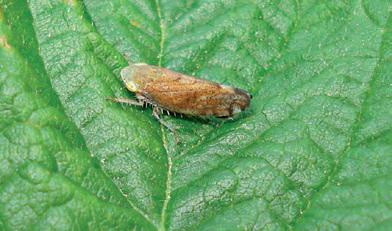
the end of the abdomen and still presenting the small black dots along their body. F. florii can live on numerous shrubs and trees among which it prefers Rosaceae plants where it also overwinters as nymph; adults occur from May to October (Swenson 1974). In North America F. florii is considered one of the most important vectors of the X-disease agent ('Ca. Phytoplasma pruni', 16SrIII group) (Gold and Sylvester 1982; van Steenwyk et al. 1990). Given its vectoring potential, at the end of the 1980s, F. florii was assumed to be a potential vector of the 'Ca. P. mali', on the basis of symptom expression and fluorescence microscopy (Krczal et al. 1988). Tedeschi and Alma (2006) confirmed the competence of F. florii in vectoring 'Ca. P. mali' through transmission trials with a 0.7-2.2 % likelihood of transmission by a single specimen. Molecular analyses of field collected specimens, revealed a natural infection rate of 5.2 % in insects collected in apple orchards and 20 % on wild plants (hawthorn, bramble, privet) (Tedeschi and Alma 2006). Thus F. florii infection rates were higher compared to C. melanoneura originating from the same area. However, the relative risk of apple tree of being infected by F. florii is considered low because of its low occurrence in apple orchards (3-9 specimens/orchard/week) and relatively inefficient transmission capability (Tedeschi and Alma 2006).
Insect vector monitoring
Accurate information gathered from insect vectors and AP form the basis to understand spatial patterns and dynamics of outbreaks and to provide recommendations of pest management strategies to apple-growers and stakeholders. For C. picta and C. melanoneura, monitoring techniques comprise visual inspection, beating tray, sticky traps and sweep net. Depending on the required information (occurrence, densities, developmental stage, generation, gender composition and spatial patterns) and on the plants (host or shelter plants) not all methods are equally suitable and therefore different techniques should be combined (Alma and Tedeschi 2010).
Figure 13 Beating tray

Visual inspection At intervals of 7-14 days, hundreds of plant organs (such as sprouts, floral rosettes and twigs) should be randomly selected to be inspected for the presence or absence of psyllids (Mattedi et al. 2008c). Visual inspection provides general information about vector population abundance and composition in terms of mobile stages (nymphs and adults) and eggs (Tedeschi et al. 2009; 2012).
Beating tray Beating tray or “limb jarring” (Burts and Retan 1973) (Fig 13) is the most common method for psyllid sampling and used to obtain absolute densities (number of psyllids per branch/shoot/leaflet) (Tedeschi et al. 2009; 2012). Psyllid sampling is recommended to be carried out at lower daytime temperatures (e.g. Horton 1999). Beating tray sampling is carried out using a white cloth-covered rectangular tray hold as close as possible below the apple limb (preferable < 20 cm) which is beaten with three medium-intense strikes (Tedeschi et al. 2012). Sequential sampling of trees in the same row should be avoided, since apple trees are often connected with wire and beating of one tree causes vibration that might in turn affect the vector presence on a neighboring tree.
Sticky traps Sticky traps have successfully been used to monitor the flight activity of insects. However, Horton (1999) showed that the interaction of different factors (e.g. environmental conditions, physiological status of the insect) influences flight activity, and thus sticky trap catches. Additional factors such as the number of trap catches, weather conditions, trap height, position, size and material of the trap are known to influence the success rate of sticky traps. Therefore, insect monitoring results gathered with sticky traps should be interpreted cautiously. Comparing several fluorescence spectra, Adams et al. (1983) found that sticky traps with a spectrum between 520-600 nm (e.g. yellow sticky traps) achieved highest pear psyllid catches, while
paring yellow sticky traps of different intensity, Hall et al. (2010) detected no significant difference in psyllid catches. Yellow sticky traps were successfully used alone (García et al. 2014; Miñarro et al. 2016) or in combination with beating trays (Tedeschi et al. 2002), to study psyllid population dynamics in apple orchards; and to sample the psyllid fauna living on hawthorn (Tedeschi et al. 2009). C. melanoneura catches by yellow sticky traps are generally male biased due to an earlier movement of males into apple orchards from overwintering sites at the beginning of the season, an increased mate searching behavior and a decreased fly activity of egg-laying females (Tedeschi et al. 2002). According to Chireceanu and Fată (2012), yellow sticky traps indicated a longer time of C. melanoneura adults’ activity, with 2-3 weeks for the overwintered and with 1-2 weeks for the spring adults, than the beating tray sampling method. For taxonomic identification or preservation for further analyzes, caught insects can be detached from sticky traps using hexane, acetone or commercial solvents such as Bio-Clear (Bio-Optica, Milan, Italy). It is important to note that data obtained by yellow sticky traps are cumulative (1 or 2 weeks of capture), while beating trays give ‘snapshot’ data and therefore direct comparison of the results is not possible.
Sweep-net Periodically sweep-net sampling has been performed for research purposes to monitor psyllids behavior on weeds under apple trees (Forno et al. 2002). The survey has been performed for three consecutive years by doing 50 sweeps per orchard and the authors reported that the psyllid species found in scrub on cover ground weeds were the same as on the neighbouring apple trees and that they disappeared from the weeds without giving offspring. Sweep-netting is also used to sample AP psyllid vectors on shelter plants during aestivation and overwintering. For this purpose, a sweep net with a telescopic handle is required in order to reach the highest parts of the plants where psyllids occur (Čermák and Lauterer 2008; Pizzinat et al. 2011) (Fig. 14). Visual inspections, beating tray and sweep-net allow to capture living specimens that can be used in further trials (e.g. transmission trails) and/or molecular analyses.
Figure 14 Use of a sweep-net in winter
Figure 15 Mortality of C. melanoneura overwintering adults (%) one hour and seven days after treatment with different insecticides; persistence was evaluated after one, three and seven days after exposure
Control strategies against the vectors
Severe outbreaks of AP across Europe and the subsequent description of the two main vectors had a high influence on the pest management strategy. The previously neglected insects C. picta and C. melanoneura moved to the center of attention of apple growers who started to treat them with pesticides (Jarausch and Torres 2014). Combatting the vectors and eradication of infected plants led to a decline of the psyllid populations and a decrease of AP occurrence in the different regions. Recently, Baldessari et al. (2017) revealed the efficacy of several insecticides against AP psyllids overwintered adults. They characterized pesticides regarding the effectiveness and persistence of their active ingredients with the aim to replace substances with an unfavorable eco-toxicological profile. For this purpose, over the years, full-field and semi-field tests have been carried out, evaluating about 15 active principles at different treatment time points, including some new pesticide formulations. Figures 15 and 16 summarize an example of the results obtained in semi-field tests in Trentino. These results show that for both AP psyllids all tested insecticides cause high mortality rate in remigrant adults even only seven days after treatment, thus demonstrating a good persistence. Furthermore, these products have also been validated in terms of crop selectivity and side effects against beneficial insects. An alternative approach to control psyllid populations is represented by wrapping particles, such as kaolin, that act by creating a film that hinders insects from feeding or moving onto the plants, and to a second extent prevent the transmission of plant pathogens by insect vectors. Processed kaolin has already been proposed as an alternative to broad-spectrum insecticides against the European pear psyllid C. pyri (Daniel et al. 2005; Pasqualini et al. 2002; Erler and Cetin 2007; Saour et al. 2009) and its efficacy was proved against C. melanoneura (Tedeschi et al. 2007a; 2007b). Late winter
[%] C. melanoneura Mortality Insert 1 hour after treatment
100
80
60
40
20
0 Fosmet Clorpirifos etile Tau fluvalinate not treated [%] C. melanoneura Mortality Insert 7 days after treatment
100
80
60
40
20
0 Fosmet Clorpirifos etile Tau fluvalinate not treated
[%] C. picta Mortality Insert 1 hour after treatment
100
80
60
40
20
0 Fosmet Clorpirifos etile Tau fluvalinate not treated [%] C. picta Mortality Insert 7 days after treatment
100
80
60
40
20
0 Fosmet Clorpirifos etile Tau fluvalinate not treated
Days of exposure Days of exposure
treatments with kaolin were able to reduce the number of laid eggs and consequently of nymphs. Since kaolin is not considered as an insecticide, but a coadiuvant it can be used in organic farming as well. In the last years there was a particular interest in innovative control strategies based on the development of species-specific traps for monitoring and mass trapping of different vector species of fruit tree phytoplasmas including C. melanoneura and C. picta (Jarausch and Torres 2014). In particular, in the AP-pathosystem, the pheromones produced by infected plants is attractive to both sexes of psyllids, so it could be possible to develop mass trapping systems for a sustainable vector control. Also, for C. melanoneura potentially behavior modifying compounds could be identified, but to date they are not species-specific. The possibility to combine attractive compounds to be used in traps as lures for monitoring and mass trapping purposes and with repellent compounds to be used in complex push-and-pull strategies is promising (Eben and Gross 2013).
Development of sustainable control strategies
Biological control: microbial symbionts of the insect vectors and their potential role in phytoplasma transmission Microorganisms are ubiquitous in insects and may influence dramatically the ecology of their host (Dale and Moran 2006). Consequences of these interactions can vary along a continuum from mutualism to parasitism. At one extreme, primary endosymbionts can positively affect the fitness of their host by providing essential nutrients or protect against parasites. Most of these symbionts are therefore required for the development of their hosts (Douglas 2016). At the other extreme, secondary symbionts can negatively affect the fitness of their hosts (Engelstädter and Hurst 2009). Being maternally inherited, they modify the reproduction of their hosts toward females to
Figure 16 Mortality of C. picta overwintering adults (%) one hour and seven days after treatment with different insecticides; persistence was evaluated at one, three and seven days of exposure
enhance their own fitness (Werren et al. 2008). These endosymbionts are usually not required by the host and in most of the cases impact its fitness negatively (Douglas 2011). The adaptation to specific host plants results in nutrition deficiencies that have to be compensated by obligate symbionts that supply lacking nutrients. Therefore, primary endosymbionts are ubiquitous in herbivorous Hemipteran species and provide essential nutrients lacking in the food source (Baumann 2005). The gammaproteobacterium Carsonella ruddii is a primary endosymbiont that appears to be present in all species of psyllids (Thao et al. 2000). As psyllids feed on phloem sap that is rich in sugars but poor in amino acids, this endosymbiont synthesizes essential nutrients missing in their diet (Baumann 2005). Moreover, many psyllids are infected by various secondary endosymbionts with unknown functional roles (Sloan and Moran 2012). The impacts of endosymbionts to their hosts offer the possibility of symbiont-based tools to control insect pests. Endosymbionts can be used to negatively influence the fitness of its host or to reduce its competence to vector diseases (Arora and Douglas 2017). For example, the endosymbiont Wolbachia has been shown to be capable to directly suppress the abundance of pest insects and to prevent viruses from replication within its vectors (McGraw and O'Neill 2013). Wolbachia-infected mosquitoes are currently being released in the field to suppress the transmission of dengue (Hoffmann et al. 2011). The diversity of endosymbionts found in vector species of phytoplasmas offers an interesting opportunity to study symbiont-based control mechanisms of phytoplasma transmission (Alma et al. 2010). Transcriptomic and metabolomic approaches have been used to comprehensively investigate interactions between 'Ca. P. mali' and its vector C. melanoneura (Weil et al., in preparation). It has been found that the pathogen likely modulates host behavior mainly by affecting the insect nervous system and rhythmic processes. Furthermore, metabolic analyses showed that carbohydrate and polyol levels are significantly altered upon 'Ca. P. mali' infection and lead to metabolic imbalance in the insect. These results suggest that infection with 'Ca. P. mali' has a major impact on the insect vector physiology and behavior, and thus on the ability to transmit the phytoplasma. Hence, deepening the knowledge of the complex dynamics behind this plant-vector-pathogen system may pave the way for the development of novel and sustainable control strategies of apple proliferation disease.
Biotechnological control: intraspecific and interspecific communication Insects can use chemical, visual and acoustic modalities to exchange information and coordinate complex courtship behaviors (Lubanga et al. 2016). It was only relatively recently that intraspecific semiochemical signalling was reported to play a role in psyllid mate attraction (Soroker et al. 2004). To-date, males of different psyllid
species have been shown to be attracted to female-produced semiochemicals, as Cacopsylla bidens (Šulc 1907), Cacopsylla pyricola (Förster 1848), Bactericera cockerelli (Šulc 1909) and Diaphorina citri (Kuwayama 1908). For instance, females of C. pyricola produce significantly larger quantities of 13-methylheptacosane than males, a compound that is known to be attractive to males (Guédot et al. 2009). This provided the first evidence of a female sex pheromone capable of attracting male psyllids from neighboring host plants. Using a similar approach, dodecanoic acid was identified as the female semiochemical attractive to male D. citri (Mann et al. 2013). After searching and recognition activity, courtship is usually brief and seems to be mediated by a combination of both epicuticular hydrocarbons and substrate-borne vibrational signals. With regard to the interspecific communication, C. picta and C. melanoneura use volatile chemical cues also for the identification of their host plants (Gross 2011). Furthermore, 'Ca. P. mali' has evolved mechanisms for the manipulation of plant physiology and indirectly vector behavior by attracting C. picta to infected plants for feeding purposes (Mayer et al. 2008a; 2008b). This coevolutionary process increases the likelihood of phytoplasma acquisition by the insect vector and subsequently its spread. In contrast, C. picta prefers to oviposit on healthy trees, maybe due to detrimental effects of phytoplasma to the offspring fitness (Mayer et al. 2011). The plant volatile mainly responsible for the vector attraction to infected trees has been identified by headspace-gas chromatography (HS-GC) analysis of infected apple trees as β-caryophyllene – a sesquiterpene. It is now under evaluation for the development of traps for monitoring and/or mass trapping (Weintraub and Gross 2013). In contrast, the hawthorn psyllid C. melanoneura did not react to this sesquiterpene (Eben and Gross 2013). Acoustic signals among insects can be divided into two categories based on the medium of transmission: air-borne signals (e.g. Cicadoidea, Grylloidea, Tettigoniidae) and substrate-borne signals (e.g. Psylloidea, Chrysopidae) (Liao and Yang 2015). In the Psylloidea, the
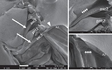
B
Figure 17 Scanning electron microscopy (SEM) investigation on C. melanoneura female A) thorax and hindwing axillary cords (axc2, axc3), mesopostnotum (pnt2); B) detail of the axillary cord B; C) detail of hindwing (A2) (R. Kostanjšek/T. Oppedisano)
A C
Figure 18 Spectrogram of C. picta vibrational signals recorded during courtship behavior. Female signal is a sequence of pulses; male signal consists in a series of pre-pulses and a buzz
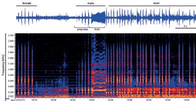
substrate-borne signals have a function only in mating and specific recognition. The male and female psyllids usually perform reciprocal duets during courtship (Tishechkin et al. 2006; Percy et al. 2006; Eben et al. 2015; Liao and Yang 2015). Psyllids are able to produce vibrations by rapidly moving their forewings that present a single row of ridges on the anal vein rubbing against similar structures on protruding ridges on the meso- and meta thorax (Taylor 1985) (Fig. 17). Signal characteristics, such as call length, pulse number and reply latency, are used for species and gender recognition (Lubanga et al. 2014). Laser vibrometer recordings of vibrational signals of the two AP vectors emitted during courtship have been recorded recently (Oppedisano et al. 2016; Oppedisano et al. 2019a). Females of C. picta were shown to initiate communication on the host plant by emitting trains of vibrational pulses, followed by a duet consisting of male call and female reply (Fig. 18). Although C. melanoneura language has not been described yet, the existence of male calls has been demonstrated as well in the same studies. This finding would open the way for testing the possibility to interfere with the mating vibrational communication of vector psyllids as an alternative pest control method as already demonstrated in application of biotechnological control of other insect pests by mating disruption (Eriksson et al. 2012; Polajnar et al. 2016; Nieri and Mazzoni 2018).
Currently, monitoring data of both AP vectors, is the basis to determine pest management strategies. Due to cost and time-consuming fieldwork and laboratory analyses, the vector monitoring is performed only at a limited number of survey sites from which management decisions are generalized. However, environmental conditions of the apple orchards are not equally suitable for the AP vectors and hence, those generalizations may result in ineffective pest management. In Valsugana (Trentino), the first appearance of the AP vector C. melanoneura is predicted using temperature thresholds (Tedes-
chi et al. 2012). Since oviposition occurs at bud burst, while egg peak and hatchings are always before the first flowering, synchrony between C. melanoneura and host-plant growth is important and assumed to be linked with temperatures (Hodkinson 2009). Thus, an immigration index to predict the progressive arrival of the overwintered adults from winter sites was defined (Tedeschi et al. 2012). In the investigated area psyllids start to reach the apple orchards when either the average of the maximum temperature of the 7 d is above 9.5 °C or the immigration index has reached the threshold. This index, based on temperatures recorded in the orchards, represents a useful tool to time insecticide treatments against C. melanoneura. Based on Tedeschi et al. (2012), a temperature based-immigration model has been developed for C. melanoneura and C. picta to predict the first presence in apple orchards in apple South Tyrol (Panassiti 2018). However, this model has several limitations and the applicability of the results needs further evaluation. Furthermore, the results of the temperature-based model for the migration prediction of C. picta and C. melanoneura showed strong differences already in a small-scale geographic comparison. This indicates that all predictions -even if enough data are available- are only valid for very small geographic areas. Geographical factors associated with the winter sites location (e.g., the regional ortography, the main air streams and distance from apple orchards) may differently affect the psyllid migration process and influence its presence or absence. Habitat models (also known as species distribution models) aim to identify and quantify the relationship between environmental variables and a response variable (Guisan and Zimmermann 2000). Examples of response variables in the AP epidemiology could include presence/absence and abundance of the vector, 'Ca. P. mali' prevalence within the insect vector, and the presence/absence of AP symptoms on apple trees. Abiotic and biotic environmental predictors are typically chosen to represent resources (e.g. host plants), disturbances (e.g. insecticides) and limiting factors (e.g. temperature) (Guisan and Thuiller 2005). The results of statistical models can be beneficial to improve AP control strategies in different ways. For example, information about vectors’ forest type preference can be helpful to narrow down overwintering sites. This would allow placing temperature logger for a better estimation of the start of the vectors’ flight activity. Further, identified species-environment relationships allow area-wide predictions and based on those, the creation of risk maps. Habitat models are not restricted to vectors, but can also be applied to the pathogen and the disease (Thébaud et al. 2006; Panassiti et al. 2015; 2017). Using Bayesian inference, Panassiti (2018) developed a joint model (i.e. simultaneously estimating the dependencies of vector, phytoplasma infection rates of the vector and AP symptoms of apple trees) for the AP epidemiology. The model allowed to account for imperfect detection of AP symptoms by estimating the detection probability conditional on the true infection status. One factor af-
fecting symptom detection are latent infections, assumable yielding an increased number of false negatives, and hence, a biased inference of the system. Therefore, Panassiti (2018) included a so called “informative prior” which takes advantage of exisisting experimental results about latent AP infections of apple trees (2.32 and 10.48 % depending on age of the apple trees, Baric et al. 2007), and allowed for a better estimation of detection probability. The model results indicated that AP vector and symptomatic tree occurrence probabilities are positively affected by increasing elevation and temperature, in contrast negatively by integrated pest management. The results of this study are however rather preliminary. In conclusion, habitat models are a useful tool to establish species-environment relationship and to create risk maps. The model results may, therefore, contribute to new insights in the AP epidemiology and allow to develop and adapt efficient management strategies.
The order of Hemiptera comprises insect groups with specific piercing-sucking mouthparts, which conferred a relevant effect in their adaptive radiation (Goodchild 1966). As phloem-limited, phytoplasmas can be acquired and transmitted only by phloem-feeding insects. Hemiptera feeding habits range from phytophagy (the majority of species) to predation, including ectoparasitism and haematophagy. Phytoplasma vectors must feed specifically and selectively on this particular plant tissue, where pathogens reside, in a nondestructive way. Weintraub and Beanland (2006) reviewed the features required by an insect species to be a successful phytoplasma vector and, according to the authors, Hemiptera are the main elicited insect group. Insects of this order are hemimetabolous and nymphs and adults besides feeding similarly, share the same physical location; often both nymphs and adults can transmit phytoplasmas. They feed specifically and selectively on certain plant tissues, which makes them efficient vectors of pathogens residing in those tissues. Furthermore, their feeding is nondestructive, promoting successful inoculation of the plant vascular system without damaging conductive tissues and eliciting defensive responses. Moreover, they have a propagative and persistent relationship with phytoplasmas. Hemiptera is a very diverse order of insects comprising e.g. the suborder Sternorrhyncha (comprising the Superfamilies: scale insects (Coccoidae), aphids (Aphidoidea), psyllids (Psylloidea) and whiteflies (Aleyrodoidea)), true bugs (Heteroptera), and Auchenorrhyncha. The latest have been traditionally divided into two suborders: Cicadomorpha (to which the families of the leafhoppers (Cicadellidae), treehoppers (Membracidae), spittlebugs (Aphrophoridae) and cicadas (Cicadidae) belong) and Fulgoromorpha (the planthopper). In the last years, the population densities and infectivity rates found
in C. picta and C. melanoneura after new AP outbreaks in Trentino-South Tyrol, were not enough to explain the levels of spreading of the disease in field. Therefore, attempts have been undertaken to identify further AP transmitting insect vectors, mainly among hemipteran species. As previously stated, transmission trials are the only proof of an insects’ phytoplasma transmission ability. Although, 'Ca. P. mali' was detected in some phloem-feeding insects, transmission trials were not carried out or yielded negative results and therefore these insects are not considered as vectors of 'Ca. P. mali'. The presence of 'Ca. P. mali' was detected in several apple aphids: Aphis pomi (De Geer 1773), Dysaphis plantaginea (Passerini 1860), Eriosoma lanigerum (Hausmann 1802), Dysaphis devecta (Walker 1849) and Rhopalosiphum insertum (Walker 1849). Aphids had a much lower 'Ca. P. mali' titer than infected psyllids and transmission trials failed. Thus, the authors concluded that aphids do not contribute to AP spreading (Cainelli et al. 2007). 'Ca. P. mali' was also detected in other Cacopsylla species, such as Cacopsylla peregrina (Förster, 1848) (Tedeschi et al. 2009), as well as in Cacopsylla mali (Schmidberger 1836) and Cacopsylla crataegi (Schrank 1801) (Baric et al. 2010b; Miñarro et al. 2016). Reported pathogen presence in these insects ranged from 21.74 % to 53.85 % for Cacopsylla peregrina, from 1 % to 10 % for Cacopsylla mali and from 1 % to 16.7 % for C. crataegi (Tedeschi et al. 2009; Baric et al. 2010b; Miñarro et al. 2016). Moreover, 'Ca. P. mali' was detected in two exotic eucalyptus psyllid pests, Ctenarytaina eucalypti (Maskell 1890) and Ctenarytaina spatulata (Taylor 1997), which both are present in apple orchards in the Asturia region in Northern Spain, with percentages of 'Ca. P. mali' positive individuals ranging from 1.4 to 3 % for C. eucalypti and from 2.3 % to 2.7 % for C. spatulata (García et al. 2014; Miñarro et al. 2016). For all these species further transmission trials need to be carried out to confirm their vector status. In the past, different insect species belonging to the family Cicadellidae (Auchenorrhyncha) occurring in the apple orchards, such as Empoasca vitis (Göethe 1875), were analyzed for the presence of 'Ca. P. mali' without success (Mattedi et al. 2008e). To investigate the vector ability of this species, Mattedi et al. (2008e) carried out transmission trials, but no positive results were obtained. Other species reported as vectors are Philaenus spumarius (Linnaeus 1758) (Homoptera: Aphrophoridae) and Artianus interstitialis (Germar 1821) (Homoptera: Cicadellidae), which were able to transmit 'Ca. P. mali' from infected celery to apple seedlings and from infected to healthy celery (Marenaud et al. 1978; Hegab and El-Zohairy 1986; Németh 1986). However, other experiments conducted with P. spumarius did not confirm the previous results (Refatti et al. 1986). Danielli et al. (1996) detected different groups of phytoplasma in the planthopper Metcalfa pruinosa (Say 1830) (Homoptera: Flatidae), including 'Ca. P. mali', but its vector status has never been confirmed. After the outbreaks of AP in Trentino South-Tyrol in 2011, researchers focused part of their work in collection and identification of
Figure 19 Species abundance in nine apple orchards surrounded by forest-dominated landscape in Valsugana (Trentino)
Figure 20 Species abundance in nine apple orchards surrounded by grass-dominated landscape in Valsugana (Trentino) Forest-dominated landscapes
Psammotettix confinis 4 % Zyginidia pullula 4 %
Empoasca vitis 5 %
Muelleraniella extrusa 11 %
Macrosteles sexnotatus 28 % Laodelphax striatella 26 %
Macrosteles cristatus 4 %
Grass-dominated landscapes
Psammotettix confinis 8 %
Macrosteles ossiannilssoni 38 % Laodelphax striatella 38 %
Figure 21 Species abundance in nine apple orchards surrounded by orchard-dominated landscape in Valsugana (Trentino) Orchard-dominated landscapes
Psammotettix confinis 6 % Empoasca vitis 13 %
Macrosteles ossiannilssoni 15 %
Laodelphax striatella 51 %
hoppers community in apple orchards, studying their distribution in the whole apple agroecosystem. Insects such as leafhoppers and planthoppers show frequent migrations (DeLong 1971; Taylor 1985, Della Giustina 2002a, 2002b) that influence their population dynamics and spatial distributions. These migrations have to be taken into account for adequate integrated pest management strategies (Matsumura and Suzuki 2003; Orestein et al. 2003; Emmen et al. 2004; Decante and van Helden 2008). As a rule, only few insects are considered as key species of any crop. However, this approach fails to explain in detail all the relationships that exist in agroecosystems, where it is the whole community that determines production and socio-economic impact. Oppedisano et al. (2017) evaluated the effects of landscapes on the presence of these communities inside the apple orchards. Moreover, in the same study, the researchers evaluated the role of the most representative species as putative vector of 'Ca. P. mali'. Preliminary results about the hoppers communities’ diversity in apple ecosystem are shown in Figure 19, 20 and 21. Molecular analyses were conducted on 1305 individuals. Two leafhoppers (Cicadellidae) were found positive regarding the presence of 'Ca. P. mali': one individual of Empoasca vitis and one of Orientus ishidae (Matsumura 1902) (Cicadellidae: Deltocephalinae). Moreover, two specimens belonging to the species Stictocephala bisonia (Kopp and Yonke 1977) (Membracidae: Membracinae) contained very low concentrations of 'Ca. P. mali'. Therefore, these results are the first step in the search of so far unknown or new vectors, albeit further investigations focused on their acquisition and transmission ability under controlled conditions are required.

4
CONCLUSIONS
During the last decades a multidisciplinary approach was adopted to deepen our understanding of the epidemiological and biological features of apple proliferation (AP). As no direct control measures are available to fight phytoplasma diseases, several projects aimed to gain knowledge about the mechanisms underlying disease spread. Many studies focused on the insect vector biology, ecology and about potential factors that predispose these insects to transmit phytoplasma. The results of the transmission trials with psyllids show that both C. picta and C. melanoneura play an important role in the disease spread, especially at high densities and in the presence of high inoculum sources, i.e. the presence of many infected apple trees. At the moment, there is no indication that other hemipteran species play a role in disease transmission. However, an involvement of other insects as vectors in AP transmission cannot be excluded. Monitoring the incidence of symptomatic apple trees and the psyllid populations is indispensable in epidemic and in endemic phases to assess the current situation and the effectiveness of the applied phytosanitary measures. In the last years in Trentino-South Tyrol very fluctuating disease incidences were recorded, thus long-term investigations are required to understand transmission dynamics in more detail. Psyllids are characterized by a univoltine biological cycle involving different (and only partially known) host plants. So far, no efficient rearing method has been established that allows the production of high numbers of insects for experimental purposes. Phytoplasma are genetically highly dynamic and up to now no efficient cell free propagation method has been established. This lack of ex vivo culture methods hampers microbiological studies and renders genetic manipulation of phytoplasma impossible. All these drawbacks and complications make any attempt of finding general conclusions even more challenging. Studies analysing the role of phytoplasmal effector proteins in disease development are emerging and important for a better understanding of the disease on the molecular level. Reliable diagnostics are furthermore essential, not only for monitoring the disease but also to determine potential new insect vectors and reservoir plants. The use of resistant rootstocks would be the most sustainable solution, but despite all efforts no apple variety was found that confers full resistance (i.e. that it cannot be infected) against 'Ca. P. mali'. All control strategies applied so far in Trentino-South Tyrol are only focusing on reducing vector densities in the orchards by applying multiple chemical treatments and uprooting infected plants. In the future novel, specific and sustainable control measures against AP must be developed that can be used in organic apple cultivation as well. The results achieved in the last projects open new intriguing possibilities for the development of such prospective strategies. The assessment of hemipteran biodiversity confirms the importance of
investing in environmentally-friendly technological advances, since also conventionally cultivated apple orchards bear a broad spectrum of different insect species. The recent studies on psyllids communication, their microbiota and habitat modelling open new perspectives for the implementation of specific, sustainable and well-timed insect vector control strategies, essential to have a low impact on human and environmental health.
AUTHORS
Gianfranco Anfora PhD, is entomologist, Associate Professor in General and Applied Entomology at Center Agriculture, Food and Environment, University of Trento/Edmund Mach Foundation. He addressed his research activity to the study of insect communication, identification of intraspecific and interspecific semiochemicals by molecular, electrophysiological and behavioural investigations, and to the development of integrated pest management and biological control programs in grape and fruit production.
Gino Angeli Is head of the Plant protection of agroforestry and apiculture in Fondazione Edmund Mach. During his career he has conducted extensive research on integrated controls of apple and grape pests. He has focused his research on side effect of agrochemicals on beneficial organisms, behavioural investigations of phytoseids and insects in field and on area-wide application of semiochemicals. He was engaged for many years in the management of the FEM test facility.
Mario Baldessari Holds a PhD in “Crop Protection” and a degree in Agricultural Sciences at Padova University. Since 2003, he deals with evaluation of PPP effectiveness (chemical or biological origin) to control several kinds of pests (including emerging pests or quarantine). As an expert in the IPM, his activity in the unit "Test Facility" concerns the development of new strategies for the integrated fruit and grape production, biological control of pests, implementation of mating disruption technique, pesticide characterization (e.g. discriminate dose, baseline, timing of spray), side-effects on beneficial organisms and research on new and emerging pests and pathogens.
Dana Barthel Dipl.-Ing., is an engineer for land use and water management and is part of the Functional Genomics group at Laimburg Research Centre. Her current research is focused on the development of new non-destructive detection methods for the AP pathogen and the physiological changes that are caused by the pathogen in the tree.
Pier Luigi Bianchedi Agronomist. Since 2003 Pier Luigi Bianchedi focusses on the problematics of the phytosanitary selection (viruses and phytoplasmas)
in apple and grape. He has more than 15 years research experience on 'Candidatus Phytoplasma mali' particularly on the plant/phytoplasma interaction aspects and in the evaluation of resistance to AP in apple genotypes. For more than ten years Pier Luigi Bianchedi manages and organizes the maintenance in vitro culture and the micropropagation of agronomically important apple and grape genotypes. Presently he is the referent for the procedure necessary for the certification of the grape material produced by the nurseries. Moreover, Pier Luigi Bianchedi is responsible of the micropropagation laboratory of the Technology Transfer Centre which is a servicing platform for the maintenance, conservation and propagation of the plant material and operates as a support for several experimental trials.
Andrea Campisano PhD, is an expert in plant-microbe associations, currently affiliated with the Italian Ministry of Education, University and Research (MIUR). His main areas of expertise are plant-microbe interactions and their dissection using the -omics approach. His research focuses on the potential of microorganisms as an alternative to pesticides and as biofertilizers, in order to achieve sustainable yet pruductive agroecosystems.
Laura Tiziana Covelli PhD, is a plant pathologist who has mostly centered her interest on the multitude of problems in agriculture related to the phytopathogen agents and the diseases they provoke with a specific attention on the new techniques of molecular biology as well as on computer-related methods and tools analysis. She also contributed to the development of two scientific software and to the implementation of the Gypsy database of mobile genetic elements (www.gydb.org). Her last research activity has been addressed to the identification of new strategies for the Apple proliferation (AP) disease’s biocontrol - in particular on the action of putative endophytic “beneficial bacteria” against 'Candidatus Phytoplasma mali' and their effects on the apple plant physiology - as well as on the identification of 'Ca. Phytoplasma mali' strains potentially involved on symptoms expression.
Gastone Dallago Is an agronomist, responsible of Test facility unit in the Technology Trasfer Centre/Fondazione Edmund Mach. He is the official delegate of the Province of Trento in the GDI (Integrated Pest Management Group) by MIPAAFT in Rome. His main activity is the experimentation of the efficacy of fungicides, insectides, herbicides, growth regulators etc. (active substances or new pesticides, registered or not), in Trentino crops. In this project he coordinates the activity of a group of advisors with the aim to control phytoplasma vectors, to monitor the presence of infected trees in apple orchards and to publish technical communications to farmers.
Stefanie Fischnaller M. Sc., biologist, is part of the Entomology group at Laimburg Research Centre. Her research interest focusses on the ecology of hemipteran insect pests (Psyllids, Auchenorrhyncha and Heteroptera). In the past years SF specialized on psyllid biology and their role in the transmission of apple proliferation.
Claudio Ioriatti PhD, director of the Technology Transfer Centre of Fondazione Edmund Mach. Entomologist and expert in the implementation of integrated plant production of apple and grape. His research activity concerning the implementation of semiochemicals, biological control of pests and insecticide resistance management, has been integrated with the coordination of the advisory service operating with local agricultural industries.
Katrin Janik Dr. rer. nat., is biologist. She is group leader of Functional Genomics and Project Leader of the apple proliferation projects at Laimburg Research Centre. Katrin Janik is specialized in molecular biology and infection biology with more than 11 years of experience. Her main research interest is in the field of host-pathogen interactions and bacterial effector protein biology. Since more than 6 years, phytoplasmoses are her main research area.
Wolfgang Jarausch PhD, is biologist at AlPlanta – Institute for Plant Research at RLP AgroScience in Neustadt an der Weinstrasse, Germany. Since the early 1990ies, his major research topics are phytoplasma diseases of European fruit trees. His studies included molecular characterisation of the phytoplasmas, development of molecular detection tools, epidemiological studies, identification and biological characterisation of insect vectors, plant tissue culture of phytoplasma-infected plants to study genetic and induced resistance and the development of resistant rootstocks.
Thomas Letschka PhD, is a molecular biologist at Laimburg Research Centre leading the area of Applied Genomics and Molecular Biology. His research activity is addressed to understand and detect specific traits of diseases, disease resistances, quality and health aspects in apples and grapevines. Translation of findings into practical or breeding efforts is his main focus.
Valerio Mazzoni PhD, is the leader of the Agricultural Entomology unit of the Research and Innovation Centre at Fondazione Edmund Mach of San Michele all’Adige. As entomolgist, his expertize includes taxonomy, behavioral ecology and IPM. Scientific activity in his Bioacoustics Lab
focuses on the field of Biotremology and he has made substantial developments in the application of this discipline for use in agricultural pest control.
Cecilia Mittelberger M. Sc., is an agronomist at Laimburg Research Centre with a scientific focus on different topics related to phytoplasmas. Currently she works on the identification and characterization of interactions between 'Candidatus Phytoplasma mali' effector proteins and their interaction partners in apple and she is enrolled as a PhD student at Martin-Luther University (Halle, Germany).
Mirko Moser Dr. rer. nat., is molecular biologist, researcher at Fondazione Edmund Mach. His research topic focus on the study of the transcriptional, post-transcriptional and epigenetic changes occurring during the plant-pathogen interaction in diseases affecting grape and apple, and changes taking place in the regulatory mechanisms underlying plant development by analyzing RNA and smallRNA levels and investigating DNA methylation and chromatin remodeling using high throughput sequencing approaches.
Sabine Öttl Dr. rer. nat., biologist, is leader of the Phytopathology group at Laimburg Research Centre. She has in-depth knowledge of South Tyrolean fruit culture and many years of experience with apple proliferation in field and applied genomics. Currently, her main research area is the biology of fungal plant pathogens with focus on resistance management.
Tiziana Oppedisano Is an entomologist and postdoctoral scholar at the Hermiston Agricultural Research and Extension Centre at Oregon State University. Her PhD research focused on the biology and ecology of insect vectors of apple proliferation disease. She is interested in studying the three-way interaction between plants, insects, and pathogens from ecosystem scales to genome-level aspects.
Bernd Panassiti Dr. rer. nat., is a quantitative ecologist particularly interested in spatial population structures, spatial population dynamics, species interactions and biodiversity. He combines field studies and experiments at different spatiotemporal scales as well as molecular analyses with statistical models to study environmental factors causing the observed spatial pattern of mostly, though not only, insects, vascular plants and diseases affecting agricultural crops.
Federico Pedrazzoli Is a biologist and works at Fondazione Edmund Mach as a technol-
ogist. He achieved his PhD in Plant Protection with a study on the role of psyllids in the transmission of 'Candidatus Phytoplasma mali'. He was further involved in the development of strategies for the biological control of chestnut insect pests and currently works in the diagnostic lab of FEM, applying molecular tools for the detection of viruses in insects and plants.
Omar Rota-Stabelli Is a permanent researcher at the Agrarian Entomology Unit of the Fondazione Edmund Mach. He leads various projects on both fundamental and applied research and he is interested in how evolution drives the biology and the ecology of organisms in particular insects of agricultural importance. He tackles this issue using various evolutionary and genomic methods, in particular comparative genomics, phylogenetics and molecular dating on a variety of model organisms including insects and fungi pests of crops, mosquitoes and their carried viruses, Wolbachia and microorganisms.
Hannes Schuler PhD, is Assistant Professor in General and Applied Entomology at the Free University of Bozen-Bolzano. Schuler has broad interests in ecology and evolution of pest insects and their associated microbes. In particular his research focuses on the the role of reproductive endosymbionts and on the ecology and evolution of their insect hosts. Moreover, he is interested in understanding the dynamics of how economically harmful pest insects are introduced into new areas and how the new environment influences their bacterial endosymbionts.
Wolfgang Schweigkofler Holds a master’s degree in microbiology from the University of Vienna (1994) and a PhD in Applied Biology from the University of Natural Resources and Applied Life Sciences, Vienna (1998). Wolfgang Schweigkofler was a Post-doctoral fellow first at the Laimburg Research Centre, Italy, from 1999-2001, working on bio-control of soil-dwelling insects, and then at UC Berkeley, USA (2002-2004), working on forest pathogens. From 2004-2011 he worked as a senior plant pathologist at the Laimburg Research Centre. He moved to the USA in 2011, working shortly at a biotech start-up before accepting a position at the Dominican University of California in San Rafael. Currently he is a Research Associate Professor and lead scientist at NORS-DUC, the National Ornamentals Research Site at Dominican University. His research interests include diseases of grapevine, apple and ornamentals, forest pathology, biological control, bio-diversity and invasive biology.
Rosemarie Tedeschi Is Associate Professor in General and Applied Entomology at the Department of Agricultural, Forest and Food Sciences (DISAFA), Univer-
sity of Torino, Italy. She holds a PhD in integrated and biological pest management and has a long lasting experience in biology, ethology, infectivity and epidemiology of insect vectors of plant pathogens, mainly phytoplasmas. Her research activity also concerns insect immunity and innovative control strategies against crop pests.
Tobias Weil PhD, is a senior researcher at the Food Quality and Nutrition Department, Fondazione Edmund Mach. He is interested in the exploration and evaluation of microscopic organisms for sustainable utilization in agriculture, industry and medicine as well as their relevance to ecosystem functions and services. This covers the ecology of microorganisms in both natural and engineered environments.
REFERENCES
Adams R. G., Domeisen C. H., Ford L. J. (1983). Visual trap for monitoring pear psylla (Homoptera: Psyllidae) adults on pears. Environmental Entomology 12 (5): 1327-1331.
Ahrens U., Seemüller E. (1992). Detection of DNA of plant pathogenic mycoplasmaike organisms by a polymerase chain reaction that amplifies a sequence of the 16S rRNA gene. Phytopathology 82 (2): 828-832.
Ait Barka E., Belarbi A., Hachet C., Nowak J., Audran J. C. (2000). Enhancement of in vitro growth and resistance to gray mould of Vitis vinifera co-cultured with plant growth- promoting rhizobacteria. FEMS Microbiology Letters 186: 91-95.
Aldaghi M., Bertaccini A., Lepoivre P. (2012). cDNA-AFLP analysis of gene expression changes in apple trees induced by phytoplasma infection during compatible interaction. European Journal of Plant Pathology 134 (1): 117-130.
Aldaghi M., Massart S., Roussel S., Dutrecq O., Jijakli M. H. (2008). Adaptation of real-time PCR assay for specific detection of apple proliferation phytoplasma. Acta Horticolturae 781: 387-393.
Aldaghi M., Massart S., Roussel S., Jijakli M. H. (2007). Development of a new probe for specific and sensitive detection of 'Candidatus Phytoplasma mali' in inoculated apple trees. Annals of Applied Biology 151: 251-258.
Alma A., Daffonchio D., Gonella E., Raddadi N. (2010). Microbial symbionts of Auchenorrhyncha transmitting phytoplasmas: a resource for symbiotic control of phytoplasmoses. In: Weintraub P. G., Jones P. (Eds.): Phytoplasmas. Genomes, Plant Hosts and Vectors. Wallingford, UK: CABI: 272-292.
Alma A., Navone C., Visentin C., Arzone A., Bosco D. (2000). Rilevamenti di fitoplasmi di “apple proliferation” in Cacopsylla melanoneura (Förster) (Homoptera Psyllidae). Petria 10: 141-142.
Alma A., Tedeschi R. (2010). Phytoplasma vectors in Italy. Knowledge, critical aspects and perspectives. Petria 20: 650-663.
Alma A., Tedeschi R., Lessio F., Picciau L., Gonella E., Ferracini C. (2015). Insect vectors of plant pathogenic Mollicutes in the Euro-Mediterranean region. Phytopathogenic Mollicutes 5 (2): 53-73.
Amici A., Refatti E., Osler R., Pellegrini S. (1972). Corpi riferibili a micoplasmi in piante di melo affette dalla malattia degli scopazzi. Rivista di Patologia Vegetale 8: 3-19.
Arora A. K., Douglas A. E. (2017). Hype or opportunity? Using microbial symbionts in novel strategies for insect pest control. Journal of Insect Physiology 103: 10-17.
Autonome Provinz Bozen / Provincia Autonoma di Bolzano (16.08.2011). Phytosanitäre Maßnahmen zur Bekämpfung der Apfeltriebsucht / Misure fitosanitarie per la lotta contro la malattia degli scopazzi del melo, vom 604/31.2. Amtsblatt / Bollettino Ufficiale (34): 16-19.
Bai X., Zhang J., Ewing A., Miller S. A., Jancso Radek A., Shevchenko D. V. Tsukerman K., Walunas T., Lapidus A., Campbell J. W., Hogenhout S. A. (2006). Living with genome instability: the adaptation of phytoplasmas to diverse environments of their insect and plant hosts. Journal of Bacteriology 188 (10): 3682-3696.
Baldessari M., Angeli G., Oppedisano T. (2017). Nuove strategie contro le psille vettori degli scopazzi del melo. L’Informatore Agrario 9: 47-51.
Baric S., Berger J., Cainelli C., Kerschbamer C., Dalla Via J. (2011a). Molecular typing of 'Candidatus Phytoplasma mali' and epidemic history tracing by a combined T-RFLP/VNTR analysis approach. European Journal of Plant Pathology 131: 573-584.
Baric S., Berger J., Cainelli C., Kerschbamer C., Letschka T., Dalla Via J. (2011b). Seasonal colonisation of apple trees by 'Candidatus Phytoplasma mali' revealed by a new quantitative TaqMan real-time PCR approach. European Journal of Plant Pathology 129: 455-467.
Baric S., Dalla Via J. (2004). A new approach to apple proliferation detection: a highly sensitive real-time PCR assay. Journal of Microbiological Methods 57 (1): 135-145.
Baric S., Kerschbamer C., Berger J., Cainelli C., Dalla Via J. (2010a). Ausbreitung der Apfeltriebsucht in Südtirol in zwei Wellen. Obstbau Weinbau (2): 70-73.
Baric S., Kerschbamer C., Dalla Via J. (2007). Detection of latent apple proliferation infection in two differently aged apple orchards in South Tyrol (northern Italy). Bulletin of Insectology 60 (2): 265-266.
Baric S., Kerschbamer C., Vigl J., Dalla Via J. (2008). Translocation of Apple Proliferation Phytoplasma via natural root grafts - a case study. European Journal of Plant Pathology 121 (2): 207-211.
Baric S., Öttl S., Dalla Via J. (2010b). Infection rates of natural psyllid populations with 'Candidatus Phytoplasma mali' in South Tyrol (Northern Italy). Julius-Kühn-Archiv 427: 189-192.
Bertaccini A. (Ed.) (2014). COST Action FA0807 Phytoplasmas and phytoplasma disease management: how to reduce their economic impact. [S. l.]: IPWG - International Phytoplasmologist Working Group.
Bertamini M., Grando M. S., Nedunchezhian N. (2003). Effects of phytoplasma infection on pigments, chlorophyll-protein complex and photosynthetic activities in field grown apple leaves. Biologia Plantantarum 47 (2): 237-242.
Bertamini M., Muthuchelian K., Grando M. S., Nedunchezhian N. (2002). Effects of Phytoplasma Infection on Growth and Photosynthesis in Leaves of Field Grown apple. Photosynthetica 40 (1): 157-160.
Bisognin C., Ciccotti A. M., Salvadori A., Moser M., Grando M. S., Jarausch W. (2008a). In vitro screening for resistance to apple proliferation in Malus species. Plant Pathology 57: 1163-1171.
Bisognin C., Schneider B., Salm H., Grando M. S., Jarausch W., Moll E., Seemüller E. (2008b). Apple proliferation resistance in apomictic rootstocks and its relationship to phytoplasma concentration and simple sequence repeat genotypes. Phytopathology 98 (2): 153-158.
Bisognin C., Seemüller E., Citterio S., Velasco R., Grando M. S., Jarausch W. (2009). Use of SSR markers to assess sexual vs apomictic origin and ploidy level of breeding progenies derived from crosses of apple proliferation-resistant Malus sieboldii and its hybrids with Malus x domestica cultivars. Plant Breeding 128: 507-513.
Blattny J. C., Seidl V., Erbenova M. (1963). The apple proliferation of various sorts and the possible strain differentiation of the virus. Phytopathologia Mediterranea 2 (3): 119-123.
Bliefernicht K., Krczal G. (1995). Epidemiological studies on apple proliferation disease in Southern Germany. Acta Horticolturae 386: 444-447.
Boonrod K., Munteanu B., Jarausch B., Jarausch W., Krczal G. (2012). An immunodominant membrane protein (Imp) of 'Candidatus Phytoplasma mali' binds to plant actin. Molecular Plant-Microbe Interactions 25 (7): 889-895.
Bormann F. H. (1966). The structure, function and ecological significance of root grafts in Pinus strobus L. Ecological Monographs 36: 1-26.
Bosco D., Tedeschi R. (2013). Insect vector transmission assays. In: Dickinson M., Hodgetts J. (Eds.): Phytoplasma. Totowa NJ: Humana Press (938): 73-85.
Bovey R. (1963). Observations and experiments on apple proliferation disease. Phytopathologia Mediterranea 2 (3): 111-114.
Bovey R. (1971). Observations sur la dissemination de la maladie des proliferations du pommier. Annual Review of Phytopathology. No. hors-serie: 387-390.
Bulgari D., Bozkurt A. I., Casati P., Cağlayan K., Quaglino F., Bianco P. A. (2012). Endophytic bacterial community living in roots of healthy and 'Candidatus Phytoplasma mali' infected apple (Malus domestica, Borkh.) trees. Antonie Van Leeuwenhoek 102 (4): 677-687.
Bulgari D., Casati P., Crepaldi P., Daffonchio D., Quaglino F., Brusetti L., Bianco P. A. (2011). Restructuring of Endophytic Bacterial Communities in Grapevine Yellows-Diseased and Recovered Vitis vinifera L. Plants. Applied and Environmental Microbiology 77: 5018-5022.
Bulgari D., Casati P., Quaglino F., Bianco P. A. (2014). Endophytic bacterial community of grapevine leaves influenced by sampling date and phytoplasma infection process. BMC Microbiology 14: 198.
Burts E. C., Retan A. H. (1973). Detection of pear psylla. Washington State University, Cooperative Extension Service Mimeo 3069.
Cainelli C., Forno F., Mattedi L., Grando M. S. (2007). Can apple aphids be vectors of 'Candidatus Phytoplasma mali'? IOBC/WPRS Bulletin 30 (4): 261-266.
Carraro L., Ermacora P., Loi N., Osler R. (2004). The Recovery Phenomenon in Apple Proliferation-Infected Apple Trees. Journal of Plant Pathology 86 (2): 141-146.
Carraro L., Ferrini F., Ermacora P., Loi N. (2008). Infectivity of Cacopsylla picta (Syn. Cacopsylla costalis), Vector of 'Candidatus Phytoplasma mali' in North East Italy. Acta Horticolturae 781: 403-408.
Carraro L., Osler R., Refatti E., Poggi P. C. (1988). Transmission of the possible agent of apple proliferation to Vinca rosea by dodder. Rivista di Patologia Vegetale 24 (4): 43-52.
Casati P., Quaglino F., Stern A. R., Tedeschi R., Alma A., Bianco P. A. (2011). Multiple gene analyses reveal extensive genetic diversity among 'Candidatus Phytoplasma mali' populations. Annals of Applied Biology 158: 257-266.
Casati P., Quaglino F., Tedeschi R., Spiga F. M., Alma A., Spadone P., Bianco P. A. (2010). Identification and Molecular Characterization of 'Candidatus Phytoplasma mali' Isolates in North-western Italy. Journal of Phytopathology 158: 81-87.
Čermák V., Lauterer P. (2008). Overwintering of psyllids in South Moravia (Czech Republic) with respect to the vectors of the apple proliferation cluster phytoplasmas. Bulletin of Insectology 61 (1): 147-148.
Chireceanu C., Fată V. (2012). Data on the hawthorn psyllid Cacopsylla melanoneura (Forster) populations in Southeast Romania. Ecologia Balkanica 4: 43-49.
Choudhary D. K., Johri B. N. (2009). Interactions of Bacillus spp. and plants-with special reference to induced systemic resistance (ISR). Microbiology Research 164: 493-513.
Ciccotti A. M., Bianchedi P. L., Bragagna P., Deromedi M., Filippi M., Forno F., Mattedi L. (2007). Transmission of 'Candidatus Phytoplasma mali' by root bridges under natural and experimental conditions. Bulletin of Insectology 60 (2): 387-388.
Ciccotti A. M., Bianchedi P. L., Bragagna P., Deromedi M., Filippi M., Forno F., Mattedi L. (2008). Natural and Experimental Transmission of Candidatus Phytoplasma mali by Root Bridges. Acta Horticolturae 781: 459-464.
Compant S., Brader G., Muzammil S., Sessitsch A., Lebrihi A., Mathieu F. (2013). Use of beneficial bacteria and their secondary metabolites to control grapevine pathogen diseases. BioControl 58 (4): 435-455.
Compant S., Nowak J., Coenye T., Clement C., Ait Barka E. (2008). Diversity and occurrence of Burkholderia spp. in the natural environment. FEMS Microbiology Reviews 32 (4): 607-626.
Compant S., Reiter B., Sessitsch A., Nowak J., Clement C., Barka E. A. (2005). Endophytic colonization of Vitis vinifera L. by plant growth promoting bacterium Burkholderia sp strain PsJN. Applied and Environmental Microbiology 71: 1685-1693.
COST FA0807 (2013). Integrated Management of Phytoplasma Epidemics in Different Crop Systems. In cooperation with Bertaccini A. (Chair), Nicolaisen M., Duduk B., Weintraub P. G., Jarausch W., Hogenhout S., Dickinson M. http://www.costphytoplasma.ipwgnet.org/, 29.09.2013.
Cubas P., Lauter N., Doebley J., Coen E. (1999). The TCP domain: a motif found in proteins regulating plant growth and development. Plant Journal 18 (2): 215-222.
Dale C., Moran N. A. (2006). Molecular interactions between bacterial symbionts and their hosts. Cell 126 (3): 453-465.
Danet J. L., Balakishiyeva G., Cimerman A., Sauvion N., Marie-Jeanne V., Labonne G., Lavina A., Batlle A., Krizanac I., Skoric D., Ermacora P., Serçe C. U., Caglayan K., Jarausch W., Foissac X. (2011). Multilocus sequence analysis reveals the genetic diversity of European fruit tree phytoplasmas and supports the existence of inter-species recombination. Microbiology 157 (2): 438-450.
Daniel C., Pfammatter W., Kehrli P., Wyss E. (2005). Processed kaolin as an alternative insecticide against the European pear sucker, Cacopsylla pyri (L). Journal of Applied Entomology 129: 363-367.
Danielli A., Bertaccini A., Vibio M., Mori N., Murari E., Posenato G., Girolami V. (1996). Detection and molecular characterization of phytoplasmas in the planthopper Metcalfa pruinosa (Say) (Homoptera: Flatidae). Phytopathologia Mediterranea 35 (1): 62-65.
De Jonghe K., De Roo I., Maes M. (2017). Fast and sensitive on-site isothermal assay (LAMP) for diagnosis and detection of three fruit tree phytoplasmas. European Journal of Plant Pathology 147 (4): 749-759.
Decante D., van Helden M. (2008). Spatial and temporal distribution of Empoasca vitis within a vineyard. Agricultural and Forest Entomology 10 (2): 111-118.
Della Giustina W. (2002a). Les cicadelles nuisibles à l’agriculture 1e partie. Insectes 126: 3-6.
Della Giustina W. (2002b). Les cicadelles nuisibles à l’agriculture 2e partie. Insectes 127: 25-28.
DeLong D. M. (1971). The Bionomics of Leafhoppers. Annual Review of Entomology 16 (1): 179-210.
Deng S., Hiruki C. (1991). Amplification of 16S rRNA genes from culturable and nonculturable Mollicutes. Journal of Microbiological Methods 14: 53-61.
Dickinson M. (2015). Loop-Mediated Isothermal Amplification (LAMP) for Detection of Phytoplasmas in the Field. In: Lacomme C. (Ed.): Plant Pathology: Techniques and Protocols. New York, NY: Springer New York: 99-111.
Douglas A. E. (2011). Lessons from studying insect symbioses. Cell Host Microbe 10 (4): 359-367.
Douglas A. E. (2016). How multi-partner endosymbioses function. Nature Reviews Microbiology 14 (12): 731-743.
Doyle J. J., Doyle J. B. (1990). Isolation of plant DNA from fresh tissue. Focus 12: 13-15.
Drénou C. (2003). Unis par les racines. Forèt Entreprise 153: 27-33.
Duffy B. K., Défago G. (1999). Environmental Factors Modulating Antibiotic and Siderophore Biosynthesis by Pseudomonas fluorescens Biocontrol Strains. Applied and Environmental Microbiology 65 (6): 2429-2438.
Eben A., Gross J. (2013). Innovative control of psyllid vectors of European fruit tree phytoplasmas. Phytopathogenic Mollicutes 3 (1), 37-39.
Eben A., Mühlethaler R., Gross J., Hoch H. (2015). First evidence of acoustic communication in the pear psyllid Cacopsylla pyri L. (Hemiptera. Psyllidae). Journal of Pest Science 88 (1): 87-95.
Emmen D. A., Fleischer S. J., Hower A. (2004). Temporal and Spatial Dynamics of Empoasca fabae (Harris) (Homoptera. Cicadellidae) in Alfalfa. Environmental Entomology 33 (4): 890-899.
Engelstädter J., Hurst G. D. D. (2009). The Ecology and Evolution of Microbes that Manipulate Host Reproduction. Annual Review of Ecology Evolution and Systematics 40 (1): 127-149.
Eriksson A., Anfora G., Lucchi A., Lanzo F., Virant-Doberlet M., Mazzoni V. (2012). Exploitation of Insect Vibrational Signals Reveals a New Method of Pest Management. PLoS ONE 7 (3), art. e32954.
Erler F., Cetin H. (2007). Effect of kaolin particle film treatment on winterform oviposition of the pear psylla Cacopsylla pyri. Phytoparasitica 35 (5): 466-473.
European and Mediterranean Plant Protection Organization (1999). EPPO certification scheme PM4/27(1), Pathogen-tested material of Malus, Pyrus and Cydonia. Certification Schemes 29: 239-252.
European and Mediterranean Plant Protection Organization (2017). PM 7/62 (2) 'Candidatus Phytoplasma mali', 'Ca. P. pyri' and 'Ca. P. prunorum'. Diagnostics 47 (2): 466-473.
Firrao G., Gobbi E., Locci R. (1994). Rapid diagnosis of apple proliferation mycoplasma-like organism using a polymerase chain reaction procedure. Plant Pathology 43: 669-674.
Fischnaller S., Parth M., Messner M., Stocker R., Kerschbamer C., Reyes-Dominguez Y., Janik K. (2017). Occurrence of different Cacopsylla species in apple orchards in South Tyrol (Italy) and detection of apple proliferation phytoplasma in Cacopsylla melanoneura and Cacopsylla picta (Hemiptera: Psylloidea). Cicadina 17: 37-51.
Fletcher J., Wayadande A., Melcher U., Ye F. (1998). The phytopathogenic mollicute-insect vector interface: a closer look. Phytopathology 88 (12): 1351-1358.
Forno F., Mattedi L., Vindimian M. E., Branz A., Forti D., Schgraffer M. (2002). Tre anni di osservazioni sulle psille del melo. Terra Trentina (3): 25-29.
Friedrich G. (1993). Handbuch des Obstbaus. In cooperation with Kegler H., Mäde A., Petzold H. Stuttgart (Germany): Eugen Ulmer Verlag.
Friedrich G., Rode H. (1996). Pflanzenschutz im integrierten Obstbau. Stuttgart (Germany), Eugen Ulmer Verlag.
Frisinghelli C., Delaiti M., Grando S., Forti D., Vindimian E. (2000). Cacopsylla costalis (Flor 1861) as a Vector of Apple Proliferation in Trentino. Journal of Phytopathology 148: 425-431.
Galetto L., Bosco D., Balestrini R., Genre A., Fletcher J., Marzachì C. (2011). The major antigenic membrane protein of 'Candidatus Phytoplasma asteris' selectively interacts with ATP synthase and actin of leafhopper vectors. PLoS ONE 6 (7), art. e22571.
Galetto L., Bosco D., Marzachi C. (2005). Universal and group-specific real-time PCR diagnosis of flavescence dorée (16Sr-V), bois noir (16Sr-XII) and apple proliferation (16SrX) phytoplasmas from field-collected plant hosts and insect vectors. Annals of Applied Biology 147 (2): 191-201.
Galetto L., Fletcher J., Bosco D., Turina M., Wayadande A., Marzachì C. (2008). Characterization of putative membrane protein genes of the 'Candidatus Phytoplasma asteris', chrysanthemum yellows isolate. Canadian Journal of Microbiology 54 (5): 341-351.
García R. R., Somoano A., Moreno A., Burckhardt D., Queiroz D., Miñarro M. (2014). The occurrence and abundance of two alien eucalypt psyllids in apple orchards. Pest Management Science 70 (11): 1676-1683.
Garcia-Chapa M., Batlle A., Laviña A., Camprubí A., Estaún V., Calvet C. (2004). Tolerance increase to pear decline phytoplasma in mychorrizal OHF-333 pear rootstock. Acta Horticolturae 657: 437-441.
Germaine K., Keogh E., Garcia-Cabellos G., Borremans B., von der Lelie D. von der, Barac T., Oeyen L., Vangronsveld J., Moore F. P., Moore E. R. B., Campbell C. D., Ryan D., Dowling D. N. (2004). Colonisation of poplar trees by gfp expressing bacterial endophytes. FEMS Microbiology Ecology 48: 109-118.
Giorno F., Guerriero G., Biagetti M., Ciccotti A. M., Baric S. (2013). Gene expression and biochemical changes of carbohydrate metabolism in in vitro micro-propagated apple plantlets infected by 'Candidatus Phytoplasma mali'. Plant Physiology and Biochemistry 70: 311-317.
Gold R. E., Sylvester E. S. (1982). Pathogen strains and leafhopper species as factors in the transmission of western X-disease agent under varying light and temperature conditions. Hilgardia 50 (3): 1-43.
Goodchild A. J. P. (1966). Evolution of the alimentary canal in the Hemiptera. Biological Reviews 41 (1): 97-139.
Grisan S., Martini M., Musetti R., Osler R. (2011). Development of a molecular approach to describe the diversity of fungal endophytes in either phytoplasma infected, recovered or healthy grapevines. Bulletin of Insectology 64 (Supplement): S207-S208.
Gross J. (2011). Chemical ecology of psyllids and their interactions with vectored phytoplasma and plants. IOBC/WPRS Bulletin 72 (72): 123-126.
Guédot C., Millar J. G., Horton D. R., Landolt P. J. (2009). Identification of a Sex Attractant Pheromone for Male Winterform Pear Psylla, Cacopsylla pyricola. Annual Review of Ecology Evolution and Systematics 35 (12): 1437-1447.
Guerriero G., Giorno F., Ciccotti A. M., Schmidt S., Baric S. (2012a). A gene expression analysis of cell wall biosynthetic genes in Malus x domestica infected by 'Candidatus Phytoplasma mali'. Tree Physiology 32 (11): 1365-1377.
Guerriero G., Spadiut O., Kerschbamer C., Giorno F., Baric S., Ezcurra I. (2012b). Analysis of cellulose synthase genes from domesticated apple identifies collinear genes WDR53 and CesA8A: partial co-expression, bicistronic mRNA, and alternative splicing of CESA8A. Journal of Experimental Botany 63 (16): 6045-6056.
Guisan A., Thuiller W. (2005). Predicting species distribution: offering more than simple habitat models. Ecology Letters 8: 993-1009.
Guisan A., Zimmermann N. E. (2000). Predictive habitat distribution models in ecology. Ecological Modelling 135: 147-186.
Gundersen D. E., Lee I. M. (1996). Ultrasensitive detection of phytoplasmas by nested-PCR assays using two universal primer pairs. Phytopathologia Mediterranea 35: 144-151.
Hall D. G., Sétamou M., Mizell R. F. (2010). A comparison of sticky traps for monitoring Asian citrus psyllid (Diaphorina citri Kuwayama). Crop Protection 29: 1341-1346.
Hardoim P. R., Overbeek van L. S., Berg G., Pirttilä A. M., Compant S., Campisano A., Döring M., Sessitsch A. (2015). The Hidden World within Plants: Ecological and Evolutionary Considerations for Defining Functioning of Microbial Endophytes. Microbiology and Molecular Biology Reviews 79: 293-320.
Hegab A. M., El-Zohairy M. M. (1985). Retransmission of mycoplasma-like bodies associated with apple proliferation disease between herbaceous plants and apple seedlings. Acta Horticolturae 193: 343-344.
Heinrich M., Botti S., Carprara L., Arthofer W., Strommer S., Hanzer V., Katinger H., Bertaccini A., Machada M. L. D. C. (2001). Improved detection methods for fruit tree phytoplasmas. Plant Molecular Biology Reporter 19: 169-179.
Herzog U., Wiedemann W., Trapp A. (2010). Apfeltriebsucht in Sachsen. Schriftenreihe des LfULG (19).
Herzog U., Wiedemann W., Trapp A. (2012). Phytoplasmen im sächsischen Obstbau. Schriftenreihe des LfULG (32).
Hilf M. E., Sims K. R., Folimonova S. Y., Achor D. S. (2013). Visualization of 'Candidatus Liberibacter asiaticus' Cells in the Vascular Bundle of Citrus Seed Coats with Fluorescence In Situ Hybridization and Transmission Electron Microscopy. Phytopathology 103 (6): 545-554.
Hodkinson I. D. (1974). The biology of the Psylloidea (Homoptera). A review. Bulletin of Entomological Research 64 (02): 325-338.
Hodkinson I. D. (2009). Life cycle variation and adaptation in jumping plant lice (Insecta. Hemiptera: Psylloidea): a global synthesis. Journal of Natural History 43 (1-2): 65-179.
Hoffmann A. A., Montgomery B. L., Popovici J., Iturbe-Ormaetxe I., Johnson P. H., Muzzi F., Greenfield M., Durkan M., Leong M. Y. S., Dong Y., Cook H., Axford J., Callahan A. G., Kenny N., Omodei C., McGraw E. A., Ryan P. A., Ritchie S. A., Turelli M., O’Neill S. L. (2011). Successful establishment of Wolbachia in Aedes populations to suppress dengue transmission. Nature 476 (7361): 454-457.
Hogenhout S. A., Oshima K., Ammar E. D., Kakizawa S., Kingdom H. N., Namba S. (2008). Phytoplasmas: bacteria that manipulate plants and insects. Molecular Plant Pathology 9 (4): 403-423.
Horton D. R. (1999). Monitoring of pear psylla for pest management decisions and research. Integrated Pest Management Reviews 4: 1-20.
Hoshi A., Oshima K., Kakizawa S., Ishii Y., Ozeki J., Hashimoto M., Komatsu K., Kagiwada S., Yamaji Y., Namba S. (2009). A unique virulence factor for proliferation and dwarfism in plants identified from a phytopathogenic bacterium. Proceedings of the National Academy of Sciences USA 106 (15): 6416-6421.
Iasur-Kruh L., Naor V., Zahavi T., Ballinger J. M., Sharon R., Robinson W. E., Perlman S. J., Zchori-Fein E. (2017a). Bacterial associates of Hyalesthes obsoletus (Hemiptera: Cixiidae), the insect vector of bois noir disease, with a focus on cultivable bacteria. Research in Microbiology 168 (1): 94-101.
Iasur-Kruh L., Zahavi T., Barkai R., Freilich S., Zchori-Fein E., Naor V. (2017b). Dyella-like bacterium isolated from an insect as a potential biocontrol agent against grapevine yellows. Phytopathology 108 (3): 336-341.
Ikeda M., Ohme-Takagi M. (2014). TCPs, WUSs, and WINDs: families of transcription factors that regulate shoot meristem formation, stem cell maintenance, and somatic cell differentiation. Frontiers in Plant Science 5:427.
Janik K., Mithöfer A., Raffeiner M., Stellmach H., Hause B., Schlink K. (2017). An effector of apple proliferation phytoplasma targets TCP transcription factors - a generalized virulence strategy of phytoplasma? Molecular Plant Pathology 18 (3): 435-443.
Janik K., Oettl S., Schlink K. (2015). Local distribution of 'Candidatus Phytoplasma mali' genetic variants in South Tyrol (Italy) based on a MLST study. Phytopathogenic Mollicutes 5 (1-Supplement): S29-S30.
Jarausch B., Fuchs A., Schwind N., Krczal G., Jarausch W. (2007). Cacopsylla picta as most important vector for 'Candidatus Phytoplasma mali' in Germany and neighbouring regions. Bulletin of Insectology 60: 189-190.
Jarausch B., Jarausch W. (2010). Psyllid vectors and their control. In: Weintraub P. G., Jones P. (Eds.): Phytoplasmas - Genomes, Plant Hosts and Vectors. Oxfordshire, UK: CABI: 250-271.
Jarausch B., Schwind N., Jarausch W., Krczal G. (2004a). Overwintering adults and springtime generation of Cacopsylla picta (synonym C. costalis) can transmit apple proliferation phytoplasmas. Acta Horticolturae (657): 409-413.
Jarausch B., Schwind N., Fuchs A., Jarausch W. (2011a). Characteristics of the spread of apple proliferation by its vector Cacopsylla picta. Phytopathology 101 (12): 1471-1480.
Jarausch B., Schwind N., Jarausch W., Krczal G., Seemüller E., Dickler E. (2003). First report of Cacopsylla picta as a vector for apple proliferation phytoplasma in Germany. Plant Disease 87 (1): 101.
Jarausch B., Weintraub P. G., Sauvion N., Maixner M., Foissac X. (2014). Diseases and insect vectors. In: Bertaccini A. (Ed.): Phytoplasmas and phytoplasma disease management: how to reduce their economic impact. Bologna: 111-121.
Jarausch W. (2007). www.apfeltriebsucht.de.
Jarausch W., Bisognin C., Grando S., Schneider B., Velasco R., Seemüller E. (2010). Breeding of apple proliferation resistant-rootstocks: where are we? Petria 20 (3): 675-677.
Jarausch W., Bisognin C., Schneider B., Grando S., Velasco R., Seemüller E. (2011b). Breeding apple proliferation-resistant rootstocks: durability of resistance and pomological evaluation. Bulletin of Insectology 64 (Supplement): S275-S276.
Jarausch W., Lansac M., Bliot C., Dosba F. (1999). Phytoplasma transmission by in vitro graft inoculation as a basis for a preliminary screening method for resistance in fruit trees. Plant Pathology 48: 238-287.
Jarausch W., Lansac M., Dosba F. (1996). Long-term maintenance of nonculturable apple-proliferation phytoplasmas in their micropropagated natural host plant. Plant Pathology 45: 778-786.
Jarausch W., Peccerella T., Schwind N., Jarausch B., Krczal G. (2004b). Establishment of a quantitative real-time PCR assay for the quantification of apple proliferation phytoplasmas in plants and insects. Acta Horticolturae 357: 415-420.
Jarausch W., Saillard C., Helliot B., Garnier M., Dosba F. (2000). Genetic variability of apple proliferation phytoplasmas as determined by PCR-RFLP and sequencing of a non-ribosomal fragment. Molecular and Cellular Probes 14 (1): 17-24.
Jarausch W., Torres E. (2014). Management of phytoplasma-associated diseases. In: Bertaccini A. (Ed.): Phytoplasmas and phytoplasma disease management: how to reduce their economic impact. Bologna: IPWG-International Phytoplasmologist Working Group: 199-208.
Kamińska M., Klamkowski K., Berniak H., Sowik I. (2010). Response of mycorrhizal periwinkle plants to aster yellows phytoplasma infection. Mycorrhiza 20 (3): 161-166.
Kartte S., Seemüller E. (1988). Variable response within the genus Malus to the apple proliferation disease. Journal of Plant Diseases and Protection 95 (1): 25-34.
Kartte S., Seemüller E. (1991). Susceptibility of grafted Malus taxa and hybrids to apple proliferation disease. Journal of Phytopathology 131: 137-148.
Kavino M., Harish S., Kumara N., Saravanakumar D., Damodaran T., Soorianathasundaram K., Samiyappan R. (2007). Rhizosphere and endophytic bacteria for induction of systemic resistance of banana plantlets against bunchy top virus. Soil Biology and Biochemistry 39: 1087-1098.
Kirkpatrick B. C., Stenger D. C., Morris T. J., Purcell A. (1987). Cloning and detection of DNA from a non culturable plant pathogenic mycoplasma-like organism. Science 238: 197-200.
Kison H., Schneider B., Seemüller E. (1994). Restriction Fragment Length polymorphisms within the Apple Proliferation mycoplasmalike organism. Journal of Phytopathology 141 (4): 395-401.
Koltunow A. M. (1994). Apomixis: Embryo Sacs and Embryos Formed without Meiosis or Fertilization in Ovules. Plant Cell 5: 1425-1437.
Krczal G., Krczal H., Kunze L. (1988). Fieberiella florii (Stål), a vector of apple proliferation agent. Acta Horticolturae 235: 99-106.
Kube M., Schneider B., Kuhl H., Dandekar T., Heitmann K., Migdoll A. M., Reinhardt R., Seemüller E. (2008). The linear chromosome of the plant-pathogenic mycoplasma 'Candidatus Phytoplasma mali'. BMC Genomics 9, art. 306.
Kube M., Mitrovic J., Duduk B., Rabus R., Seemüller E. (2012). Current view on phytoplasma genomes and encoded metabolism. The Scientific World Journal 2012: ID185942.
Kunze L. (1976). Spread of apple proliferation in a newly established apples plantation. Acta Horticolturae 67: 121-128.
Kunze L. (1989). Apple proliferation. In: Virus and Virus-like Diseases of Pome Fruits and Simulating Noninfectious Disorders, Cooperative Extension College of Agriculture and Home Economics. Pullmann, WA. Washington State University: 99-113.
Lauterer P. (1999). Results of the investigations on Hemiptera in Moravia, made by the Moravian museum (Psylloidea). Acta Musei Moraviae, Scientiae biologicae: 71-151.
Lee I. M., Bertaccini A., Vibio M., Gundersen D. E. (1995). Detection of multiple phytoplasmas in perennial fruit trees with decline symptoms in Italy. Phytopathology 85 (6): 728-735.
Lee I. M., Davis R. E., Gundesen-Rindal D. E. (2000). Phytoplasma: phytopathogenic mollicutes. Annual Review of Microbiology 54: 221-55.
Lee I. M., Gundersen-Rindal D. E., Bertaccini A. (1998). Phytoplasma: ecology and genomic diversity. Phytopathology 88 (12): 1359-1366.
Lee I. M., Hammond R. W., Davis R. E., Gundersen D. E. (1993). Universal Amplification and Analysis of Pathogen 16S rDNA for Classification and Identification of Mycoplasmalike Organisms. Phytopathology 83 (8): 834-842.
Lemoine R., La Camera S., Atanossova R., Dédaldéchamp F., Allario T., Pourtau N., Bonnemain J. L., Laloi M., Coutos-Thévenot P., Maurousset L., Faucher M., Girousse C., Lemmonier P., Parrilla J., Durand M. (2013). Source-to-sink transport of sugar and regulation by environmental factors. Frontiers in Plant Science 4: 272.
Lepka P. Stitt M., Moll E., Seemüller E. (1999). Effect of phytoplasmal infection on concentration and translocation of carbohydrates and amino acids in periwinkle and tobacco. Physiological and Molecular Plant Pathology 55: 59-68.
Lesnik M., Brzin J., Mehle N., Ravnikar M. (2008). Transmission of 'Candidatus phytoplasma mali' by natural formation of root bridges in M9 apple. Agricultura 5: 43-46.
Liao Yi-Chang, Yang Man-Miao (2015). Acoustic Communication of Three Closely Related Psyllid Species. A Case Study in Clarifying Allied Species Using Substrate-borne Signals (Hemiptera: Psyllidae: Cacopsylla). Annals of the Entomological Society of America 108 (5): 902-911.
Liebenberg A., Wetzel T., Kappis A., Herdemertens M., Krczal G., Jarausch W. (2010). Influence of apple stem grooving virus on Malus-sieboldii-derived apple proliferation resistant rootstock. Julius-Kühn-Archiv 427: 186-188.
Lingua G., D’Agostino G., Massa N., Antosiano M., Berta G. (2002). Mycorrhiza-induced differential response to a yellows disease in tomato. Mycorrhiza 12: 191-198.
Loi N., Ermacora P., Carraro L., Osler R., & Chen T. A. (2002). Production of monoclonal antibodies against apple proliferation phytoplasma and their use in serological detection. European Journal of Plant Pathology 108 (1): 81-86.
Lopez J. A., Sun Y., Blair P. B., Mukhtar M. S. (2015). TCP three-way handshake: linking developmental processes with plant immunity. Trends in Plant Science 4: 238-245.
Lòpez-Fernàndez S., Sonego P., Moretto M., Pancher M., Engelen K., Pertot I., Campisano A. (2015). Whole-genome comparative analysis of virulence genes unveils similarities and differences between endophytes and other symbiotic bacteria. Frontiers in Microbiology 6, art. 419.
Lorenz K. H., Schneider B., Ahrens U., Seemüller E. (1995). Detection of the Apple Proliferation and Pear Decline Phytoplasmas by PCR Amplification of Ribosomal and Nonribosomal DNA. Phytopathology 85 (7): 771-776.
Lubanga U. K., Drijfhout F. P., Farnier K., Steinbauer M. J. (2016). The Long and the Short of Mate Attraction in a Psylloid: do Semiochemicals Mediate Mating in Aacanthocnema dobsoni Froggatt? Journal of Chemical Ecology 42 (2): 163-172.
Lubanga U. K., Guedot C., Percy D. M., Steinbauer M. J. (2014). Semiochemical and Vibrational Cues and Signals Mediating Mate Finding and Courtship in Psylloidea (Hemiptera): A Synthesis. Insects 5 (3): 577-595.
Luge T., Kube M., Freiwald A., Meierhofer D., Seemüller E., Sauer S. (2014). Transcriptomics assisted proteomic analysis of Nicotiana occidentalis infected by 'Candidatus Phytoplasma mali' strain AT. Proteomics 14 (16): 1882-1889.
MacLean A. M., Sugio A., Makarova O. V., Findlay K. C., Grieve V. M., Tóth R., Nicolaisen M., Hogenhout S. A. (2011). Phytoplasma effector SAP54 induces indeterminate leaf-like flower development in Arabidopsis plants. Plant Physiology 157 (2): 831-841.
Maejima K., Iwai R., Himeno M., Komatsu K., Kitazawa Y., Fujita N., Ishikawa K., Fukuoka M., Minato N., Yamaji Y., Oshima K., Namba S. (2014). Recognition of floral homeotic MADS domain transcription factors by a phytoplasmal effector, phyllogen, induces phyllody. The Plant Journal 78 (4): 541-554.
Maixner M., Ahrens U., Seemüller E. (1995). Detection of the German grapevine yellows (Vergillbungskrankheit) MLO in grapevine, alternative hosts and vector by a specific PCR procedure. European Journal of Plant Pathology 101: 241-250.
Malagnini V., Pedrazzoli F., Papetti C., Cainelli C., Zasso R., Gualandri V., Pozzebon A., Ioriatti C. (2013). Ecological and Genetic Differences between Cacopsylla melanoneura (Hemiptera, Psyllidae) Populations Reveal Species Host Plant Preference. PLoS ONE 8 (7), art. E69663.
Mann R. S., Rouseff R. L., Smoot J., Rao N., Meyer W. L., Lapointe S. L., Robbins P. S., Cha D., Linn C. E., Webster F. X., Tiwari S., Stelinski L. L. (2013). Chemical and behavioral analysis of the cuticular hydrocarbons from Asian citrus psyllid, Diaphorina citri. Insect Science 20 (3): 367-378.
Marcone C., Gibb K. S., Streten C., Schneider B. (2004). 'Candidatus Phytoplasma spartii', 'Candidatus Phytoplasma rhamni' and 'Candidatus Phytoplasma allocasuarinae', respectively associated with spartium witches’-broom, buckthorn witches’-broom and allocasuarina yellows diseases. International Journal of Systematic and Evolutionary Microbiology 54 (4): 1025-1029.
Marcone C., Ragozzino A., Seemüller E. (1996). Association of phytoplasmas with the decline of European hazel in southern Italy. Plant Pathology 45: 857-863.
Marenaud C., Mazy K., Lansac, M. (1978). La prolifération du pommier: une maladie curieuse et dangereuse. P.H.M. Revue Horticole 188: 41-50.
Martini M., Lee I. M. (2013). PCR and RFLP analyses based on the ribosomal protein operon. Methods in Molecular Biology 938: 173-188.
Marwitz R., Petzold H., Özel M. (1974). Untersuchungen zur Übertragbarkeit des möglichen Erregers der Triebsucht des Apfels auf einen krautigen Wirt. Phytopathologische Zeitung 81: 85-91.
Maszkiewicz J., Blaszczak W., Millikan D. F. (1979). Investigation on the apple proliferation disease I. Increased susceptibility of affected leaf tissue to Podosphaera leucotricha. Phytoprotection (60): 47-54.
Matsumura M., Suzuki Y. (2003). Direct and feeding-induced interactions between two rice planthoppers, Sogatella furcifera and Nilaparvata lugens. Effects on dispersal capability and performance. Ecological Entomology 28 (2): 174-182.
Mattedi L., Ciccotti A. M., Bianchedi P. L., Bragagna P., Deromedi M., Filippi M., Forno F., Pedrazzoli F. (2008a). Trasmissione di Apple proliferation attraverso anastomosi radicali. In: Ioriatti C., Jarausch W. (Eds.): Scopazzi del melo - Apple proliferation. San Michele all’Adige (TN): Fondazione Edmund Mach: 76-91.
Mattedi L., Forno F., Branz A., Bragagna P., Battocletti I., Gualandri V., Pedrazzoli F., Bianchedi P. L., Deromedi M., Filippi M., Dallabetta N., Varner M., Ciccotti A. M. (2008f). Come riconoscere la malattia in campo: novità sulla sintomatologia. In: Ioriatti C., Jarausch W. (Eds.): Scopazzi del melo - Apple proliferation. San Michele all’Adige (TN): Fondazione Edmund Mach: 41-50.
Mattedi L., Forno F., Branz A., Piffer I., Gualandri V., Pedrazzoli F., Salvadori A., Stoppa G., Schneider B., Jarausch W. (2008c). Analisi della diffusione della malattia in regioni modello. In: Ioriatti C., Jarausch W. (Eds.): Scopazzi del melo - Apple proliferation. San Michele all’Adige (TN): Fondazione Edmund Mach: 63-75.
Mattedi L., Forno F., Cainelli C., Grando M. S., Jarausch W. (2007). Transmission of “Candidatus Phytoplasma mali” by psyllid vectors in Trentino. IOBC/WPRS Bulletin 30 (4): 267-272.
Mattedi L., Forno F., Cainelli C., Grando M. S., Jarausch W. (2008d). Research on 'Candidatus Phytoplasma mali' transmission by insect vectors in Trentino. Acta Horticolturae 781: 369-374.
Mattedi L., Forno F., Cainelli C., Grando M. S., Jarausch W. (2008e). Psille del melo da curiosità a temibili parassiti. L’Informatore Agrario. 64 (4): 109-116.
Mayer C. J., Jarausch B., Jarausch W., Jelkmann W., Vilcinskas A., Gross J. (2009). Cacopsylla melanoneura has no relevance as vector of apple proliferation in Germany. Phytopathology 99 (6): 729-738.
Mayer C. J, Vilcinskas A., Gross J. (2008a). Phytopathogen Lures Its Insect Vector by Altering Host Plant Odor. Journal of Chemical Ecology 34: 1045-1049.
Mayer C. J., Vilcinskas A., Gross J. (2008b). Pathogen-induced Release of Plant Allomone Manipulates Vector Insect Behavior. Journal of Chemical Ecology 34 (12): 1518-1522.
Mayer C. J., Vilcinskas A., Gross J. (2011). Chemically mediated multitrophic interactions in a plant-insect vector-phytoplasma system compared with a partially nonvector species. Agricultural and Forest Entomology 13 (1): 25-35.
McGraw E. A., O’Neill S. L. (2013). Beyond insecticides: new thinking on an ancient problem. Nature Reviews Microbiology 11 (3): 181-193.
Miñarro M., Somoano A., Moreno A., Garcia R. R. (2016). Candidate insect vectors of apple proliferation in Northwest Spain. SpringerPlus 5 (1): 1240.
Ministero delle Politiche Agricole e Forestali (23.02.2006). Misure per la lotta obbligatoria contro il fitoplasma Apple Proliferation Phytoplasma. http://www.provinz.bz.it/ landwirtschaft/download/Decreto_ministeriale_23_febbraio_2006.pdf.
Minucci C., Navone P., Boccardo G. (1996). Presenza di scopazzi del melo (apple proliferation) in frutteti del Piemonte. Informatore Fitopatologico 6: 47-49.
Mittelberger C., Mitterrutzner E., Fischnaller S., Kerschbamer C., Janik K. (2016). Populationsdichten der Apfeltriebsucht- vektoren 2012 - 2014 im Burggrafenamt. Obstbau Weinbau (4): 17-20.
Mittelberger C., Obkircher L., Oettl S., Oppedisano T., Pedrazzoli F., Panassiti B., Kerschbamer C., Anfora G., Janik K. (2017a). The insect vector Cacopsylla picta vertically transmits the bacterium 'Candidatus Phytoplasma mali' to its progeny. Plant Pathology 66 (6): 1015-1021.
Mittelberger C., Pichler C., Yalcinkaya H., Erhart T., Gasser J., Schumacher S., Janik K., Robatscher P., Kräutler B., Oberhuber M. (2017b). Pathogen-Induced Leaf Chlorosis: Products of Chlorophyll 2 Breakdown Found in Degreened Leaves of Phytoplasma-Infected 3 Apple (Malus × domestica Borkh.) and Apricot (Prunus armeniaca L.) 4 Trees Relate to the Pheophorbide a Oxygenase/Phyllobilin Pathway. Journal of Agricultural and Food Chemistry 65: 2651-2660.
Monti M., Martini M., Tedeschi R. (2013). EvaGreen real-time PCR protocol for specific 'Candidatus Phytoplasma mali' detection and quantification in insects. Molecular and Cellular Probes 27 (3-4): 129-136.
Morvan G., Castelain C. (1975). Nouvelles observations sur la sensibilitè de Malus x dawsoniana Rehd. à la maladie de la proliferation du pommier et sur son utilisation comme indicateur. Acta Horticolturae (44): 175-180.
Musetti R., Buxa S. V., De Marco F., Loschi A., Polizzotto R., Kogel K. H., van Bel A. J. E. (2013a). Phytoplasma-triggered Ca2+ influx is involved in sieve-tube blockage. Molecular Plant-Microbe Interactions 26 (4): 379-386.
Musetti R., De Marco F., Farhan K., Polizzotto R., Santi S., Ermacora P., Osler R. (2011b). Phloem-specific protein expression patterns in apple and grapevine during phytoplasma infection and recovery. Bulletin of Insectology 64 (Supplement): S211-S212.
Musetti R., Farhan K., De Marco F., Polizzotto R., Paolacci A., Ciaffi M., Ermacora P., Grisan S., Santi S., Osler R. (2013b). Differentially-regulated defence genes in Malus domestica during phytoplasma infection and recovery. European Journal of Plant Pathology 136: 13-19.
Musetti R., Grisan S., Polizzotto R., Martini M., Paduano C., Osler R. (2011a). Interactions between 'Candidatus Phytoplasma mali' and the apple endophyte Epicoccum nigrum in Catharanthus roseus plants. Journal of Applied Microbiology 110 (3): 746-756.
Musetti R., Marabottini R., Badiani M., Martini M., Sanità di Toppi L., Borselli S., Borgo M., Osler R. (2007). On the role of H2O2 in the recovery of grapevine (Vitis vinifera cv. Prosecco) from Flavescence dorée disease. Functional Plant Biology 34: 750-758.
Musetti R., Paolacci A., Ciaffi M., Tanzarella O. A., Polizzotto R., Tubaro F., Mizzau M., Ermacora P., Badiani M., Osler R. (2010). Phloem cytochemical modification and gene expression following the recovery of apple plants from apple proliferation disease. Phytopathology 100 (4): 390-399.
Musetti R., Sanità di Toppi L., Ermacora P., Favali M. A. (2004). Recovery in apple trees infected with the apple proliferation phytoplasma: an ultrastructural and biochemical study. Phytopathology 94 (2): 203-208.
Musetti R., Sanità di Toppi L., Martini M., Ferrini F., Loschi A., Favali M. A., Osler R. (2005). Hydrogen peroxide localisation and antioxidant status in the recovery of apricot plants from European Stone Fruit Yellows. European Journal of Plant Pathology 112: 53-61.
Namba S., Kato S., Iwanami S., Oyaizu H., Shiozawa H., Tsuchizaki T. (1993). Detection and Differentiation of Plant-Pathogenic Mycoplasmalike Organsims Using Polymerase Chain Reaction. Phytopathology 83 (7): 786-791.
Naor V., Ezra D., Zahavi T. (2011). The use of Spiroplasma melliferum as a model organism to study the antagonistic activity of grapevine endophytes against phytoplasma. Bulletin of Insectology 64 (Supplement): S265-S266.
Naor V., Iasur-Kruh L., Barkai R., Bordolei R., Rodoy S., Harel M., Zahavi T., Zhori-Fein E. (2015). Introduction of beneficial bacteria to grapevines as a possible control of phytoplasma associated diseases. Phytopathogenic Mollicutes 5 (1s): S111-S12.
Naor V., Iasur-Kruh L., Zahavi T., Kapulnik Y., Bahar O., Lidor O., Zchori-Fein E. (2017). The potential use of endosymbiont/endophytic bacteria to reduce yellows disease symptoms in wine grapes. In: Future IPM 3.0 towards a sustainable agriculture (Conference). IOBC-WPRS general assembly - Meeting of the WGs Integrated protection in viticulture, induced resistance in plants against insects and diseases and Multitrophic interactions in soil. 214-215.
Németh M. V. (1986). Virus, Mycoplasma and Rickettsia Diseases of Fruit Trees. Lancaster, Boston, USA/Dordrecht, Netherlands: M. Nijhoff Publishers.
Neumüller M., Siemonsmeier A., Hadersdorfer J., Treutter D. (2014). Blue LAMP - Neues Verfahren erleichtert den Nachweis von Apfeltriebsucht and Birnenverfall. Obstbau Weinbau (1): 14-17.
Nieri R., Mazzoni V. (2018). The reproductive strategy and the vibrational duet of the leafhopper Empoasca vitis. Insect Science 25 (5): 869-882.
Notomi T. (2000). Loop-mediated isothermal amplification of DNA. Nucleic Acids Research 28 (12), art. e63.
Oettl S., Schlink K. (2015). Molecular Identification of Two Vector Species, Cacopsylla melanoneura and Cacopsylla picta (Hemiptera: Psyllidae), of Apple Proliferation Disease and Further Common Psyllids of Northern Italy. Journal of Economic Entomology 108 (5): 2174-2183.
Oppedisano T., Panassiti B., Pedrazzoli F., Mittelberger C., Bianchedi P. L., Angeli G., De Cristofaro A., Janik K., Anfora G., Ioriatti C. (2019b). Importance of psyllids’ life stage in the epidemiology of apple proliferation phytoplasma. Journal of Pest Science 93: 49-61.
Oppedisano T., Pedrazzoli F., Cainelli C., Franchi R., Gubert F., Marini L., Mazzoni V., De Cristofaro A., Ioriatti C. (2017). Investigation of the biodiversity and landscape ecology of apple orchards to investigate potential new vectors of apple proliferation. IOBC/WPRS Bulletin 123: 104-105.
Oppedisano T., Polajnar J., Kostanjšek R., De Cristofaro A., Ioriatti C., Virant Doberlet M., Mazzoni V. (2019a). Substrate-borne vibrational communication in the vector of apple proliferation disease Cacopsylla picta (Hemiptera: Psyllidae). Journal of Economic Entomology 113 (2): 596-603.
Oppedisano T., Polajnar J., Kostanjšek R., Ioriatti C., De Cristofaro A., Virant-Doberlet M., Mazzoni V. (2016). Substrate-borne vibrational communication in the vectors of Apple Proliferation Cacopsylla picta and C. melanoneura (Homoptera: Psyllidae). Proceedings of 1st International Symposium on Biotremology - San Michele all’Adige, Italy: 47.
Orestein S., Zahavi T., Nestel D., Sharon R., Barkalifa M., Weintraub P. G. (2003). Spatial dispersion patterns of potential leafhopper and planthopper (Homoptera) vectors of phytoplasma in wine vineyards. Annals of Applied Biology 142 (3): 341-348.
Oshima K., Ishii Y., Kakizawa S., Sugawara K., Neriya Y., Himeno M., Minato N., Miura C., Shiraishi T., Yamaji Y., Namba S. (2011). Dramatic transcriptional changes in an intracellular parasite enable host switching between plant and insect. PLoS ONE 6 (8), art. e23242.
Oshima K., Maejima K., Namba S. (2013). Genomic and evolutionary aspects of phytoplasmas. Frontiers in Microbiology 4, art. 230.
Osler R., Loi N., Carraro L., Ermacora P., Refatti E. (2000). Recovery in plants affected by phytoplasmas. Proceedings of the 5th Congress of the European Foundation for Plant Pathology, Taormina, Italy: 589-592.
Ossianilsson F. (1992). The Psylloidea (Homoptera) of Fennoscandia and Denmark. In: Kristensen N. P., Michelsen V. (Eds.): Fauna Entomologica Scandinavica. Leiden (NL), New York (USA), Cologne (D): Brill: 26-347.
Österreicher J., Thomann M. (2003). Apfeltriebsucht in Südtirol. Obstbau Weinbau (11): 305-307.
Österreicher J., Thomann M. (2015a). Apfeltriebsuchtbefall in Südtirol. Bozen (IT), 17.07.2015. E-Mail (comunicazione personale / persönliche Kommunikation) a/an K. Janik.
Österreicher J., Thomann M. (2015b). Wirtschaftliche Ausfälle durch Apfeltriebsucht in Südtirol. Bozen (IT), 17.07.2015. E-mail (comunicazione personale / persönliche Kommunikation) a/an K. Janik.
Österreicher J., Unterthurner M. (2014). Starker Anstieg von Apfeltriebsucht im Burggrafenamt and Vinschgau. Obstbau Weinbau (2): 52-54.
Öttl S., Baric S., Dalla Via J. (2008). Teilweise Rotfärbung weist nicht auf Apfeltriebsucht hin. Obstbau Weinbau (2): 58-59.
Ouvrard D. (2017). Psyl’list - The World Psylloidea Database. http://www.hemipteradatabases.com/psyllist - Accesso / Suche vom 14.04.2017.
Pagliari L., Buoso S., Santi S., Furch A. C. U., Martini M., Degola F., Loschi A., van Bel A. J.E., Musetti R. (2017). Filamentous sieve element proteins are able to limit phloem mass flow, but not phytoplasma spread. Journal of Experimental Botany 68 (13): 3673-3688.
Panassiti B. (2018). Using Bayesian Inference to Investigate the Influence of Environmental Factors on a Phytoplasma Disease Using Bayesian Inference to Investigate the Influence of Environmental Factors on a Phytoplasma Disease. In: Nezhad M. S. F. (Ed.): New Insights into Bayesian Inference: IntechOpen: 166-172.
Panassiti B., Hartig F., Breuer M., Biedermann R. (2015). Bayesian inference of environmental and biotic factors determining the occurrence of the grapevine disease ʼbois noirʼ. Ecosphere 6 (8), art. 143.
Pasqualini E., Civolani S., Grappadelli L. C. (2002). Particle film technology: approach for biorational control of Cacopsylla pyri (Rhynchota Psyllidae) in north Italy. Bulletin of Insectology 55: 39-42.
Patui S., Bertolini A., Clincon L., Ermacora P., Braidot E., Vianello A., Zancani M. (2013). Involvement of plasma membrane peroxidases and oxylipin pathway in the recovery from phytoplasma disease in apple (Malus domestica). Physiologia Plantarum 148 (2): 200-213.
Pažoutová S., Šrůtka P., Holuša J., Chudíčková M., Kubátová A., Kolařík M. (2012). Liberomyces gen. nov. with two new species of endophytic coelomycetes from broadleaf trees. Mycologia 104: 198-210.
Pedrazzoli F., Ciccotti A. M., Bianchedi P. L., Salvadori A., Zorer R. (2008). Seasonal colonisation behaviour of Candidatus Phytoplasma mali in apple trees in Trentino. Acta Horticolturae 781: 483-489.
Pedrazzoli F., Gualandri V., Forno F., Mattedi L., Malagnini V., Salvadori A., Stoppa G., Ioriatti C. (2007). Acquisition capacities of the overwintering adults of the psyllid vectors of 'Candidatus Phytoplasma mali'. Bulletin of Insectology 60 (2): 195-196.
Percy D. M., Taylor G. S., Kennedy M. (2006). Psyllid communication. Acoustic diversity, mate recognition and phylogenetic signal. Invertebrate Systematics 20 (4): 431-445.
Pflanzenschutzdienst Baden-Württemberg (2003). Reaktion von Apfelsorten auf Infektion mit Apfeltriebsucht (Infektion: 2000). Jahresbericht des Pflanzenschutzdienstes Baden-Württemberg: 55.
Pinna M., Gamba U., Spagnolo S., Zaccara P., Tedeschi R., Gallo S. (2003). Monitoraggio dei meleti del Canavese colpiti da fitoplasmi agenti causali di AP (Apple Proliferation). Bollettino di Agricoltura biologica a cura del CRAB (Reference Centre for Organic Farming, Turin Province) 1: 9-20.
Pizzinat A., Tedeschi R., Alma A. (2011). Cacopsylla melanoneura (Foerster): aestivation and overwintering habitats in Northwest Italy. Bulletin of Insectology 64 (Supplement): S135-S136.
Polajnar J., Eriksson A., Virant-Doberlet M., Mazzoni V. (2016). Mating disruption of a grapevine pest using mechanical vibrations. From laboratory to the field. Journal of Pest Science 89 (4): 909-921.
Quaglino F., Zhao Y., Casati P., Bulgari D., Bianco P. A., Wei W., Davis R. E. (2013). 'Candidatus Phytoplasma solani', a novel taxon associated with stolbur- and bois noir-related diseases of plants. International Journal of Systematic and Evolutionary Microbiology 63 (Pt 8): 2879-2894.
Rashidi M., Galetto L., Bosco D., Bulgarelli A., Vallino M., Veratti F., Marzachì C. (2015). Role of the major antigenic membrane protein in phytoplasma transmission by two insect vector species. BMC Microbiology 15 (193), art. 193.
Razin S., Tully J. G. (Eds.) (1995). Molecular and Diagnostic Procedures in Mycoplasmology. Elsevier (1).
Refatti E., Osler R., Loi N., Roggero P. (1986). Research on transmission of apple proliferation. Acta Horticolturae 193: 345-350.
Richter S. (2003). Achtung auf Apfeltriebsucht and Flachästigkeit. Besseres Obst 12: 4-6.
Rid M., Mesca C., Ayasse M., Gross J. (2016). Apple Proliferation Phytoplasma Influences the Pattern of Plant Volatiles Emitted Depending on Pathogen Virulence. Frontiers in Ecology and Evolution 3 (67), art. 152.
Romanazzi G., Musetti R., Marzachì C., Casati P. (2009). Induction of resistance in the control of phytoplasma diseases. Petria 19: 113-129.
Rui D. (1950). Una malattia inedita: la virosi a scopazzi del melo. Humus 6 (11): 7-10.
Saour G., Ismail H., Hashem A. (2010). Impact of kaolin particle film, spirodiclofen acaricide, harpin protein, and an organic biostimulant on pear psylla Cacopsylla pyri (Hemiptera: Psyllidae). International Journal of Pest Management 56: 75-79.
Schaper U., Seemüller E. (1982). Condition of the phloem and the persistence of mycoplasmalike organisms associated with apple proliferation and pear decline. Phytopathology 72 (7): 736-742.
Schaper U., Seemüller E. (1984). Recolonization of the stem of apple proliferation and pear decline-diseased trees by the causal organisms in spring. Zeitschrift Für Pflanzenkrankheiten Und Pflanzenschutz / Journal of Plant Diseases and Protection 91: 608-613.
Schmid G. (1975). Prolonged observations on spread and behaviour of proliferation disease in apple orchards. Acta Horticolturae (44): 183-192.
Schmidt S., Baric S., Massenz M., Letschka T., Vanas V., Wolf M., Kerschbamer C., Zelger R., Schweigkofler W. (2015). Resistance inducers and plant growth regulators show only limited and transient effects on infection rates, growth rates and symptom expression of apple trees infected with 'Candidatus Phytoplasma mali'. Journal of Plant Diseases and Protection 122 (5/6): 207-214.
Schmidt S., Vanas V., Schweigkofler W., Öttl S., Berger J., Cainelli C., Baric S. (2009). Das Schwerpunktprojekt Apfeltriebsucht am Versuchszentrum Laimburg. Obstbau Weinbau (7): 272-277.
Schneider B., Ahrens U., Kirkpatrick B. C., Seemüller E. (1993). Classification of plant-pathogenic mycoplasma-like organisms using restriction-site analysis of PCR-amplified 16s rDNA. Journal of general microbiology 139: 519-527.
Schneider B., Gibb K. S., Seemüller E. (1997). Sequence and RFLP analysis of the elongation factor Tu gene used in differentiation and classification of phytoplasmas. Microbiology 143 (10): 3381-3389.
Schouten A., van der Berg G., Edel-Hermann V., Steinberg C., Gautheron N., Alabouvette C., Vos C. H. de, Lemanceau P., Raaijmakers J. M. (2004). Defense responses of Fusarium oxysporum to 2,4-diacetylphloroglucinol, a broad-spec- trum antibiotic produced by Pseudomonas fluorescens. Molecular Plant-Microbe Interactions 17: 1201-1211.
Seemüller E. (1990). Apple proliferation. Jones, A. L., Aldwinkel, H. S. (Eds.): Compendium of apple and pear diseases. St. Paul, MN, USA: APS Press: 67-68.
Seemüller E. (2002). Apple proliferation: Etiology, epidemiology and detection. In: Brunelli A., Canova A. (Eds.): Proceedings Giornate Fitopatologiche, Vol. 1. Baselga di Piné (TN): 3-6.
Seemüller E., Bisognin C., Grando S., Schneider B., Velasco R., Jarausch W. (2010a). Breeding of rootstocks resistant to apple proliferation disease. Julius-Kühn-Archiv 427: 183-185.
Seemüller E., Carraro L., Jarausch W., Schneider B. (2011a). Apple Proliferation Phytoplasma. In: Hadidi A., Barba M., Candresse T., Jelkmann W. (Eds): Virus and Virus-like diseases or Pome and Stone fruits: The American Phytopathological Society: 67-73.
Seemüller E., Dickler E., Berwarth C., Jelkmann W. (2004). Occurrence of Psyllids in Apple Orchards and Transmission of Apple Proliferation by Cacopsylla picta (syn. C. costalis) in Germany. Acta Horticolturae 657: 533-537.
Seemüller E., Gallinger J., Jelkmann W., Jarausch W. (2018a). Inheritance of apple proliferation resistance by parental lines of apomictic Malus sieboldii as donor of resistance in rootstock breeding. European Journal of Plant Pathology 151, (3): 767-779.
Seemüller E., Harries H. (2010). Plant Resistance. In: Weintraub P. G., Jones P. (Eds.): Phytoplasmas - Genomes, Plant Hosts and Vectors. Oxfordshire, UK: CABI: 147-169.
Seemüller E., Kampmann M., Kiss E., Schneider B. (2011b). HflB gene-based phytopathogenic classification of 'Candidatus phytoplasma mali' strains and evidence that strain composition determines virulence in multiply infected apple trees. Molecular Plant-Microbe Interactions 24, (10): 1258-1266.
Seemüller E., Kartte S., Kunze L. (1992). Resistance in established and experimental apple rootstocks to apple proliferation disease. Acta Horticolturae 309: 245-251.
Seemüller E., Kison H., Lorenz K. H. (1998). On the geographic distribution and prevalence of the apple proliferation phytoplasma in low-intensity orchards in Germany. Journal of Plant Diseases and Protection 105: 404-410.
Seemüller E., Kiss E., Sule S., Schneider B. (2010b). Multiple infection of apple trees by distinct strains of 'Candidatus Phytoplasma mali' and its pathological relevance. Phytopathology 100 (9): 863-870.
Seemüller E., Kunze L., Schaper U. (1984a). Colonization behavior of MLO, and symptom expression of proliferation-diseased apple trees and decline-diseased pear trees over a period of several years. Zeitschrift für Pflanzenkrankheiten und Pflanzenschutz / Journal of Plant Diseases and Protection 91: 525-532.
Seemüller E., Schaper U., Zimbelmann F. (1984b). Seasonal variation in the colonization patterns of mycoplasmalike organisms associated with apple proliferation and pear decline. Zeitschrift für Pflanzenkrankheiten und Pflanzenschutz / Journal of Plant Diseases and Protection 91: 371-382.
Seemüller E., Schneider B. (2007). Differences in Virulence and Genomic Features of Strains of 'Candidatus Phytoplasma mali', the Apple Proliferation Agent. Phytopathology 97 (8): 964-970.
Seemüller E., Sule S., Kube M., Jelkmann W., Schneider B. (2013). The AAA+ ATPases and HflB/FtsH proteases of 'Candidatus Phytoplasma mali': phylogenetic diversity, membrane topology, and relationship to strain virulence. Molecular Plant-Microbe Interactions 26 (3): 367-376.
Seemüller E., Zikeli K., Furch A. C. U., Wensing A., Jelkmann W. (2018b). Virulence of 'Candidatus Phytoplasma mali' strains is closely linked to conserved substitutions in AAA+ ATPase AP460 and their supposed effect on enzyme function supposed effect on enzyme function. European Journal of Plant Pathology 150 (3): 701-711.
Seemüller E., Moll E., Schneider B. (2008). Apple proliferation resistance of Malus sieboldii-based rootstocks in comparison to rootstocks derived from other Malus species. European Journal of Plant Pathology 121 (2): 109-119.
Seemüller E., Schneider B. (2004). 'Candidatus Phytoplasma mali', 'Candidatus Phytoplasma pyri' and 'Candidatus Phytoplasma prunorum', the causal agents of apple proliferation, pear decline and European stone fruit yellows, respectively. International Journal of Systematic and Evolutionary Microbiology 54: 1217-1226.
Seidl V. (1980). Some results of several years’ on apple proliferation disease. Acta Phytopathologica Academiae Scientiarum Hungaricae 15: 241-245.
Seidl V., Komarkova V. (1974). Studies on natural spread of proliferation disease of apple. Phytopathology 81 (4): 301-313.
Šeruga Musić M. S., Skorić D. (2013). Single-strand conformation polymorphism analysis for differentiating phytoplasma strains. Methods in Molecular Biology 938: 217-222.
Siewert C., Luge T., Duduk B., Seemüller E., Büttner C., Sauer S., Kube M. (2014). Analysis of expressed genes of the bacterium 'Candidatus Phytoplasma mali' highlights key features of virulence and metabolism. PLoS ONE 9 (4), art. e94391.
Sloan D. B., Moran N. A. (2012). Genome reduction and co-evolution between the primary and secondary bacterial symbionts of psyllids. Molecular Biology and Evolution 29 (12): 3781-3792.
Smart C. D., Schneider B., Blomquist C. L., Guerra L. J., Harrison N. A., Ahrens U., Lorenz K. H., Seemüller E., Kirkpatrick B. C. (1996). Phytoplasma-specific PCR primers based on sequences of the 16S-23S rRNA spacer region. Applied and Environmental Microbiology 62 (8): 2988-2993.
Soroker V., Talebaev S., Harari A., Wesley D. (2004). The Role of Chemical Cues in Host and Mate Location in the Pear Psylla Cacopsylla bidens (Homoptera: Psyllidae). Journal of Insect Behavior 17 (5): 613-626.
Spagnolo S., Gamba U., Pinna M., Zaccara P., Tedeschi R., Gallo S. (2005). Monitoraggio dei meleti del Pinerolese colpiti da Apple Proliferation. Bollettino di Agricoltura biologica a cura del CRAB (Reference Centre for Organic Farming, Turin Province) 2: 21-36.
Springhetti M., Ianes P., Dallago G. (2002). Diffusione degli scopazzi (AP) nel melo nelle Valli del Noce. In: Brunelli A., Canova A. (Eds.): Proceedings Giornate Fitopatologiche, Vol. 2. Baselga di Piné, Trento: 599-606.
Strauss E. (2009). Microbiology. Phytoplasma research begins to bloom. Science 325 (5939): 388-390.
Sugio A., Hogenhout S. A. (2012). The genome biology of phytoplasma: modulators of plants and insects. Current Opinion in Microbiology 15 (3): 247-254.
Sugio A., Kingdom H. N., MacLean A. M., Grieve V. M., Hogenhout S. A. (2011a). Phytoplasma protein effector SAP11 enhances insect vector reproduction by manipulating plant development and defense hormone biosynthesis. Proceedings of the National Academy of Sciences USA 108 (48): E1254-E1263.
Sugio A., MacLean A. M., Kingdom H. N., Grieve V. M., Manimekalai R., Hogenhout S. A. (2011b). Diverse targets of phytoplasma effectors: from plant development to defense against insects. Annual Review of Phytopathology 49: 175-195.
Suzuki S., Oshima K., Kakizawa S., Arashida R., Jung H. Y., Yamaji Y., Nishigawa H., Ugaki M., Namba S. (2006). Interaction between the membrane protein of a pathogen and insect microfilament complex determines insect-vector specificity. Proceedings of the National Academy of Sciences USA 103 (11): 4252-4257.
Swenson K. G. (1974). Host plants and seasonal cycle of a leafhopper Fieberiella florii in Western Oregon. Journal of Economic Entomology 67 (2): 299-300.
Tan C. M., Li C. H., Tsao N. W., Su L. W., Lu Y. T., Chang S. H., Lin Y. Y., Liou J. C., Hsieh L-C., Yu J. Z., Sheue C. R., Wang S. Y., Lee C. F, Yang J. Y. (2016). Phytoplasma SAP11 alters 3-isobutyl-2-methoxypyrazine biosynthesis in Nicotiana benthamiana by suppressing NbOMT1. Journal of Experimental Botany 67 (14): 4415-4425.
Tarroux E., Des Rochers A., Tremblay F. (2014). Molecular analysis of natural root grafting in jack pine (Pinus banksiana) trees: how does genetic proximity influence anastomosis occurrence? Tree Genetics & Genomes 10: 667-677.
Taylor K. L. (1985). A possible stridulatory organi in some psylloidea (Homoptera). Australian Journal of Entomology 24 (1): 77-80.
Tedeschi R., Alma A. (2004). Transmission of apple proliferation phytoplasma by Cacopsylla melanoneura (Homoptera: Psyllidae). Journal of Economic Entomology 97 (1): 8-13.
Tedeschi R., Alma A. (2006). Fieberiella florii (Homoptera: Auchenorrhyncha) as a Vector of “Candidatus Phytoplasma mali”. Plant Disease 90 (3): 284-290.
Tedeschi R., Alma A. (2007). 'Candidatus Phytoplasma mali': the current situation of insect vectors in northwestern Italy. Bulletin of Insectology 60 (2): 187-188.
Tedeschi R., Baldessari M., Mazzoni V., Trona F., Angeli G. (2012). Population dynamics of Cacopsylla melanoneura (Hemiptera: Psyllidae) in northeast Italy and its role in the apple proliferation epidemiology in apple orchards. Journal of Economic Entomology 105 (2): 322-328.
Tedeschi R., Bertignono L., Alma A. (2007a). Il caolino: buone prospettive per la difesa della Cacopsylla melanoneura (Förster) (Homoptera Psyllidae). Proceedings XXI Congresso Nazionale Italiano di Entomologia. XXI Congresso Nazionale Italiano di Entomologia. Campobasso, 11-16 giugno 2007, Tipografia Coppini: 235.
Tedeschi R., Bosco D., Alma A. (2002). Population dynamics of Cacopsylla melanoneura (Homoptera: Psyllidae), a vector of apple proliferation phytoplasma in northwestern Italy. Journal of Economic Entomology 95 (3): 544-551.
Tedeschi R., Ferrato, V., Rossi, J., Alma, A. (2006). Possible phytoplasma transovarial transmission in the psyllids Cacopsylla melanoneura and Cacopsylla pruni. Plant Pathology 55: 18-24.
Tedeschi R., Gamba U., Pinna M., Spagnolo S., Alma A. (2007b). Il caolino: una nuova opportunità nella difesa biologica ed integrata. Un esempio di applicazione nel contenimento di Cacopsylla melanoneura (Forster) in meleti del Canavese affetti da AP. Bollettino di Agricoltura biologica a cura del CRAB 3: 9-21.
Tedeschi R., Jarausch B., Delic D., Weintraub P. G. (2013). Actual distribution of fruit tree and grapevine phytoplasma diseases and their vectors in Europe and neighboring regions. Phytopathogenic Mollicutes 3 (1): 3-4.
Tedeschi R., Lauterer P., Bertignono L., Alma A. (2008). Hawthorn psyllid fauna in northwestern Italy. Bulletin of Insectology 61 (1): 143-144
Tedeschi R., Lauterer P., Brusetti L., Tota F., Alma A. (2009). Composition, abundance and phytoplasma infection in the hawthorn psyllid fauna of northwestern Italy. European Journal of Plant Pathology 123: 301-310.
Tedeschi R., Nardi F. (2010). DNA-based discrimination and frequency of phytoplasma infection in the two hawthorn-feeding species, Cacopsylla melanoneura and Cacopsylla affinis, in northwestern Italy. Bulletin of Entomological Research 100 (6): 741-747.
Tedeschi R., Visentin C., Alma A., Bosco D. (2003). Epidemiology of apple proliferation (AP) in northwestern Italy: evaluation of the frequency of AP-positive psyllids in naturally infected populations of Cacopsylla melanoneura (Homoptera: Psyllidae). Annals of Applied Biology 142 (3): 285-290.
Thakur P., Handa A. (1999). Phytoplasma Diseases of Temperate Fruits. In: Verma L. R., Sharma R. C. (Eds.) Diseases of Horticultural Crops: Fruits. New Dehli: Indus: 291-315.
Thao M. L., Moran N. A., Abbot P., Brennan E. B., Burckhardt D. H., Baumann P. (2000). Cospeciation of psyllids and their primary prokaryotic Endosymbionts. Applied and Environmental Microbiology 66 (7): 2898-2905.
Thébaud G., Sauvion N., Chadœuf J., Dufils A., Labonne G. (2006). Identifying risk factors for European stone fruit yellows from a survey. Phytopathology 96 (8): 890-899.
Thomann M., Tumler E. (2000). Der Besenwuchs des Apfels. Obstbau Weinbau (10): 293-294.
Tishechkin D. Y., Drosopoulos S., Claridge M. F. (2006). Vibratory communication in Psylloidea (Hemiptera). In: Drosopoulos S., Claridge M. F. (Eds.): Insect sounds and communication: physiology, behaviour, ecology and evolution. Boca Raton, Florida: CRC Press: 357-363.
Torres E., Bertolini E., Cambra M., Montón C., Martín M. P. (2005). Real-time PCR for simultaneous and quantitative detection of quarantine phytoplasmas from apple proliferation (16 SrX) group. Molecular and Cellular Probes 19 (5): 334-340.
Trivedi P., Spann T., Wang N. (2011). Isolation and characterization of beneficial bacteria associated with citrus roots in Florida. Microbial Ecology 62 (2): 324-336.
Turner T. R., James E. K., Poole P. S. (2013). The plant microbiome. Genome Biology 14 (6): 209.
Unterthurner M., Baric S. (2011). Sechs Jahre Erfahrungen in einer Modellanlage. Obstbau Weinbau (3): 77-78.
Valiunas D., Jomantiene R., Davis R. E. (2013). Evaluation of the DNA-dependent RNA polymerase β-subunit gene (rpoB) for phytoplasma classification and phylogeny. International Journal of Systematic and Evolutionary Microbiology 63 (10): 3904-3914.
van Steenwyk R. A., Havens D. M., and Freeman R. (1990). Evaluation of trap types for two vectors of western X-Disease: Colladonus montanus and Fieberiella florii (Homoptera: Cicadellidae). Journal of Economic Entomology 83: 2279-2283.
van Wees S.C., de Swart E.A., van Pelt J. A., van Loon L. C., Pieterse C.M. (2000). Enhancement of induced disease resistance by simultaneous activation of salicylate- and jasmonate-dependent defense pathways in Arabidopsis thaliana. Proceedings of the National Academy of Sciences USA 97 (15): 8711-8716.
Verhagen B. W. M., Trotel-Aziz P., Couderchet M., Hofte M., Aziz A. (2010). Pseudomonas spp. induced systemic resistance to Botrytis cinerea is associated with induction and priming of defence responses in grapevine. Journal of Experimental Botany 61 (1): 249-260.
Vindimian M. E. (2002). Scopazzi del melo: diffusione in Trentino e ricerche in atto. Atti Giornate fitopatologiche 1: 7-12.
Vindimian M. E., Ciccotti A. M., Filippi M., Springhetti M., Deromedi M. (2002). Trasmissione di Apple Proliferation (AP) tramite anastomosi radicale. Petria 12 (3): 375.
Vindimian M. E., Delaiti L. (1996). Indagine sistematica sugli scopazzi del melo. Terra Trentina 8: 30-33.
Weber R. W. S., Zahn V. (2013). Die Apfeltriebsucht an der Niederelbe. Mitteilungen des Obstbauversuchsringes des Alten Landes 68: 283-288.
Weintraub P. G., Beanland L. (2006). Insect vectors of phytoplasmas. Annual Review of Entomology 51: 91-111.
Weintraub P., Gross J. (2013). Capturing insect vectors of phytoplasmas. Methods in Molecular Biology. 938: 61-72.
Werren J. H., Baldo L., Clark M. E. (2008). Wolbachia: master manipulators of invertebrate biology. Nature Reviews Microbiology 6 (10): 741-751.
Wolf M., Zelger R. (2006). Auftreten von Sommerapfelblattsauger in Südtirol. Obstbau Weinbau (1): 14-17.
Zawadzka B. J. (1976). Reaction of apple cultivars to infection by apple proliferation disease. Acta Horticolturae (67): 113-120.
Zimmermann M. R., Schneider B., Mithöfer A., Reichelt M., Seemüller E., Furch C. U. (2015). Implications of 'Candidatus Phytoplasma mali' infection on phloem function of apple trees. Endocytobiosis and Cell Research 26: 67-75.
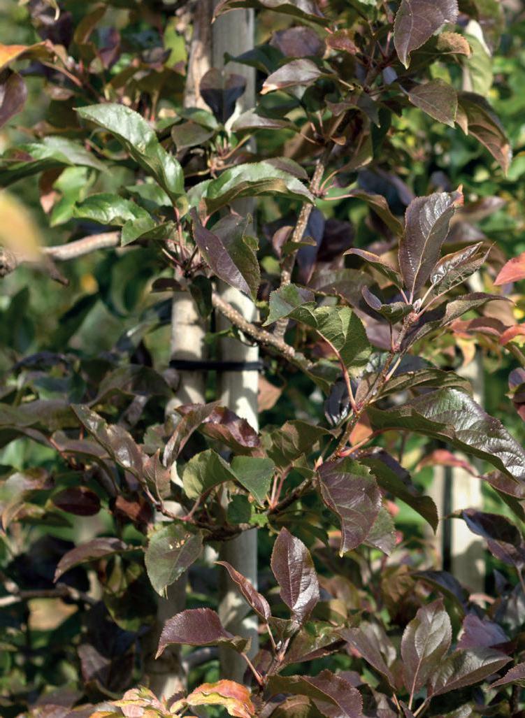
ISBN 978-88-784-3054-9


