10 minute read
Feline Pruritus

by FVMA

F LINE PRURITUS
Amelia G. White, DVM, MS, DACVD
Advertisement
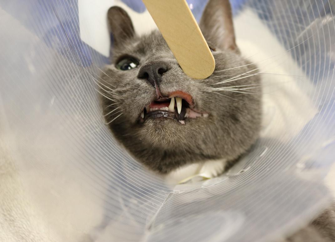
Prominent lesions of the eosinophilic granuloma complex. Rodent ulcer on the upper lip. Image courtesy of Dr. Amelia G. White.
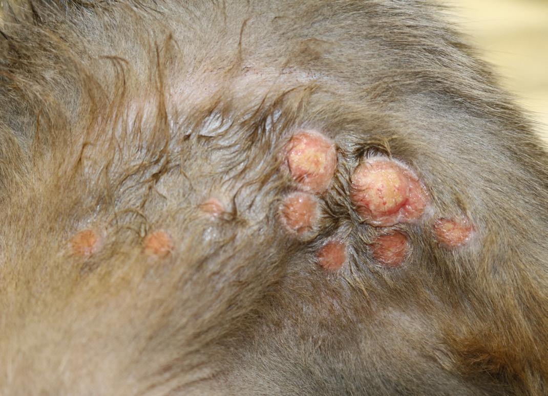
Prominent lesions of the eosinophilic granuloma complex. Multiple eosinophilic granulomas on the medial pelvic limb. Image courtesy of Dr. Amelia G. White.
INTRODUCTION
Allergies in cats are one of the most challenging and frustrating medical conditions we deal with as veterinarians. This is largely due to the severity of dermatological lesions, significantly decreased quality of life, limited medical therapy options and difficulty administering medications to cats. Cats tend to develop significant psychogenic components to pruritic behaviors that need to be addressed in order to maintain adequate control of the clinical signs.
Types of Allergy in Cats
There are several types of allergies in cats and the most common include flea bite hypersensitivity, mosquito bite hypersensitivity, atopic dermatitis and adverse food reaction (food allergy). Each of these conditions creates significant pruritus and dermatitis that can lead to self-mutilation and severe secondary bacterial and fungal skin infections. These allergies are not curable, but, with a thorough understanding of which allergies are problematic for the cat, they can be well-managed long term.
The Eosinophilic Granuloma Complex/ Syndrome
Cats are unique in that they tend to develop similar dermatological lesions despite a differing causative allergy. In general, cats tend to develop lesions infiltrated with eosinophils. Lesions are highly pruritic to the point of self-mutilation. The classic lesions that make up this complex are: 1) miliary dermatitis (tiny millimeter sized crusted papules), 2) eosinophilic granulomas (mouth, lateral pelvic limbs or interdigital), 3) rodent/indolent ulcer (eosinophilic ulcer on upper maxillary lip margin) and 4) eosinophilic plaque (ventral abdomen or dorsal head). Identifying these lesions on a cat is helpful in understanding that an allergy is most likely driving the dermatological abnormalities. Additional workup is required to determine which allergy or allergies play a role.
FLEA BITE HYPERSENSITIVITY
Pathogenesis
Flea bite hypersensitivity, or flea allergy, is the most common type of allergy in the cat (and in the dog). It occurs when the cat is sensitized to flea salivary proteins. While in dogs it has been demonstrated that intermittent flea exposure is associated with
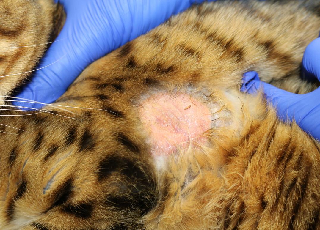
Prominent lesions of the eosinophilic granuloma complex. Large eosinophilic plaque on the lateral thorax. Image courtesy of Dr. Amelia G. White.
a higher risk of developing flea allergy, the opposite is true for cats. Commonly, cats with flea bite hypersensitivity have other concurrent allergies.
Clinical Signs
There is no age, breed or sex predilection for a flea allergy in the cat. Cats are extremely pruritic and develop miliary dermatitis, pustules, alopecia, erythema, and excoriations on the head, neck, caudal dorsum, abdomen, and distal extremities. Lesions of the eosinophilic granuloma complex develop overtime including eosinophilic granuloma, eosinophilic plaques and rodent ulcers.
Diagnosis
Cats receiving routine flea control can still display clinical signs consistent with flea allergy if a heavy flea burden exists within their environment. If lesions are distributed in the caudal half of the cat, this can be helpful in making a diagnosis when coupled with historical information. Finding fleas or flea dirt on the cat shows evidence of flea exposure but does not rule in/out the diagnosis of a flea allergy. Likewise, high serum IgE or positivity for intradermal flea antigen does not confirm the diagnosis of a flea allergy.
Other parasites and allergies, along with bacterial and fungal infections, must be excluded as these diseases cause identical dermatological lesions. Several diagnostics can help work through these differentials, including cytology to evaluate for bacterial and fungal infections, skin scrapings to evaluate for parasites, and bacterial and/or fungal cultures. Histopathology can support the diagnosis of an allergic etiology but is not a diagnostic for a flea allergy.
Treatment
Avoidance of flea bites is the most effective treatment for a flea allergy. This can be very challenging, especially in households with numerous animals or outdoor cats. If you suspect a flea allergy in a cat, all animals in the household (even if they are indoor-only animals) and the environment should be routinely treated for fleas. Typically, animals allergic to fleas will receive two forms of flea control, most commonly combining a topical agent with an oral agent. One of these agents should contain a repellent to discourage flea bites. It generally is best to hire a professional exterminator to treat the environment.
Anti-inflammatory and antimicrobial therapies may be required in some cats according to clinical and cytological findings. Glucocorticoids are fast and highly effective for flea-allergic cats. Remember that cats have fewer steroid receptors and generally require a slightly higher anti-inflammatory dose as compared to dogs. Oral prednisolone (2mg/kg/day) on a tapering schedule over a few weeks will provide short-term relief. Methylprednisolone
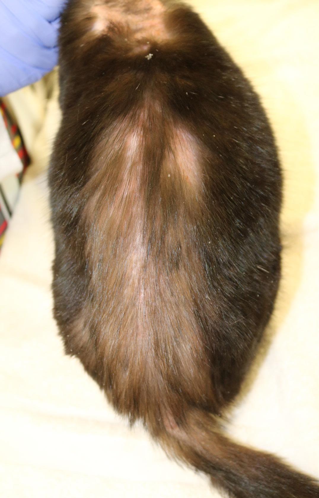
Well-demarcated, self-induced alopecia on the dorsum extending to the tail base in a cat with flea allergy dermatitis. Image courtesy of Dr. Amelia G. White.
Mild alopecia, erythema and edema of the dorsal muzzle in a cat with mosquito bite hypersensitivity. Similar lesions were present on the lip margins, chin, ear pinnae and paw pads. Image courtesy of Dr. Amelia G. White.

acetate (DepoMedrol) (10–20 mg per cat, once every 8–12 weeks) may be a better choice for cats that are difficult to medicate. Antihistamines and omega fatty acids will decrease inflammation in mildly affected cats, but it may not provide enough relief for most cats. Allergen-specific immunotherapy is not effective.
Prognosis
Overall, prognosis is good if the flea bite can be avoided. Oftentimes, this disease will wax and wane with changes in environmental flea burden, acquisition of new pets, boarding/ grooming, etc.
MOSQUITO BITE HYPERSENSITIVITY
Pathogenesis
Similar to flea bite hypersensitivity, mosquito bite hypersensitivity is an allergic response to a mosquito bite. It is thought to be a Type I reaction with most lesions developing within 20 minutes.
Clinical Signss There is no breed, age or sex predilection. This disease is variably pruritic. Lesions originate at the site of the mosquito bite most commonly on the head, pinna and bridge of the nose. Lesions may extend to include eyelids, footpads, chin and lips. Outdoor cats are more severely affected, but indoor cats can be affected especially in geographical locations with heavy mosquito populations (e.g. humid, temperate regions). Dermatological lesions begin as pruritus, wheals, papules and plaques. Over time, lesions become nodules, crusts, excoriations, alopecia and changes in pigmentation.
Diagnosis
Diagnosis is achieved through history, physical exam and ruling out other diseases. Other diseases to consider include other allergies, pyoderma, fungal dermatitis, demodicosis, Cryptococcus infection, ulcerative herpes virus dermatitis, pemphigus foliaceus and contact reaction. Histopathology does not provide a definitive diagnosis, but does support an allergic etiology. High serum IgE or positivity for intradermal mosquito antigen does not confirm the diagnosis of mosquito bite allergy.
Treatment
Confining cats to an indoor lifestyle, treating the environment for mosquitoes, removal of mosquito breeding sites (e.g. standing fresh water) and treating the cat with a flea preventative with repellent activity (e.g. Seresto® collar) will reduce the clinical signs. Clinical signs can wax and wane based on the environmental burden and time of year. Use of glucocorticoids as above for flea allergy is highly effective to control pruritus and dermatitis associated with mosquito bite hypersensitivity.
Prognosis
Prognosis is usually good if mosquito bites can be avoided and if cats adapt to the indoor lifestyle.
ATOPIC DERMATITIS
Pathogenesis
Atopic dermatitis is a hypersensitivity typically characterized by high levels of IgE to environmental allergens such as plant pollens, molds, insects and mites. Sensitization occurs primarily transcutaneously through recognition of environmental antigens via epidermal dendritic cells (e.g. Langerhans cells) and presentation of naïve T cells in nearby lymphoid tissue. Upon presentation, these naïve T cells skew towards a Th2 response and lead to overproduction of pro-inflammatory cytokines (e.g. IL-2, IL-4, IL-13, IL-31). Typical presentation of disease occurs in young adulthood, unless a cat has recently moved to a new geographic location.
Clinical Signs
The most common clinical signs are extreme pruritus originating on the head and neck and progressing throughout the body especially on the ventrum, lateral thorax and distal extremities. Miliary dermatitis, eosinophilic plaques/nodules, rodent ulcers, pustules, alopecia and excoriations.
Diagnosis
Much like the other feline allergies, atopic dermatitis is a diagnosis of exclusion. The diagnosis based on history, physical exam findings and ruling out other pruritic diseases. Other diseases to consider include other allergies, pyoderma, fungal infections and parasitic infections. Serum IgE concentrations and
intradermal reactivity are not diagnostic for atopic dermatitis. Histopathology does not provide a definitive diagnosis but does support an allergic etiology.
Treatment
Treatment is aimed at reducing inflammation since allergen avoidance is nearly impossible. Allergen-specific immunotherapy remains the gold standard and can be formulated from serological or intradermal test results. Intradermal testing in cats is very difficult because cats have less profound and long-lasting wheal and flare reactions as compared to dogs. For this reason, serology is frequently used to formulate immunotherapy in cats. Effective anti-inflammatory medications include glucocorticoids, antihistamines, omega fatty acids and cyclosporine (Atopica®). Apoquel® (oclacitinib) has been attempted off-label in cats but is not very effective unless dosed twice daily, and one study has suggested that methylprednisolone is more effective than Apoquel to manage allergic disease in cats. The safety and efficacy of oclacitinib is unknown in the cat. Cytopoint® is a monoclonal canine antibody and should not be used in the cat due to the possibility of a severe anaphylaxis in response to foreign canine antigens.
Prognosis
Prognosis is poor for a cure but good for long-term control. Refractory cases of atopic dermatitis can carry a poor prognosis if they fail to respond to traditional therapies. These cases are less common in occurrence and typically warrant referral to a veterinary dermatologist.
ADVERSE FOOD REACTION: FOOD ALLERGY
Pathogenesis
A food allergy is typically a hypersensitivity to one or more food ingredients; dietary proteins are most commonly implicated. Other ingredients less commonly implicated include cereals/ grains, dyes and preservatives. The mean age of onset in the cat is two-five years of age with Siamese cats overrepresented.
Clinical Signs
Pruritus is the most common clinical sign along with eosinophilic granuloma complex lesions, alopecia, excoriations and pustules. Gastrointestinal disturbance occurs in 10–15% of cases including vomiting, diarrhea, weight loss and picky appetites.
Diagnosis
The only effective diagnostic test is a dietary elimination trial. Typically, a novel protein or hydrolyzed veterinary-formulated diet is preferred and is fed exclusively for eight-12 weeks. Flavored medications, supplements, treats and other food additives must be avoided. Novel protein diets are less commonly used in patients with unknown dietary histories or cats that have been exposed to many diets already. Resolution of clinical signs is suggestive of the diagnosis, but a dietary challenge is required to confirm the diagnosis. Single ingredient challenges can be performed to better understand which food antigens are a problem so that an over-the-counter diet can be selected. Oftentimes, clients continue to feed the diagnostic diet for life in order to avoid recurrence of clinical signs. Most of the veterinary-formulated, diagnostic diets for food allergies are approved for long-term feeding.
Treatment
The avoidance of problematic food ingredients will prevent clinical signs. It can be difficult to prevent all clinical signs if owners are unable to maintain a restrictive diet (e.g. outdoor cats). Glucocorticoids or cyclosporine (Atopica®) are initially helpful in reducing inflammation and itchiness.
Prognosis
If food allergen avoidance is possible, prognosis is good.
Amelia White, DVM, MS, DACVD
Dr. Amelia White received her DVM from the University of Georgia College of Veterinary Medicine in 2010, followed by a one-year internship in small animal medicine and surgery at Auburn University. She was accepted to a dermatology residency at the University of Illinois at ChampaignUrbana the following year, where she completed three years of specialty training and a master’s degree in 2014. Dr. White completed her board certification exam in veterinary dermatology in fall 2014 and became a diplomate of the American College of Veterinary Dermatology. She currently is an associate clinical professor of dermatology at Auburn University College of Veterinary Medicine. She has authored or contributed to various abstracts, case reports, primary research and a book chapter in her short career. Dr. White has been a consultant for the Veterinary Information Network (VIN) since 2015. She teaches the communications skills course at Auburn University and enjoys opportunities to provide continuing education seminars to veterinary professionals. Her research interests include allergic skin and ear diseases, infectious diseases, and student mental health and well-being.


