13 minute read
Core Facilities
PROVIDE GATES CENTER MEMBERS WITH ACCESS TO STATE-OF-THE-ART EQUIPMENT, TECHNOLOGY AND EXPERTISE
In last year’s annual report, we introduced the new Organoid Core, to which Gates Center members will have discounted access beginning in 2021. Drs. Bruce Appel and Peter Dempsey, co-directors of this core, anticipate that the core facility will be fully operational by late spring 2021.
During 2020, the Gates Center leadership team began discussions to provide Gates Center members with discounted access to two additional core facilities on campus, the Genomics Core and the Human Immune Monitoring Shared Resource. Both these cores are well established; the Genomics Core was launched in 1999 as one of the original shared resources in the University of Colorado Cancer Center, and the Human Immune Monitoring Shared Resource (HIMSR), was created in 2016 as part of the Human Immunology Immunotherapy Initiative supported by the University of Colorado School of Medicine. The Gates Center is very pleased to be able to leverage existing infrastructure so that members can have subsidized access to existing cutting-edge equipment and technology beyond what is now available in other Gates Center subsidized cores. For example, the Genomics Core recently purchased Visium Spatial Gene Expression technology from 10X Genomics for its facility. Visium Spatial Gene Expression is a next-generation molecular technology for classifying tissue based on total mRNA. This technology will allow Gates Center members to map the whole transcriptome with morphological context to discover novel insights into normal development, disease pathology and clinical translational research. Similarly, the HIMSR leveraged resources from across campus to obtain one of five IonPath’s Multiple Ion Beam Imaging (MIBI) beta units in operation worldwide. Two commercial MIBI units have now been released, and the HIMSR unit received a commercial upgrade in January 2021. The MIBI allows single cell analysis in situ using antibodies tagged with isotopically pure metal reporters to image up to 100 target proteins or RNAs in fresh-frozen and fixed tissue, with a five-log dynamic range and 100 nanometer cellular resolution.
Gates Center Core Facilities were not “immune” from being impacted by the COVID-19 pandemic. To illustrate this, we have included some reflections from Lester Acosta, new manager of the Flow Cytometry Core:
My first official day as manager was March 15, 2020 — a Sunday. My first day on the job was March 16, 2020, and my first official act was closing the core for the pandemic.
We were able to re-open the lab in an abbreviated form to offer our services again on May 18, 2020. We used Zoom to interact with our customers, and we also started using TeamViewer software to allow remote access of our instruments by our trained selfrun clients. Self-run clients could operate instruments from any computer, even from home.
All Gates Center Core Facilities were forced to close on March 16, 2020 but were gradually allowed to open at reduced capacity by May 18, 2020.
Gates Center cores and shared cores include the following, and their descriptions are below:
CORE FACILITIES
Flow Cytometry Core (FCC)
Genomics Core (GC)
Human Immune Monitoring Core (HIMSR)
Histology (Morphology And Phenotyping) Core
Organoid Core (OTMSR)
Stem Cell Biobank And Disease Modeling Core (SCB&DM)
CORE DIRECTORS
Eric Clambey, Ph.D.
Bifeng Gao, Ph.D., MBA
Jill Slansky, Ph.D. Kim Jordan, Ph.D.
Igor Kogut, Ph.D
Peter Dempsey, Ph.D. Bruce Appel, Ph.D.
Ganna Bilousova, Ph.D Igor Kogut, Ph.D.
CORE LAB MANAGERS
Lester Acosta
Katrina Diener
N/A
Laura Hoaglin
Sean McGrath, Ph.D.
Michael Ferreyros
Christine Childs, Sr PRA, operating the Helios in the Flow Cytometry Core.

FLOW CYTOMETRY CORE
With the retirement of Karen Helm, the Flow Cytometry Core (FCC) is now being operated under new leadership, with Eric Clambey, Ph.D., continuing to serve as director and Lester Acosta as the new manager. The Flow Cytometry Core was established in 1999 as one of the original shared resources in the University of Colorado Cancer Center. Flow cytometry is an essential tool for stem cell research, allowing the examination of cells at the single-cell level by using cell surface, internal, and nuclear labels. The FCC has specialized equipment, which can rapidly isolate and collect unique types of cells. Specific cell types can be identified by cell surface antigen expression, cell cycle status, or by physiological properties of the cell that are unique amongst the general cell population. Often, these and other identification criteria can be combined simultaneously in a single experiment to yield a very powerful means of identifying and quantifying very specific cell populations. Historically, the FCC has only offered cell sorting done by staff during standard work hours. However, annual client surveys identified a growing need for cell sorting services that could be done by individual researchers, without staff assistance. Therefore, the FCC identified the Sony MA900 as a robust cell sorter, designed specifically with the intention to offer client-operated walkup cell sorting. The FCC applied for and received institutional funds, along with additional funds provided by the Cancer Center Support Grant and the Gates Center, to purchase the Sony MA900. To ensure consistent training of researchers, all users interested in this service receive formal training by FCC staff. Once training is completed, users are granted increasing independence to perform self-run cell sorting. With these procedures in place, the FCC now offers cell sorting services on a 24/7 basis. The response to this new service has been robust and extremely positive, especially during the pandemic since self-run clients could operate instruments from any computer – even from home. In the past 12 months, nine clients booked 37 sorting appointments for 63 billable hours.
Traditional flow cytometers use laser beams and fluorescent tags to identify the presence or absence of cell markers, however the number of easily identifiable labels is limited to 10 to 15 in conventional systems. In 2018, with funding provided by the Gates Center and other campus sources, the core purchased a mass cytometer, the CyTOF (Helios). The Helios uses rare-earth metal tags to identify up to 45 different markers on each cell.

183 Years of Combined Biomedical/Genomics Laboratory Experience

Bifeng Gao, Ph.D., MBA, Director Katrina Diener, B.S. Manager Monica Ransom, Ph.D., Experimental Consultation, New Protocol Development Brian Woessner, B.S., NGS, Single Cell Genomics Okyong Cho, M.S., Single Cell Genomics, Arrays An Doan, B.S., NGS, Spacial Genomics, Arrays Colin Larson, B.S., NGS Ted Shade, M.S., Bioinformatics Support Megan Zapel, B.S., NGS David Farrell, IT Support
GENOMICS CORE
Beginning in 2021, the Gates Center will offer members discounted access to cutting-edge next generation sequencing (NGS), single cell and spatial multi-omics and microarray technologies offered by the Genomics Core (GC). The GC was established in 1999 as one of the original shared resources in the University of Colorado Cancer Center under the direction of Mark Geraci, M.D., and Bifeng Gao, Ph.D., MBA, and has since provided nationally and internationally utilized next generation sequencing (NGS) and microarray research support and services. The GC initially offered microarray methodologies to investigate changes in gene expression. As microarray-based technology advanced, the GC quickly adapted by offering a broader range of microarray capacity including gene expression, genotyping, cytogenomics, and methylation analysis. In 2010, the GC gained its first NGS platform, Illumina HiSeq 2000, and continued to expand its services and expertise in NGS methodologies. In 2015, Dr. Geraci relocated to Indiana University to become the Chair of the Department of Medicine. With his departure, Dr. Gao assumed the directorship of the GC and has been instrumental in growing and developing the GC over the past 20 years. Since 2015, the GC has secured institutional support to upgrade its NGS technology and advance its genomic analysis to the single cell level, including spatial gene expression and single cell immune profiling. The GC also expanded its services and expertise in single-cell multi-omics solution that simultaneously detects SNV, CNV, and protein data from the same cell, providing Gates Center members with a platform to reveal biomarkers that help stratify patients, signal resistance, and predict relapse.
Recently, the GC purchased Visium Spatial Gene Expression technology from 10X Genomics for its facility. Visium Spatial Gene Expression is a next-generation molecular technology for classifying tissue based on total mRNA. This technology will allow Gates Center members to map the whole transcriptome with morphological context to discover novel insights into normal development, disease pathology, and clinical translational research.
HUMAN IMMUNE MONITORING CORE

Beginning in 2021, the Gates Center will offer members discounted access to the Human Immune Monitoring Shared Resource (HIMSR), which was established in 2016 as part of the Human Immunology Immunotherapy Initiative supported by the University of Colorado School of Medicine. The HIMSR is operated under the leadership of Director Jill Slansky, Ph.D., and Assistant Director Kim Jordan, Ph.D. The HIMSR leveraged resources from across campus to obtain one of five IonPath’s Multiple Ion Beam Imaging (MIBI) beta units in operation worldwide. Two commercial MIBI units have now been released, and the HIMSR unit received a commercial upgrade in January 2021. The MIBI allows single cell analysis in situ using antibodies tagged with isotopically pure metal reporters to image up to 100 target proteins or RNAs in freshfrozen and fixed tissue with a five-log dynamic range, and 100 nanometer cellular resolution. The HIMSR also operates state-of-the-art multiplex imaging instruments to visualize and quantify tissue microenvironments in human, humanized mouse, and mouse model tissues on the Vectra 3 and Vectra Polaris multispectral fluorescence imaging systems. The HIMSR has extended the capability of these detection systems beyond the standard commercially-available reagents to include goat, rat, biotinylated and humanized antibodies. Further, the HIMSR has developed novel assays for co-staining with RNA probes and proteins on the same tissue slide. The HIMSR’s extensive IHC experience with the Vectra platform has also translated into new innovations on the MIBI platform, in which up to 27 antibodies have thus far been successfully imaged on a single slide. The HIMSR offers a standardized panel of 34 antibody targets for human tissue, with six metal channels available for customization for Gates Center members. The HIMSR offers full-service metal isotope antibody conjugations, titrations, and optimization of new antibody targets.

The HIMSR has also made many advancements in image analysis. It has developed novel scripts that pair with commercially available imaging analysis software for handling large whole-slide imaging datasets generated on the Vectra platform. Lacking commercially available software
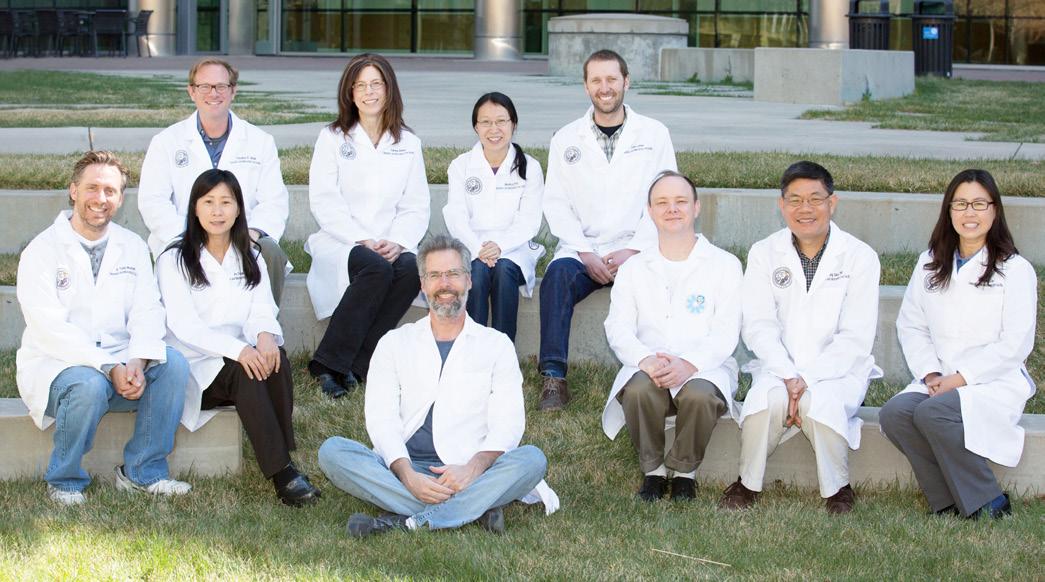
The Human Immune Monitoring Shared Resource Team
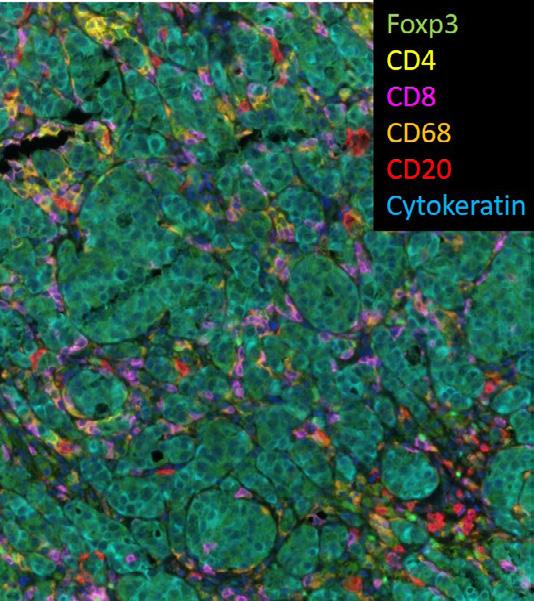
Multiple Ion Beam Imaging (MIBI) imaging of immune cells detected by antibodies specific for Foxp3 (Regulatory T Cells), CD4 (CD4 T Cells), CD8 (CD8 T Cells), CD68 (Macrophages) and CD20 (B Cells) infiltrating a metastases of metastatic breast cancer detected by Cytokeratin antibodies (specific for epithelial tumor cells).
for the analysis of MIBI images, the HIMSR has additionally developed a novel workflow for cell segmentation and high parameter phenotyping algorithms to quantify cells in complex tumor microenvironments. Once MIBI data is converted to single cell data, it is compatible with the high parameter flow cytometry analysis software offered by the FCC for the analysis of CyTOF data. The HIMSR will continue to innovate and improve analysis solutions for MIBI data, including development of tools for spatial analysis, scoring, and tissue region comparisons.
HISTOLOGY (MORPHOLOGY AND PHENOTYPING) CORE
The Histology Core is now operated under new leadership with Igor Kogut, Ph.D., serving as director and a certified technician, Laura Hoaglin, as core manager. The core provides a full set of histology services including the following: • Paraffin and OCT embedding • Sectioning of frozen and paraffin blocks • Routine (H&E) and special staining for all types of tissues • Consultation to optimize tissue isolation and fixation procedures
In 2020, the Histology Core updated its automated equipment, which greatly improved the turnaround time and quality of provided services. Due to high demand, the core will enhance its service portfolio in 2021 by including immunohistochemistry staining.
Laura Hoaglin operating the microtome in the Histology (Morphology and Phenotyping) Core. (Photo courtesy of Nicole Diette)

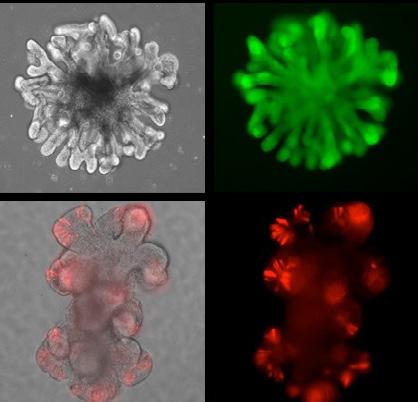
Intestinal Organoids Generated in the Organoid Core
ORGANOID CORE
Organoid and Tissue Modeling Shared Resource (OTMSR) is directed by Dr. Peter Dempsey and Dr. Bruce Appel from the Section of Developmental Biology in the Department of Pediatrics on the Anschutz Medical campus. The newly established OTMSR core facilities are located in research space on the third floor of the Barbara Davis Center for Diabetes, and is anticipated to be fully operational by late spring 2021. OTMSR looks forward to providing Gates Center members with discounted access to a variety of organoid technologies and to synergize with other core facilities and services provided by the Gates Center. The Organoid Core will provide the following: 1) access and training in current and emerging organoid technologies for in vitro disease modeling; 2) access, training and implementation of genome editing technology and gene expression systems; 3) critical key quality control and assurance measures to assure the validation and authenticity of core-derived resources to rigorously meet and exceed emerging NIH guidelines; and 4) data sharing of all Organoid Core-related resources, protocols and technologies to Gates Center members and the Anschutz Medical Campus community. The overall mission of the Organoid Core is to facilitate access, generation and usage of novel mouse and human organoid in vitro models to promote innovative basic and translational research. In January 2021, Sean McGrath, Ph.D., was recruited as the new OTMSR Lab Manager, and he is joined by PRAs Monica Brown and Claire Levitt, who are responsible for dayto-day operations.

The Stem Cell Biobank and Disease Modeling Core was established in 2017 on the basis of the development of a more efficient approach for reprogramming a patient’s diseased skin cells into stem cells by a team of scientists at the Gates Center, Ganna Bilousova, Ph.D., associate professor of dermatology, Igor Kogut, Ph.D., assistant professor of dermatology, and Gates Center Director Dennis Roop, Ph.D. The process, which was described in a paper published in Nature Communications in February 2018, reports a clinically safe approach that consistently reprograms healthy and disease-associated patients’ skin cells into induced pluripotent stem cells (iPSCs) with an unprecedented efficiency.
This core is co-directed by Drs. Bilousova and Kogut and offers complete services related to the production of high-quality human iPSCs from patient-derived somatic cells at a low cost. The core can reprogram multiple cell types, including dermal fibroblasts, urine-derived epithelial cells, freshly isolated and previously frozen peripheral blood mononuclear cells, etc. In addition to reprogramming services, the core provides genome engineering services using CRISPR/Cas to modify genes of interest in human iPSCs including the following: • The development of iPSC-based lineage tracing models by the introduction of gene-specific fluorescent reporters • The correction and introduction of disease-associated mutations in human iPSCs • The generation of isogenic pairs of genetically corrected and unmodified iPSCs by simultaneous reprogramming and gene editing of patient’s somatic cells • The production of custom-made modified mRNAs encoding a variety of factors for transient transfection into cells
This core continues to provide services for numerous clients on the Anschutz Medical Campus and at CU Boulder, as well as for national and international external clients. Additionally, the core works on several projects that have been initiated and generously underwritten by community benefactors. These include using iPSCs to determine the underlying causes and specific treatments of neurogenetic diseases such as epilepsy funded by Rick and Janie Stoddard, as well as using iPSCs and gene editing approaches to identify novel mutations that cause Ehlers-Danlos Syndrome funded by support from The Sprout Foundation, a Denver-area foundation funded by Suzanne and Bob Fanch, and Annalee and Wag Schorr.


