
3 minute read
Map in dairy goat colostrum and milk


2 Department of Bacteriology and Epidemiology, Wageningen Bioveterinary Research, Lelystad, The Netherlands
3 Department of Farm Animal Health, Faculty of Veterinary Medicine, Utrecht University, The Netherlands
Objective
Mycobacterium avium subsp. paratuberculosis (Map) infection is estimated to be present in 78% of Dutch dairy goat herds. To reduce transmission, it is advocated to snatch kids at birth and feed them cow- and/or artificial colostrum. This research evaluated the potential risk of colostrum and milk feeds to goat kids within Map test-positive dairy goat herds.
Materials and methods

Convenient colostrum (117 goats, six herds) and milk samples (202 goats, one herd), as well as colostrum (22 goats) from herds and/or individuals with clinical paratuberculosis were tested. Furthermore twenty nine clinically Map infected cull goats (from three herds) were post-mortemed and bilateral milk as well as tissue sampled. Tissues pools consisted of ilium, jejunum, ileocecal valve, mesenteric lymph nodes as well as bilateral mammary tissue and mammary lymph nodes. All milk and colostrum samples were tested by ELISA (ID Vet) and PCR (IS900). Tissue was tested by PCR.
Results
Of the 202 milk samples 47% (95% confidence interval (CI): 40-55%) were antibody positive, but only 4% ( 95% CI: 2-8%) of the samples were PCR positive. A large proportion of the colostrum from all six herds was positive for map-antibodies in the ELISA (95%, 95% CI: 90-98%), although the antibody levels differed depending on herd vaccination with the Gudair® vaccine. As shown in figure 1a here were significant differences between three out of the four fully vaccinating (A p<0.0001, B p<0.01, and D p<0,05) and non-vaccinating herds (Cnv), meanwhile not with the fifth (E). One herd had some purchased vaccinated goats within their unvaccinated herd (F), and antibody levels did not differ significantly with the non-vaccinating herd, but did with the others (A p<0.0001, B p<0.0001, D p<0.001 and E p<0,01). There was no difference depending on time post vaccination, being less than one year age, between one and two years, or more than two years ago (figure 1b). Even so there was no significant difference in antibodies depending on age at vaccination (as kids <6 months of age, or as young stock >6 months of age) (figure 1c). In the initial 117 colostrum samples there was no Map signal (0%, 95% CI: 0-3%), but two of the 22 colostrum samples from goats with clinical paratuberculosis were PCR-positive (9%, 95% CI: 1-29% (figure 2)). There was no oneon-one match between antibody and PCR positives. Results from sacrificed goats showed a PCR signal in 72% (95% CI: 53-87%) in the intestine pools and 83% (95% CI: 64-94%) in the mesenteric lymph nodes. Mammary lymph nodes and mammary tissue had a PCR signal in 34% (95% CI: 18-54%) and 45% (95% CI: 26-64%) of the goats, respectively (figure 3). Of those mammary tissue PCR positives, only 15% were PCR positive in their milk plus three doubtful PCR (Ct>36) signals in milk from both udder halves.
Discussion and conclusion
Figure 3. Map in tissues (pools of ilium, jejunum, ileocecal valve, mesenteric lymph nodes), as well as bilateral mammary tissue and mammary lymph nodes and milk samples (PCR (IS900)) from dairy goats with clinical paratuberculosis
1a. Comparison of paratuberculosis antibodies (ID Vet MAP ELISA) in colostrum samples from six commercial dairy goat farms (A, B, D, E Gudair® vaccinating, C non vaccinating, and F partially vaccinating. Total n=121). *Dunn’s multiple comparisons test p= <0,0001 - <0,05.

1b. Comparison of paratuberculosis antibody (ID Vet MAP ELISA) in colostrum samples from four Gudiar® vaccinating commercial dairy goat farms. Young stock, between the first and second year of life, and older goats.
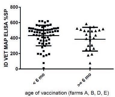
1C. Comparison of paratuberculosis antibody (ID Vet MAP ELISA) in colostrum samples from four Gudiar® vaccinating commercial dairy goat farms. Vaccinated as kids younger than six month of age, and older than six months of age.
In milk and colostrum samples low numbers are PCR positive. The antibody responses in a high proportion of milk and colostrum samples suggested that the selected goats had been in contact with Map and/or been vaccinated. It was unknown if they actually carried Map. Despite vaccination most of the sacrificed goats had one or more PCR positive tissue samples. The potential risk for Map transmission of Map via milk and colostrum seems to be low.

Acknowledgement contact: k.lievaart-peterson@gdanimalhealth.com
This work has been made possible by the Public-Private Partnership (PPS) Small ruminants Paratuberculosis in the milking goat industry a cooperation between the Ministry of Economic Affairs and Goat Dairy Produce Organisation & Dairy Goat Industry Platform. The authors would like to thank all the farmers that participated in this study.
