12 minute read
Featured Research
Summer 2020 Vol. 27, No. 2
CONTENTS
Featured Research Invasion Patterns of External Auditory Canal Squamous Cell Carcinoma:
A Histopathology Study....................1
Liquid Biopsy of the Human Inner Ear and Interpretation of Novel Molecular Biomarkers in Patients with Vestibular Schwannomas: Measuring Mediators of Hearing Loss and Predicting
Surgical Outcomes.............................6
Registry News Otopathology Mini-Travel
Fellowship.......................................11
Order Form for Temporal Bone
Donation Brochures........................12
MISSION STATEMENT
The NIDCD National Temporal Bone, Hearing and Balance Pathology Resource Registry was established in 1992 by the National Institute on Deafness and Other Communication Disorders (NIDCD) of the National Institutes of Health (NIH) to continue and expand upon the former National Temporal Bone Banks (NTBB) Program. The Registry promotes research on hearing and balance disorders and serves as a resource for the public and scientific communities about research on the pathology of the human auditory and vestibular systems.
Omer J. Ungar 1 , Felipe Santos 2 , Joseph B. Nadol, Jr. 2 , Gilad Hrowitz 1 , Dan M. Fliss 1 , William C. Faquin 3 , Ophir Handzel 1
1 Departments of Otolaryngology Head and Neck Surgery and Maxillofacial Surgery, Tel-Aviv Sourasky Medical Center, Sackler School of Medicine. Tel-Aviv University, Tel-Aviv, Israel 2 Department of Otolaryngology, Massachusetts Eye and Ear, Boston, Massachusetts, U.S.A., and the Department of Otolaryngology and Harvard Medical School, Boston, Massachusetts, U.S.A. 3 Department of Pathology, Massachusetts Eye and Ear, Boston, Massachusetts, U.S.A., and the Department of Pathology Massachusetts General Hospital and Harvard Medical School, Boston, Massachusetts, U.S.A.
Temporal bone (TB) malignant neoplasms are rare, occurring in 1:5000-20,000 ear disorders 1 . These malignant tumors can be categorized as primary or secondary. Primary TB malignant tumors originate in the TB, most commonly in the external auditory canal (EAC). Secondary tumors arise from extra-TB tissue and invade it by means of local extension or metastatic spread. The most common advanced stage malignancy is squamous cell carcinoma (SCC) of the EAC 2 .
EAC SCC tends to expand through consistent patterns such as invasion of compact bone, along blood vessels, cranial nerves and areas of osseous weaknesses like sutures and along the TB air cells tracks.
The aim of this study is to examine the invasion patterns of advanced stage SCC of the EAC and to compare the histological findings to clinical data. To the best of our knowledge, this is the first analysis of a series of TBs of patients with primary SCC of the TB.
Nine TBs from 9 patients were included in the cohort. Patients were diagnosed from 1968 to 2010. All cases presented with T4 stage disease according to the modified Pittsburgh
THE
DIRECTORS
Joseph B. Nadol, Jr., MD M. Charles Liberman, PhD Alicia M. Quesnel, MD Felipe Santos, MD
COORDINATOR
Csilla Haburcakova, PhD
ADMINISTRATIVE STAFF
Kristen Kirk-Paladino
EDITOR
Medical: Felipe Santos, MD
DESIGNER
Garyfallia Pagonis
NIDCD National Temporal Bone, Hearing and Balance Pathology Resource Registry
Massachusetts Eye and Ear
243 Charles Street Boston, MA 02114
(800) 822-1327 Toll-Free Voice (617) 573-3711 Voice (617) 573-3838 Fax
Email: tbregistry@meei.harvard.edu Web: www.tbregistry.org
The reports in the Registry Newsletter are not peer reviewed. staging system 3 (Figure 1). The demographics and clinical presentation are found in table 1. The most common presenting symptom was hearing loss (7 patients), the majority of which was severe to profound (5 patients). The most common otoscopic finding was obstructing mass EAC (5 patients).
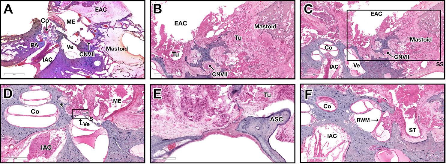
Several soft tissue elements within the TB were found to serve as barriers, limiting tumor invasion. The tympanic membrane was found to limit tumor extension medially in 4 patients. In these patients, the pathway of spread from the EAC to the middle ear (ME) was through the bony posterior EAC wall, to the mastoid air cells system (MACS) and the antrum. In five patients the tumor invaded the ME from the EAC directly, through the TM. The vestibulo-stapedial (annular) ligament was found to be a significant anatomic barrier for tumor spread from the middle to the inner ear. The resistance against tumor spread was so effective, that otic capsule invasion was seen adjacent to intact vestibulostapedial ligament. The round window membrane (RWM) was not invaded, although its niche was filled with tumor in 3 subjects. Examples of the relationship between these soft tissue barriers and tumor extension are shown in figure 2.
Several patterns of tumor spread were identified: besides serving as a route to the ME, the MACS was found to serve as a tumor conduit to the tegmen mastoideum and overlying dura, the posterior fossa dura, the vertical segment of the facial nerve, the ME and the lateral semicircular canal. The supra and infra labyrinthine pneumatization patterns allowed direct routes of tumor spread to the petrous apex (PA), leaving the otic capsule intact and most easily demonstrated in the vertical oriented histological preparations. The petromastoid canal served as an additional route to the PA via the subarcuate route (Figure 3). Trans labyrinthine PA invasion was seen in 2 patients. Once in the ME, tumors tended to spread to the anterior and posterior attic. We found no evidence that surgical modification of the TB anatomy created iatrogenic pathways for tumor spread. The ottic capsule itself was involved in 6 patients. The most common locus of otic capsule invasion was the cochlea, followed by the lateral semicircular canal (LSCC) and vestibule.
The tympanic segment of facial nerve was involved in 5 subjects and the vertical segment was involved in 3, resulting in varying degrees of facial nerve weakness. Wallerian degeneration was present distal to the proximal-most site of neural invasion (Figure 4).
Primary TB SCC is a rare, aggressive tumor, comprising 80% of primary TB malignancies 4, 5 . This tumor carries a substantial risk for morbidity and mortality because of its aggressive nature, location at the skull base and the common delay in diagnosis 6 . Early symptoms can be easily attributed to more common conditions such as otitis media and externa. This delay is likely to have a detrimental effect on treatment and outcome.
FIGURE 1: Malignant invasion throughout the temporal bone, indicating stage T4. (A-I) These H&E horizontal oriented histological preparations belong to patient #2 (A) Middle ear and petrous apex air cells are filled with the tumor. Stapes is partially subluxated. The otic capsule is invaded near the basal turn of the cochlea and internal auditory canal (asterisk). (B) High power magnification of figure 1A. The vestibular-stapedial (annular) ligament is intact, limiting tumor invasion to the vestibule (arrow). The tumor invades the otic capsule towards the cochlea and vestibule. (C) Low power magnification of the same TB. The tumor fills the external ear canal, mastoid and petrous apex. The posterior fossa dura over the mastoid is partially dehiscent. (D) The tumor invades the round window niche and the otic capsule overlying the posterior semicircular canal and cochlea (arrows). (E) Low power magnification of the petrous apex. The tumor encases the carotid canal, which is partially dehiscent. (F) High power magnification of figure 1E. The tumor invades the tunica externa of the petrous segment of the carotid artery. (G) Petrous apex air cells and internal auditory canal are filled with tumor. (H) High power magnification of figure 1G. The internal auditory canal is partially invaded by the tumor, as well as the posterior fossa dura. (I) Pure tone audiogram of the same patient showing profound conductive hearing loss, with reduced discrimination.

FIGURE 2: Soft tissue barriers limit SCC spread throughout the TB. (A) H&E horizontal oriented histological preparations of patient #1. This patient was treated with high dose of XRT (120 Gy) to the TB, replacing the SCC by fibrotic mass. EAC is filled with fibrotic mass. The TM is intact, effectively preventing medial invasion. Mastoid is invaded through the EAC posterior wall. The facial nerve is intact. (B-F) H&E preparation from the temporal bone described in figure 1, belong to patient #2 (B) The mastoid is invaded by the tumor, totally destroying the air cells system. (C) Low power magnification of figure 2B. The dura is invaded, as well as the otic capsule around the vestibule, which is partially dehiscent. (D) The vestibular-stapedial ligament limits medial tumor extension to the vestibule. The tumor invades the otic capsule around the basal and medium cochlear turns (asterisk). (E) High power magnification of figure 2D. (F) The tumor fills the round window niche. The round window is intact, preventing tumor extension to the inner ear spaces.

FIGURE 3: Hematoxylin and eosin horizontally oriented histological preparations of patient #2 throughout this article. Tumor extension to the petrous apex between the superior semicircular canal crura is seen, in the subarcuate route.

KEY for Figures 1–4
(Co) Cochlea, (ME) Middle Ear, (Ve) Vestibule, (IAC) Internal Auditory Canal, (PA) Petrous Apex, (Ma) Mastoid, (PSCC) Posterior Semicircular Canal, (SS) Sigmoid Sinus, (PFD) Posterior Fossa Dura, (CNVII) 7th Cranial Nerve, (S) Stapes, (Tu) Tumor, (EAC) External Auditory Canal, (ST) Sinus Tympani, (RWM) Round Window Membrane, (Ty) Tympanic Segment, (LSCC) Lateral Semicircular Canal, (A) Ampulary End, (NA) Non Ampulary End, (SSCC) Superior Semicircular Canal, (SMF) Stylomastoid Foramen, (GSPN) Greater Superficial Petrosal Nerve.
Symptoms can point to TB sites with tumor spread. The triad of pain, bleeding and otorrhea is the classical presentation of TB cancer 7 . Obstructing mass in the EAC is probably the most specific sign, especially when combined with facial nerve palsy.
Several anatomical routes and barriers for tumor spread were identified. Routes of cancer spread through the TB are highly dependent on TB pneumatization. Most of the published literature relates to cholesteatoma patterns of spread. However, there are several major differences between SCC and cholesteatoma, including cancer propensities to spread along nerves and the site of origin of the disease. Cholesteatoma originates from the TM (except when congenital, iatrogenic and blast-induced), whereas SCC may originate anywhere in the EAC 8,9 . From the EAC tumor approaches the ME via the TM, whether intact or perforated, or by means of invasion the MACS through the posterior EAC wall. The intact TM was found to serve as a reliable tumor expansion barrier. In several specimens, it was impossible to determine if the tumor invaded and perforated a previously intact TM, or via a previously perforated TM. Another soft tissue barrier was found to be the vestibulo-stapedial ligament (VSL). An intact VSL was seen adjacent to otic capsule (promontory) invasion. The RWM was the second soft tissue barrier for inner ear cavity penetration.
The clinical importance of these anatomical barriers is the difference in surgical extent indicated in case of intact versus invaded barriers. Tumor confined to the EAC with some bone erosion (T1-T2) can be treated effectively with, a lateral TB resection (LTBR) 10 . In these cases LTBR with free pathological margins can save a patient from the need to be irradiated. Historically extensive en-bloc surgery (subtotal and total temporal bone resections) have been offered to patients with more advanced stage disease. However, en-bloc resection has not proven to have superior results as compared to procedures involving piecemeal dissection. These advanced stage disease ears usually requires the addition of radiation therapy. Once the TM is invaded and tumor extends to the ME, LTBR may be considered and subtotal TB resection (STTBR) or extended

FIGURE 4: Facial nerve invasion. (A+B) Hematoxylin and eosin horizontally oriented histological preparations belong to patient #2. The meatal segment of the facial nerve is invaded by the tumor, distal valerian degeneration is seen. (C) Schematic drawing of the facial nerve of the same patient. The perineural invasion occurred in the internal auditory canal (black), resulting in distal Wallerian degeneration (gray).
canal wall down tympano-mastoidectomy may be used as a more radical alternative, with similar cure rates 11,12 . While inner ear is involved, through otic capsule invasion or through one of the inner ear windows, STTBR is indicated. Total TB resection (TTBR) is rarely indicated or performed nowadays. Defining the typical routes of SCC spread in the TB can help plan surgery, and direct efforts to TB subsites involved and anticipate possible location of disease not depicted by pre-operative imaging.
The neurotrophic nature of SCC can make the FN an additional route for tumor spread. The vertical (mastoid) segment is the most commonly involved followed by the tympanic segment. l
Take home messages:
• TB histopathology can elucidate the extension routes of primary SCC carcinoma. • The TM may serve as an incomplete barrier for tumor extension from the EAC to the ME. • SCC does not tend to extended from the ME to the inner ear through the round window and vestibule-stapedial ligament. • Tumors do tend to spread along the pre-existing TB air-tract routes. • Well aerated TB may facilitate easier extension to the petrous apex.
REFERENCES
1. Lewis, J. S. (1983). Surgical management of tumors of the middle ear and mastoid. The Journal of Laryngology & Otology, 97(4), 299-312.
2. Koriwchak, M., 1993. Temporal bone cancer. The American Journal of Otology, 14(6), pp.623-626. 3. Moody, S.A., Hirsch, B.E. and Myers, E.N., 2000. Squamous cell carcinoma of the external auditory canal: an evaluation of a staging system. Otology & Neurotology, 21(4), pp.582-588.
4. Morton, R.P., Stell, P.M. and Derrick, P.P., 1984. Epidemiology of cancer of the middle ear cleft. Cancer, 53(7), pp.1612-1617.
5. Kuhel, W.I., Hume, C.R. and Selesnick, S.H., 1996. Cancer of the external auditory canal and temporal bone. Otolaryngologic Clinics of North America, 29(5), pp.827-852.
6. Zhang, T., Dai, C. and Wang, Z., 2013. The misdiagnosis of external auditory canal carcinoma. European Archives of Oto-rhino-laryngology, 270(5), pp.1607-1613
7. Moffat DA, Wagstaff SA, Hardy DG. The outcome of radical surgery and postoperative radiotherapy for squamous carcinoma of the temporal bone. Laryngoscope. 2005 Feb;115(2):341-7. PubMed PMID: 15689763.
8. Ouaz, K., Robier, A., Lescanne, E., Bobillier, C., Moriniere, S. and Bakhos, D., 2013. Cancer of the external auditory canal. European Annals of Otorhinolaryngology, Head and Neck Diseases, 130(4), pp.175-182.
9. Breau, R.L., Gardner, E.K. and Dornhoffer, J.L., 2002. Cancer of the external auditory canal and temporal bone. Current Oncology Reports, 4(1), pp.76-80.
10. Pensak, M.L., Gleich, L.L., Gluckman, J.L. and Shumrick, K.A., 1996. Temporal bone carcinoma: contemporary perspectives in the skull base surgical era. The Laryngoscope, 106(10), pp.1234-1237.
11. Moffat, D.A., Grey, P., Ballagh, R.H. and Hardy, D.G., 1997. Extended temporal bone resection for squamous cell carcinoma. Otolaryngology–Head and Neck Surgery, 116(6), pp.617-623.
12. Prasad, S. and Janecka, I.P., 1994. Efficacy of surgical treatments for squamous cell carcinoma of the temporal bone: a literature review. Otolaryngology—Head and Neck Surgery, 110(3), pp.270-280.
ACKNOWLEDGEMENTS
We are grateful to Meng Yu Zhu, Barbara Burgess, Diane Jones, and Jennifer T. O’Malley for technical assistance. This work was supported by NIH-NIDCD (U24DC013983-01)


