3 minute read
A ‘martian’ solution for a mineral problem: chalcocite coating on pyrite


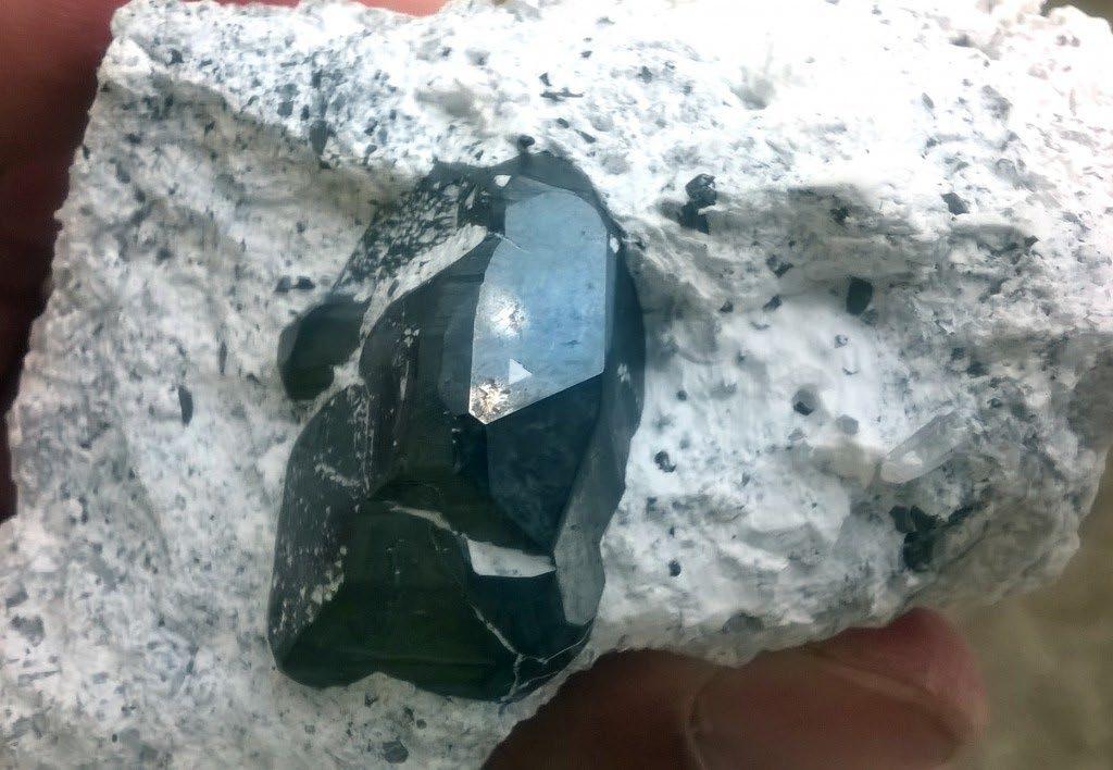
Our friends from Fabre Minerals a sked us an interesting mineralogical question: what is the species present in the beautiful specimens of copper sulfide - coated pyrite from Milpillas mine (Mexico)?
Initial analyses performed by other researchers, using SEM- EDS, suggested that the specimens are bornite (copper-iron sulfide)-coated pyrite, but the optical microscopy study of a polished section confirmed that the pyrite is coated by chalcocite (copper sulfide). This is mineralogically consistent, as chalcocite coatings are relatively common, protecting pyrite in secondary enrichment zones. The reflected light optical microscopy the right tool for the species identification in this case. The microscopy require sample preparation and this is a potential problem if all specimens (as the one depicted above) are expensive and should be preserved. Is possible, then, to observe the elemental composition in a non-destructive way?.
Advertisement
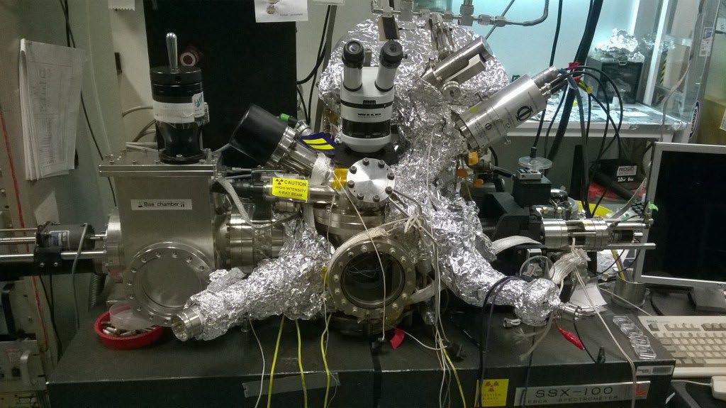
The problem with coatings is the penetration depth of the excitation radiation used in the spectrometric analysis. The incident radiation used in EDS (electrons) or XRF (Xray) reach the inner pyrite, with emission of the iron characteristic lines. Hence, iron appears as an artifact in the analysis and its presence in the spectrum of the coating do not imply the presence of Fe in the material. If a precise and specific analysis of the coating mineral is needed, XPS (X-ray photoelectron spectroscopy) should be used; in XPS, the photoelectrons emitted by the surface of material irradiated by a focused X-ray beam, allow the identification of elements and precise determination of the composition of material even with information of oxidation states. The disadvantages are that is an expensive technique available only in advanced research laboratories and requires sample preparation.
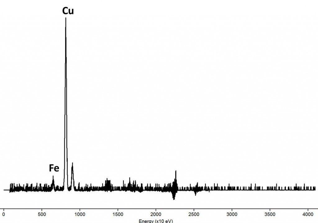
If we use APXS (alpha particle induced X-ray spectroscopy), a technique designed for the Mars exploration rovers, we can turn the disadvantage into an useful tool for the thickness estimation of the coating and to infer its composition. We irradiated the above sample with 5.86 MeV alpha particles and obtained this spectrum:

Using the Bragg - Kleeman rule for the estimation of penetration, we get that the coating should have a thickness lower than 29 µm. The alpha particles travel through the copper sulfide coating and transfer its energy to the iron sulfide beneath the thin layer, which fluoresces, showing the iron K lines and the characteristic spectrum of pyrite. Part of the alpha particles also exc ites the copper coating, showing the copper K lines in the spectrum. Also, the Cu X-ray could excite the X-ray fluorescence of iron, so iron will be always present.
What happens if we use less energy in the irradiation? we expect, then, an increase in the copper emission if the particles are stopped by the coating. To test this, we can use an outside the box complex trick: recently purified uranium-238 as excitation source. The U-238 is a weak, low energy alpha emitter. We made a source of u ranium oxide purified few days before the analysis, because one year after purification, the thorium-234 and protactinium-234 formed in the uranium decay chain enters in secular equilibrium; in this case, the beta emission of Pa - 234 overwhelms the alpha emission of U-238. The problem using U-238 is its very low activity (people usually fear the radioactivity of uranium, but actually is a very weak radioactive source), that requires a long time for analysis (24 hours of spectrum accumulation plus the same time to create a background spectrum, against a few minutes with our regular APXS system). After a long analysis and cleaning of the resulting spectrum, we showed this:
This result imply that the major element in coating is copper, with iron present as the expected artifact, coherent with the optical microscopy result. The calculations using the irradiation energy gave a thickness estimation of 10-15 µm for the chalcocite coating, again in coherence with the optical microscopy.
The showed experiments only suggests that we have an iron rich mineral coated by copper sulfide and possibly not by bornite. The easiest way for identification is by means of the petrographic or metallographic microscope and a polished section, although is difficult to differentiate chalcocite from blaubleinder covellite, covellite or djurleite. For a precise characterization, is necessary a surface characterization technique, as XPS or a type of XRF designed for coating analysis.
XPS system of the School of Chemistry at Georgia Tech (GA, USA). The ideal tool for exact quantitative analysis and surface characterizations.