
5 minute read
IMAGING Technology advances breast cancer detection


MRI technology advances breast cancer detection
BY AMI FELKER
DorothyStrauss breathed a sigh of relief when the lump removed from her left breast was found to be benign. She was only 19.
Over the next several years, she had three additional lumps removed — all benign. Later, in her mid-40s, the frequency of her masses increased. She had six to eight biopsies during that time, again all benign. But last September, Strauss received devastating news — doctors found a cancerous mass.
“It was a shock because I’d had so many benign masses,” said Strauss, now 52. “Because of my medical history, I had to undergo mammograms every six months. Having mammograms, ultrasounds and biopsies just became another one of my routines.”
Strauss’ September test was unlike her previous tests, and it may have saved her life. Rather than a conventional mammogram, she underwent a breast magnetic resonance imaging (MRI) test at the Methodist Breast Center.
“I have very fibrous breasts, and I’ve been told my mammograms are extremely difficult to read,” she said.
Dense breast tissue, which is made up of less fat and more glands and ligaments, appears as white areas on a mammogram. The problem is that tumors also show up as white areas on film.
“In many cases, dense breast tissue or breast implants obscure the standard mammogram, making it difficult to pick up small cancers,” said Dr. Luz A. Venta, medical director of the Methodist Breast Center.
“With an MRI, you get a 3-dimensional image of the breast, which allows a physician to see the detailed anatomy, down to the arteries and veins of the breast. It’s a more precise view of the breast tissue,” she said. MRI also differs from the standard mammogram in that a woman lies on her stomach instead of stands. There is less compression, which means less discomfort.
Venta and her team found a 9 mm mass (about the circumference of a AAA battery) in Strauss’ breast and after a biopsy, determined it was cancerous.
“I am so very thankful I underwent the MRI,” Strauss said. “With the density of my breasts, they probably would not have seen the mass on a mammogram. The MRI was a tool that worked and was very successful for me.”
Thankfully, Strauss’ tumor was small enough to remove without her undergoing radiation or chemotherapy. However, she did decide to undergo a bilateral mastectomy (removal of both breasts) and reconstruction.
She still undergoes regular screening because there is always a chance for another mass to show up in the chest cavity or remaining breast tissue, but she said, “I’m very blessed to have the experience I did.”
In addition to women with dense breast tissue, Venta says MRI also is recommended for women who have a higher risk for breast cancer. “It can distinguish the tumor from the tissue around it,” she said. “This technology can prevent a potentially devastating misdiagnosis.”
Who should have one
MRI is a relatively new tool for breast cancer. Most doctors only recommend it for women with a higher risk or a strong family history of breast cancer. In most cases, insurance will cover the cost of an MRI if the woman has dense breast tissue or is high risk.
Breast Cancer 5-Year Relative Survival Rates 1996-2003
88.6% 98.0%
83.5%
56.9%
20 26.7%
0 All stages Localized Regional Distant Unstaged
Source: SEER Cancer Statistics Review 1975-2004,National Cancer Institute Localized: cancer that is limited to the organ in which it began without evidence of spread Regional: cancer that has spread beyond the original (primary) site to nearby lymph nodes or organs and tissues Distant: cancer that has spread from the primary site to distant organs or distant lymph nodes Unstaged: cancer for which there is not enough information to indicate a stage
METHODISTHEALTH.COM
Dorothy Strauss underwent a breast MRI test at the Methodist
Breast Center. The test revealed a small cancerous mass. Because it was detected early, she avoided radiation and chemotherapy.

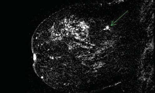
Mammogram and breast MRI of a 46-year--old woman. Her physical exam was normal. The mammogram (left) is normal, showing dense breast tissue and a benign (noncancerous) calcification. An MRI (right) of the same breast was obtained in the same week and shows a small cancer (arrow).
A three-year research study, conducted at 17 different sites throughout the United States, Canada and Germany, found that MRI was twice as effective as a regular mammogram in detecting tumors in more than 1,000 women with suspicious lesions on their mammograms.
“We have seen several high risk patients, some with implants and dense breast tissue, whose mammograms were normal, but MRI showed cancer,” Venta said. “For these women, we were able to start treatment more quickly and give them a better chance at survival.”
MRI’s ability to better pinpoint small cancers gives women a chance to explore treatment options other than mastectomy, including lumpectomy with radiation. “The goal is to save as many lives as possible, and this gives us another tool with which to do that,” Venta said.
As with most cancers, early detection is the key. “Not all mammograms or breast MRIs are created equal and not all medical institutions can detect and test masses with the same accuracy,” said Venta, adding, “women should do their homework. Make sure you can get your screening and biopsy at the same place so you’re not shifted from one place to the next.”
To learn more about the Methodist Breast Center, visit methodisthealth.com. To schedule an appointment or request a second opinion, call 713.441.PINK (7465).
Guidelines for the early detection of breast cancer in average-risk, asymptomatic women
AGES 40 AND OLDER
Annual mammogram Annual clinical breast examination
Monthly breast self-examination (optional)
AGES 20-39
Clinical breast examination every three years Monthly breast self-examination (optional)
Source: American Cancer Society: Breast Cancer Facts & Figures 2007-2008


