
7 minute read
HINDLIMB LAMENESS

by hqmagazine

FIVE COMMON CAUSES
Whilst forelimb lamenesses are relatively common findings in our horses, hindlimb lamenesses or issues are often less evident and, thus, underdiagnosed. This article looks at the most common causes of hindlimb lameness and how to diagnose and treat them.
Identifying a hindlimb lameness
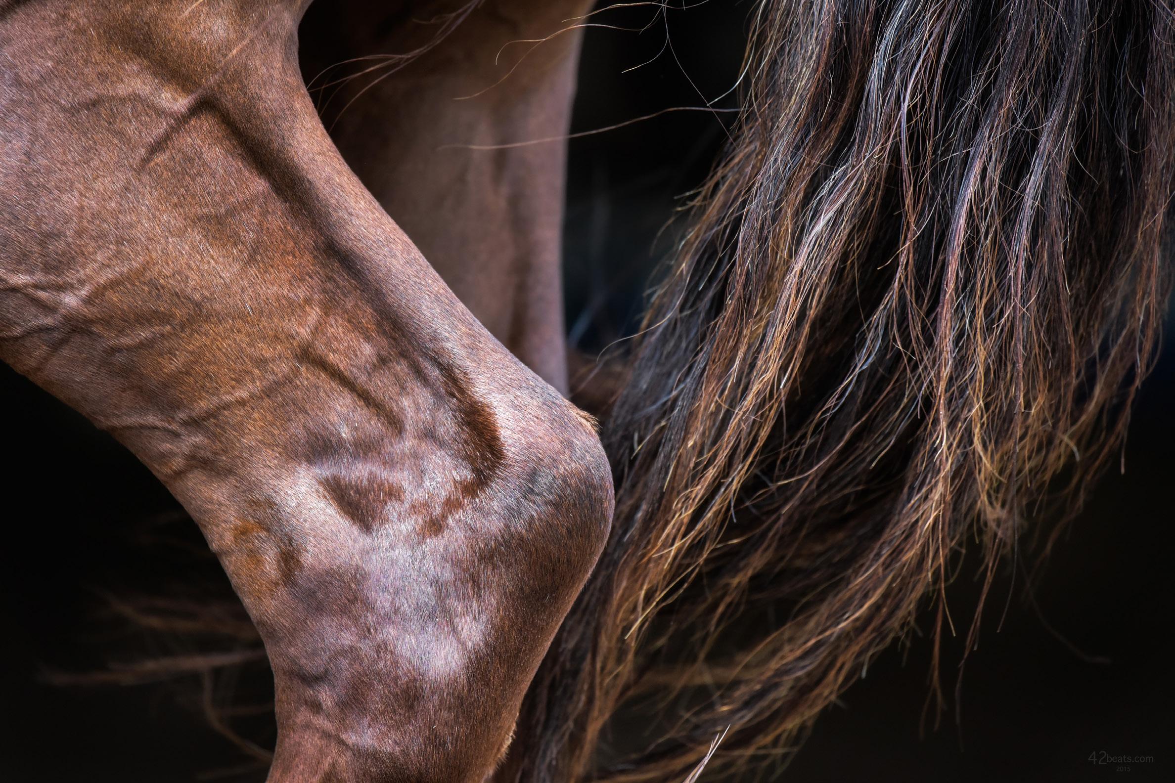
One of the first questions we need to answer is what exactly a hindlimb lameness looks like. Most owners can spot front limb lamenesses with the naked eye. An uneven gait, shorter steps or a head bob are telltale signs of forelimb lameness, but what are the indicators of a hindlimb issue?
Lameness in the hindlimb can present in a couple of ways. When doing a lameness work-up, a vet will typically need to incorporate both longeing on a circle and trotting in a straight line to determine precisely where the lameness is most apparent. Sometimes vets will ask to see the horse under saddle, as this can accentuate subtle issues that are less obvious in hand or on the longe. In all these scenarios, vets are looking out for excessive hip movements or 'hip hikes', an unwillingness to swing a leg forwards, toe dragging or a 'bunny hopping' canter. Yet, each presentation of hindlimb lameness is slightly different, and the compensatory patterns employed by the horse to alleviate the discomfort can confuse the picture.
DID YOU KNOW? With distal hock arthritis, it is common for both limbs to be affected, which means that clinical signs are often less obvious and tend to reveal themselves over time.
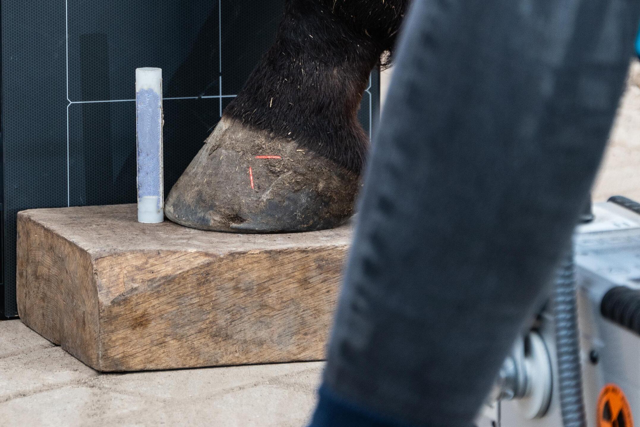
Imaging of the hindlimb
Most vets, before imaging, will perform nerve and joint blocking to isolate the lameness to a specific area of the leg. Once this has been done, X-Rays are generally the modality of choice for evaluating any bone or joint in this setting. However, X-Rays do not reveal many details of the soft tissue. Ultrasound is needed in these cases to evaluate tendons, ligaments or other soft tissue structures. More advanced imaging such as MRI and CT are only available at large veterinary hospitals, and whilst they can better pinpoint the issue and detect more subtle issues like proximal suspensory desmitis, navicular bursitis and sesamoidean ligament issues, they are out of the price range of most ordinary equestrians.
The most common findings
There are five conditions that most commonly cause hindlimb lameness, and we will examine these here in turn:
1. OSTEOARTHRITIS
Osteoarthritis (OA) is a prevalent equine condition. Whilst it's most common in older horses, osteoarthritis can have devastating effects on any equine athlete and dramatically affect performance. The condition starts with joint inflammation and progresses to arthritis when the cartilage becomes involved. It is termed osteoarthritis when the bone surrounding the joint is also involved. While OA can develop in any joint, it is a common cause of hock pain in horses. The hock is made up of four joints. The lowest two, the distal intertarsal and tarsometatarsal joints, are especially susceptible.
Thankfully, vets have lots of OA management and treatment options available to them. The least invasive products are neutraceuticals, better known as oral joint health supplements, containing glucosamine, hyaluronic acid (HA) and other compounds. Prescribed non-steroidal anti-inflammatories can also be helpful in the short term, and systemic injectable treatments, like Legend (intravenously administered HA), can help to restore synovial fluid to its original state. Adequan (intramuscularly administered polysulfated glycosaminoglycan) is incorporated more directly into cartilage, and in horses with multiple joints affected, Legend and Adequan can both be very effective. Outside of systemic options, hock injections can be performed. These are one of the most common joint injections vets perform, but not all injected drugs are created equal. Steroids are often used to reduce the inflammation in osteoarthritis cases and minimise damage to the cartilage, but newer joint treatments, such as platelet-rich-plasma (PRP) and IRAP (interleukin-1 receptor antagonist protein), help the body's own immune system to treat itself.
2. OSTEOCHONDROSIS DISSECANS
Osteochondrosis dissecans (OCD) is a disease predominantly affecting younger horses, although the exact cause is not fully understood. Owners will often notice it just as horses start under saddle. In OCD, the bone and cartilage in the joints don't form normally. Subsequently, the cartilage and bone underneath it become irregular in thickness and weaker than in normal joints. This can lead to the formation of cartilage and bone flaps that may remain partially attached to the bone or break off and float around in the joint. Loose flaps and abnormal cartilage and bone cause inflammation in the joint and, over time, may lead to the development of arthritis. OCD can occur in any joint but most often presents in the fetlocks, hocks and stifles. While owners can use conservative therapies such as oral medications and joint injections, surgery remains prognosis. However, large fragments or those sitting directly on weight-bearing surfaces are a little more complex to work with.
3. SACROILIAC JOINT DISEASE

Sacroiliac pain is increasingly being recognised as a cause of hindlimb discomfort, particularly in performance horses. Common signs of pain in this region include a reluctance to go forward, a lack of impulsion and an uncoordinated 'bunny hop' canter, among others.
Subluxation (misalignment) and osteoarthritis are the leading causes of discomfort in the sacroiliac joint. However, these issues can be exacerbated by more acute sources of pain, such as problems in the hock or stifle that force the sacroiliac joint to compensate.
Because the sacroiliac joint is beneath layers and layers of muscle, imaging can be difficult. Treatment options are also somewhat limited and include corticosteroid injections and chiropractic adjustments. If these therapies resolve the lameness (and any other sources of hindlimb lameness have been dealt with), it's important to maintain the horse's comfort with a good fitness regime that allows the gluteal muscles to develop and properly support this area of weakness.
4. UPWARD FIXATION OF THE PATELLA
Commonly known as 'locking stifle', upward fixation of the patella (UFP) is a relatively common condition in which a horse's hind leg gets stuck in extension. UFP can manifest in young horses in early training, but it can also be associated with straight hindlimb conformation or a lack of proper musculature in all ages of horses. It is caused by a horse's inability to release the stifle's passive stay apparatus (a network of muscles, tendons and ligaments that allows a horse to stand with minimal effort). Treatment options range from conservative to radical. The easiest treatment option involves alterations in your horse's training to increase the development of the quadriceps muscle. Increasing this muscle's strength will effectively 'tighten' the patella ligaments, thereby correcting the upward fixation of the patella in some horses. You can help strengthen the quadriceps by trotting for extended periods, doing hill work and working in deep footing.
If changes to the exercise regime fail, surgical options include medial patella ligament splitting, which involves making several small incisions in the medial patella ligament to cause the ligament to thicken and help the stifle to stabilise.
5. SOFT TISSUE INJURIES
Soft tissue strains and tears and resulting lameness are all too common. Vets frequently diagnose tendonitis, suspensory ligament injuries and degenerative suspensory ligament desmitis as causes of hindleg lameness. Once the degree of damage is established, you can decide what the horse can do going forward. Rest is the mainstay of treatment, but it will not fix the issue. Controlled exercise is important for this, as you need there to be function, so you want enough exercise to enhance the rehabilitation of the soft tissue structures.
Bringing a horse back from soft tissue injury involves returning him to work slowly, usually by hand walking. This must, however, be done carefully, as bringing a horse back to work too quickly after a soft tissue injury can be catastrophic. Once you overdo it, you start breaking down tendon or ligament fibres, and it can be difficult to get back on track.
Final thoughts
Hindlimb lameness can be a real issue for all horses. Whether the condition develops suddenly or insidiously, you stand the best chance of diagnosing it and getting the proper treatment with your veterinary team's guidance.


