
HSOA Journal of Clinical Studies and Medical Case Reports

Research Article
Utility of Scanning Electron Microscopy (SEM) for Suspected Microbial Keratoconjunctivitis
Unresponsive to BroadSpectrum Antibiotic Therapy
Mario Troisi1*, Salvatore Del Prete2, Salvatore Troisi3, Maria Vittoria Turco4, Vito Turco3, Antonio Del Prete1, Gravina Alessandro5, Gravina Antonio6 and Daniela Marasco2
1Ophthalmologic Unit, Department of Neurosciences, Reproductive and Dentistry sciences, University of Naples Federico II, 80131 Naples, Italy
2Service Biotech s.r.l., Naples, Italy
3Ophthalmologic Unit, Department of Head-Neck, Salerno Hospital University, 84100 Salerno, Italy
4Ophthalmology Unit, Department of Experimental Medicine, University of Rome “Tor Vergata”, Rome, Italy
5Department of Medicine and Surgery, Università Cattolica del Sacro Cuore, Rome, Italy
6Antonio Gravina Opthalmic Center, 81040 Curti, Italy
Abstract
The aim of this work is to evaluate the sensitivity and efficacy of a diagnostic approach based on scrapings of the superior tarsal conjunctiva examined by scanning electron microscopy (SEM). We examined 30 eyes of 26 consecutive patients with presumed microbial keratitis, who had no improvement after local broad-spectrum antibiotic therapy performed for at least 7 days at an effective dosage. 13 patients had negative culture tests for bacteria and fungi obtained from corneal curettage. After 72 hours of wash-out, scrapings of the upper tarsal conjunctiva for cytological examination of cell morphology by scanning electron microscope (SEM) were performed. With
*Corresponding author: Mario Troisi, Ophthalmologic Unit, Department of Neurosciences, Reproductive and Dentistry sciences, University of Naples Federico II, 80131 Naples, Italy, Email: troisi165@gmail.com
Citation: Troisi M, Del Prete S, Troisi S, Turco MV, Turco V, et al.(2023) Utility of Scanning Electron Microscopy (SEM) for Suspected Microbial Keratoconjunctivitis Unresponsive to Broad-Spectrum Antibiotic Therapy. J Clin Stud Med Case Rep 10:155.
Received: May 01, 2023; Accepted: May 16, 2023; Published: May 23, 2023
Copyright: © 2023 Troisi M, et al. This is an open-access article distributed under the terms of the Creative Commons Attribution License, which permits unrestricted use, distribution, and reproduction in any medium, provided the original author and source are credited.
this method, the presence of pathogenic microorganisms was detected in 26/30 eyes and specific therapy was carried out on the basis of these results. Clinical improvement and eradication of organisms previously detected by SEM were observed in all positive patients within 1-7 weeks. No organisms were detected in four patients; in two of them the presence of inflammatory cells indicative of an allergic reaction (eosinophils and mast cells) was detected, in the other two there were signs of severe dry eye.
The efficacy of this diagnostic technique is confirmed by the clinical response to treatment based on the microorganisms identified by SEM and by the eradication of previously detected microorganisms, verified by subsequent scanning electron microscopic (SEM) control examination. The results obtained support the validity of the proposed method and we believe that it constitutes a very useful tool for distinguishing, on the basis of the inflammatory infiltrate, the infectious forms from those of other nature and for detecting the presence of microorganisms resistant to the usual therapies.
Keywords: Microbial keratitis; Conjunctival scraping examination; Correlative microscopy; SEM conjunctival diagnosis; Corneal ulcer diagnosis; Corneal infection
Introduction
Infectious keratitis is an infection of the cornea caused by a wide range of microorganisms, including bacteria, fungi, virus, parasites and polymicrobial infection, which can cause serious visual impairment [1].Globally, it is the fifth leading cause of blindness, affecting about 6 million of the world population[2].The main predisposing factors include contact lens wear, ocular injury and ocular surface disease[3].The incidence of microbial keratoconjunctivitis varies greatly by geographic area and socioeconomic status, ranging from 11/100,000/year in the United States [4] to 799/100,000/year in developing countries [5]. Early diagnosis and appropriate therapy, aimed at the rapid eradication of infectious germs, are the key factors in the prevention of corneal ulceration. Identification of the infecting agent, therefore, plays a key role in infection management. The options available for etiological diagnosis are essentially clinical examination and microbiological analysis. A detailed anamnesis and bio microscopic examination are fundamental steps to arrive at the etiological diagnosis. However, the clinical features of microbial keratitis can vary considerably and no clinical sign can be considered absolutely pathognomonic of a particular type of etiologic agent [6]; so clinical examination alone cannot be the base for treatment decision. However, microbiological identification of specific microbes can more reliably guide the patient with personalized treatment of the corneal infection. Gram and Giemsa stains provide instant results and help in initiating treatment plan. Gram stain was reported to have a sensitivity of 56.6% and specificity of 97.8% in bacterial keratitis [7]. Culture of corneal scrapings is considered the gold standard method in diagnosing bacterial keratitis [8]. Blood and chocolate agar are the most commonly used culture plates for bacterial cases. Fluid thioglycollate medium is used to culture aerobic or facultative anaerobic bacteria. Microbiological analysis requires taking corneal scrapings using a
surgical blade or Kimura spatula from the base and edges of the ulcer and using the scrapings to prepare smears for wet viewing and direct inoculation on various culture media [9]. Other methods used for etiological diagnosis are confocal microscopy and determination of microbial antigens by Polymerase Chain Reaction [10].
PCR requires specific equipment and a clinical suspicion, particularly for specific viruses, fungi or Chlamydia, to select which type of test to perform [11]. Diagnostic accuracy using confocal microscopy depends on observer experience. Intraobserver repeatability is generally better than interobserver reproducibility. The difficulty in distinguishing host cells from pathogenic organisms and the inability to identify small microbes limit the value of confocal microscopy as a standalone tool in the diagnosis of microbial keratitis [12].
In recent years, scanning electron microscopy has also been used to identify the microbial agents responsible for keratitis. The method offers the advantage of detecting microorganisms of various kinds, even of small dimensions, and of highlighting the cellular alterations of the host and the inflammatory infiltrate, which provide further valuable elements for the diagnosis [13].
The aim of this study is to evaluate the efficacy of a scanning electron microscopic (SEM) examination of the scraping of the upper tarsal conjunctiva for the identification of the pathogenic microorganisms in case of suspected microbial keratitis in which broad-spectrum antibiotic therapies had not a positive result and in which any culture tests for bacteria and fungi were negative.
Materials and Methods
We examined 26 patients hospitalized at the Ocular Surface Pathology Center of the Salerno Hospital between January 1 and September 30, 2021 affected by unilateral or bilateral keratoconjunctivitis of suspected microbial etiology, unresponsive to broad-spectrum topical antibiotic therapy carried out for at least 7 days.
The exclusion criteria of the study are corneal ulceration with diameter > 3 mm; depth > 80% of corneal thickness; age < 18 years and > 80 years; pregnant or nursing women; severe dry eyes; Vernal or atopic keratoconjunctivitis.
Ophthalmologic evaluation was performed by a corneal specialist using a slit-lamp bio microscope and fluorescein staining; the results were recorded in a predefined format. The antibiotic therapy already carried out and the results of any culture tests performed were also reported. Detailed schematic documentation of the ulcer was recorded at first observation and at follow-up. After at least 48 hours of washout, conjunctival sampling was performed in eyes affected by keratitis by scraping the tarsal conjunctiva with a smooth spatula [9, 14]. Cells and secretions scraped from the conjunctiva were placed on glass slide (Super Frost Plus Menzel - Gläser, Thermo Scientific, Milan, Italy) and stained according to the panoptic method (3 min in pure May-Grunwald dye [Carlo Erba, Milan, Italy], 6 min in May-Grunwald dye 50%; 1 min in bidistilled water [Carlo Erba, Milan, Italy]; and 30 min in Giemsa solution [Carlo Erba, Milan, Italy] diluted 1:10 v/v). The slide was then covered with a glass cover with dimensions of 24 x 50 mm and observed under an optical microscope (Nikon Eclipse 50i) at 100x magnification by immersion in oil. The images were recorded using a Nikon DS1 camera and digitized using a NIS-D elements computer support. The observation with the SEM method was carried out by placing the mucosal secretion on a DIA 13mm
round slide (agar scientific). The round slide sample was fixed in 2% glutaraldehyde, then washed in PBS pH 7.4 for 15 minutes 3 times; then treated with Osmium 4% for 2 hours. Then the samples were washed twice in PBS pH 7.4 [Carlo Erba, Milan, Italy], 30 minutes each time; finally the samples thus treated were dehydrated in alcohol at increasing concentrations: 30% at 25 min, 50% at 25 min, 70% at 20 min and 96% at 20 min twice. Once dehydrated, they were placed in the CO2 critical point (critical point at 31°C and 73 atm) (Leica EM CPD300). The preparation was observed under a scanning microscope with a JEOL microscope supplied by the University of Naples Federico II [15, 16].
The treatment of the samples made it possible to visualize at various magnifications the microbial species that colonize the ocular mucosa and the typical inflammatory cells. Attention was paid to all pathogens involved in the inflammation of the conjunctival mucosa of the examined patient. The use of correlative microscopy has highlighted the correlation between inflammatory cells highlighted with classical cytology and pathogens identified with scanning electron microscopy [17-19]. In the presence of corneal dendritic ulcers or a history of a previous herpes infection in the same eye, the herpes virus rapid test was also performed [10, 11].
The therapeutic strategy was based on clinical data and on the results of the related SEM cytological examination. For the treatment of micrococcal, mycobacterial and chlamydia infections the following have been used: 1% tetracycline hydrochloride in eye drops, 1% tetracycline + 5% sulfamethylthiazole in ophthalmic ointment, antiseptics and, in the most serious cases, 100 mg doxycycline or 500 mg azithromycin tablets [7, 20]. In case of fungal infections, fluconazole 0.2% or voriconazole 1 or 2% were used in eye drops and possibly in tablets [21]. In the case of Acanthamoeba, the therapy was based on PHMB 0.2% and Chlorhexidine 0.2%[18, 21]. Patients were monitored every 2-7 days, depending on the severity of clinical manifestations, by slit-lamp examination and fluorescein staining until recovery. Once a significant clinical improvement was obtained or in the presence of worsening of the corneal and inflammatory conditions, the cytological examination of the cell morphology in Scanning Electron Microscopy was repeated, after at least 48 hours of washout. The dosages and duration of therapy were established according to the severity of the clinical picture, the outcome and the result of the subsequent SEM examination of tarsal conjunctival scraping. In case of further detection of pathogenic microorganisms, the therapy was remodulated on the basis of the results of the cytological examination. Treatment regimen, treatment response, and final outcome were recorded in all cases.
Results
30 eyes of 26 patients, 10 male and 16 female, were examined. Average age 45.3 ±14.5 years (range 19-73). Two or more topical antibiotics were used in 8 cases; in the other cases, only one broad-spectrum antibiotic was used for at least seven days at full dosage, without any clinical improvement. 13 patients had undergone culture tests for bacteria and fungi, with negative results. Cytological examination of cell morphology by scanning electron microscope (SEM) allowed the identification of pathogenic microorganisms in 26 eyes (86.7%); in 11 of them two or more pathogens were present. In four eyes (13.3%), the presence of pathogenic microorganisms was not detected; two of them had an allergic inflammatory infiltrate (Figure 1); in the other two signs of squamous metaplasia, resulting from keratoconjunctivitis sicca(Figure 2).
Citation: Troisi M, Del Prete S, Troisi S, Turco MV, Turco V, et al.(2023) Utility of Scanning Electron Microscopy (SEM) for Suspected Microbial Keratoconjunctivitis Unresponsive to Broad-Spectrum Antibiotic Therapy. J Clin Stud Med Case Rep 10:155.

The age and gender of each patient, the therapies previously carried out, the results of any culture tests carried out, the microorganisms identified by the SEM, the inflammatory infiltrate present and the duration of the therapy cycle carried out until the negative result of the SEM are listed in Table 1.
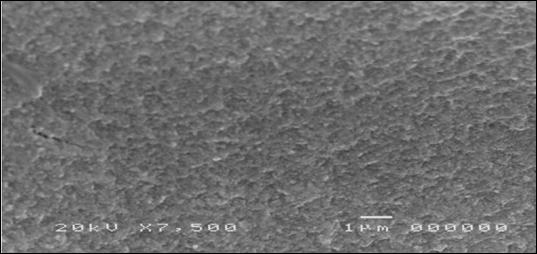
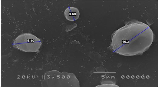
The germs found are reported in order of frequency in Figure 3: Candida (13)(Figure 4), Mycobacteria (8) (Figure 5), Acanthamoeba (7)(Figure 6), Micrococci (4), Mycoplasma (2)(Figure 7), Chlamydia (2)(Figure 8), Cocci (2)(Figure 9), Aspergillus (2), Cladosporium (1) (Figure 10); in most cases of Candida and Acanthamoeba other microorganisms were associated (Figure 11).
The negative result of the cytological examination of the cell morphology in the SEM report and a significant clinical improvement were obtained in a period of time between 1 and 7 weeks (mean: 4.03). In 6 eyes of patients suffering from infectious keratitis, the examination performed after the improvement of the clinical conditions highlighted the persistence of pathogenic microorganisms, for which
it was necessary to prolong or modify the originally fixed therapeutic regimen, which proved to be insufficient to eradicate the germs found. Intolerance to the prescribed drugs was highlighted in 3 patients, such as to require the replacement of the products initially used.
Discussion
Corneal infections represent an ophthalmological emergency due to the risk of perforating the eye or forming permanent scars with reduced vision after healing. The onset can be underestimated when only punctate keratopathy is present, which can also be due to non-infectious causes, such as dry eye disease, toxic phenomena or allergies. For these reasons, in many cases, the therapy initially implemented may not be suitable for counteracting the pathogens. It is therefore necessary, first of all, to recognize an infectious cause in corneal damage and then identify the microorganism that has caused the inflammatory process. The gold standard for the diagnosis are considered microbiological culture tests for the correct identification of the etiological agent [8, 22].
Citation: Troisi M, Del Prete S, Troisi S, Turco MV, Turco V, et al.(2023) Utility of Scanning Electron Microscopy (SEM) for Suspected Microbial Keratoconjunctivitis Unresponsive to Broad-Spectrum Antibiotic Therapy. J Clin Stud Med Case Rep 10:155.


10.24966/CSMC-8801/1000155

The culture test of the corneal or conjunctival scraping is a necessary study in severe or moderate forms, but also in those of a milder entity that have not responded to treatment based on clinical data or broad-spectrum therapy. Cultures also allow for antimicrobial susceptibility testing, which is essential for detection of resistance to antimicrobial drugs, and subsequent culture-guided targeted therapy [22].
Unfortunately it is not always possible to isolate the etiological agent with culture tests, because some pathogens do not grow in normal culture media or are present in very low concentrations due to previous therapies or as a consequence of the immune response. In some studies it has been observed that standard culture tests allow the identification of pathogenic microorganisms only in about 60% of cases [8, 23]. Firstly, the results of the present study indicate that the clinical evaluation proved to be very reliable in relation to the hypothesis of microbial origin of the keratitis, as 26 out of 30 cases were actually related to the presence of microorganisms. Secondly, the proposed method showed high sensitivity, as it allowed to correctly identify the pathogens in 26/30 eyes, 11 of which had already been swabbed for standard culture tests with negative results. In most cases microorganisms considered rare were found, such as Acanthamoeba, Candida, Chlamydia, mycobacteria and mycoplasma or a combination of these. The pathogenic potential of the microorganisms detected is confirmed by the presence of an inflammatory infiltrate consistent with this finding and by the disappearance of the symptoms after therapy ascertained on the basis of the data provided by the SEM examination. This result indicates that, in case of keratitis that does not heal with broad-spectrum antibiotic therapies, the diagnosis of infection, often attributable to the pathogens mentioned, must still be suspected. These microorganisms, in fact, do not grow in normal culture media and require specific therapies to be eradicated. In these forms of keratitis further diagnostic tests should be therefore performed with methods suitable for the identification of infectious agents other than Gram positive and Gram negative bacteria. An additional advantage of cytological examination of cell morphology in scanning electron microscopy is the possibility of suspecting other pathogenetic mechanisms, such as allergy, if present eosinophils or mast cells [24]. In the four eyes, with no pathogenic microorganisms at SEM, the characteristics of the inflammatory infiltrate and lining epithelium actually indicated other inflammatory causes [25]. The proposed method also shows good tolerability, as it does not require corneal scraping, but only the tarsal conjunctiva next to the corneal surface. The limit of the SEM exam consists instead in the lack of an antibiogram or antimycogram to set up the therapy in a targeted manner. These data are usually available in case of germ isolation from plate culture [26].
Conclusion
The absence of clinical improvement after broad-spectrum local antibiotic therapy indicates a possible non-bacterial etiology or the presence of germs resistant to the drug used. For this second hypothesis, a conjunctival brushing culture test for bacteria and fungi was performed on 11 patients, which resulted negative for all cases examined.
Cytological examination of cell morphology in Scanning Electron Microscopy made it possible to detect the presence of microbial agents in 26/30 patients who had not shown clinical improvement with broad-spectrum therapies. The detection of pathogenic microorganisms in patients with clinical suspicion of ocular surface infection confirmed the diagnostic hypothesis and allowed to implement a specific therapy based on the type of germ. This therapy proved effective in all patients examined over a period ranging from 1 to 7 weeks.
In the cases subjected to culture examination, the nature of the germ did not allow its isolation with the normal culture tests for bacteria and fungi, being mycoplasmas, mycobacteria, chlamydia or Acanthamoeba. It should also be considered that the culture test, carried out before or after a cycle of ineffective local therapy, may not detect the pathogen, due to a reduced microbial load or to previously performed therapies.
The proposed method, based on the recognition of the different microbial species based on their size and morphological characteristics, has proved to be very useful and effective for the diagnosis of infections that do not respond to broad-spectrum therapies or negative standard culture tests.
Ultimately, the results of the study indicate a high sensitivity of the microbiological diagnostic method based on SEM examination and the possibility of obtaining further information deriving from the evaluation of the inflammatory cells present. Finally, good tolerability is noted, linked to the sampling methods, which do not involve direct scraping of the corneal lesion.
The Authors suggest to perform this test in all cases of keratitis of suspected microbial origin in which the pathogen has not been identified by standard tests and which have not shown clinical improvement after topical broad-spectrum antibiotic therapy.
Author’s Contribution
Conceptualization: M.T., S.T. and S.D.P.; Methodology: S.T., M.T., D.M., V.T., M.V.T., A.G. and S.D.P.; Validation: A.D.P., M.T. and S.D.P.; Formal analysis: M.T.; Investigation: M.T., S.D.P., S.T. and D.M.; Resources: S.D.P., D.M., M.T., M.V.T. and S.T.; Data curation: M.T., D.M., A.G., A.D.P. and S.T.; Writing-original draft preparation: M.T., S.T. and S.D.P.; Writing-review and Editing: S.T., M.T., V.T., M.V.T., A.D.P. and D.M..; Visualization: M.T., S.D.P. and D.M.; Supervision: M.T, S.T. and S.D.P. All authors have read and agreed to the published version of the manuscript.
Funding
This research received no external funding.
Institutional Review Board Statement
“The study was conducted in accordance with the Declaration of Helsinki, and approved by the Ethics Committee of Campania Sud (prot. n. 16, 2.12.2019).”
Citation: Troisi M, Del Prete S, Troisi S, Turco MV, Turco V, et al.(2023) Utility of Scanning Electron Microscopy (SEM) for Suspected Microbial Keratoconjunctivitis Unresponsive to Broad-Spectrum Antibiotic Therapy. J Clin Stud Med Case Rep 10:155.
Informed Consent Statement
Informed consent was obtained from all subjects involved in the study.
Conflicts of Interest
The authors declare no conflict of interest.
References
1. Caruso C, Eletto D, Tosco A, PannettaM, Scarinci F, et al. (2022) Comparative Evaluation of Antimicrobial, Antiamoebic, and Antiviral Efficacy of Ophthalmic Formulations. Microorganisms, 10:1156.
2. Ting DSJ, Ho CS, Deshmukh R, et al. (2021) Infectious keratitis: an update on epidemiology, causative microorganisms, risk factors, and antimicrobial resistance. Eye, 35:1084-1101.
3. Durand ML, Barshak MB, Chodosh J (2021) Infectious keratitis in 2021. JAMA, 326:1319-1320.
4. Erie JC, Nevitt MP, Hodge DO, Ballard DJ (1993) Incidence of Ulcerative Keratitis in a Defined Population From 1950 Through 1988. Arch Ophthalmol, 111: 1665-167.
5. Upadhyay MP, Karmacharya PC, Koirala S,Shah DN, Shakya S, et al. (2001) The Bhaktapur eye study: ocular trauma and antibiotic prophylaxis for the prevention of corneal ulceration in Nepal. Br J Ophthalmol, 85:388392.
6. Thomas PA, Leck AK, Myatt M (2005) Characteristic clinical features as an aid to the diagnosis of suppurative keratitis caused by filamentous fungi. Br J Ophthalmol, 89:1554-1558.
7. Gopinathan U, Sharma S, Garg P, Rao GN (2009) Review of epidemiological features, microbiological diagnosis and treatment outcome of microbial keratitis: experience of over a decade. Ind J Ophthalmol, 57:273-279.
8. McLeod SD, Kolahdouz-Isfahani A, Rostamian K, Flowers CW, Lee PP, et al. (1996) The role of smears, cultures, and antibiotic sensitivity testing in the management of suspected infectious keratitis. Ophthalmology, 103:23-28.
9. Garg P (2010) Diagnosis of microbial keratitis. Br J Ophthalmol, 94:961962.
10. Satpathy G, Mishra AK, Tandon R, Sharma MK, Sharma A, et al. (2011) Evaluation of tear samples for herpes simplex virus 1 (HSV) detection in suspected cases of viral keratitis using PCR assay and conventional laboratory diagnostic tools. Br J Ophthalmol, 95:415-418.
11. Madhavan HN, Priya K, Anand AR, Therese KL (1999) Detection of herpes simplex (HSV) genome using polymerase chain reaction (PCR) in clinical samples comparison of PCR with standard laboratory methods for detection of HSV. J Clin Virol, 14:145-151.
12. Hau SC, Dart JKG, Vesaluoma M,Parmar DN, Claerhout I, et al. (2010) Diagnostic accuracy of microbial keratitis with in vivo scanning laser confocal microscopy. Br J Ophthalmol, 94:982-987.
13. Troisi M, Del Prete S, Troisi S, Marasco D, Costagliola C (2023) Scanning Electron Microscopy of Conjunctival Scraping: Our Experience in the Diagnosis of Infectious Keratitis with Negative Culture Tests. Reports, 6:10.
14. Dahlgren MA, Lingappan A, Wilhelmus KR (2007) The Clinical Diagnosis of Microbial Keratitis. Am. J. Ophthalmol, 143:940-944.
15. Cennamo GL, Del Prete A, Forte R, Del Prete S, Marasco D (2008) Impression cytology with scanning electron microscopy: a new method in the study of conjunctival microvilli. Eye, 22:138-143.
16. Cennamo GL, Forte R, Del Prete S, Cardone D (2013) Scanning Electron Microscopy Applied to Impression Cytology for Conjunctival Damage From Glaucoma Therapy. Cornea, 32:1227-1231.
17. Del Prete S, Marasco D, Del Prete A, Meloni M, Grumetto L, et al. (2019) Scraping cytology and scanning electron microscopy in diagnosis and therapy of corneal ulcer by mycobacterium infection. Arch Case Rep, 3:050-053.
18. Dart JKG, Saw VPJ, Kilvington S (2009) Acanthamoeba Keratitis: Diagnosis and Treatment Update. Am. J. Ophthalmol, 148:487-499.
19. Raimondo F, Cennamo G, Del Prete S, Cesarano I, Del Prete A (2010) Scanning Electron Microscopy of Corneal Epithelium in Soft Contact Lens Wearers. Cornea, 29:732-736.
20. Thanathanee O, O’Brien TP (2011) Conjunctivitis: Systematic Approach to Diagnosis and Therapy. Curr Infect Dis Rep, 13: 141-148.
21. Gopinathan U, Sharma S, Garg P, Rao GN (2009) Review of epidemiological features, microbiological diagnosis and treatment outcome of microbial keratitis: experience of over a decade. Indian J Ophthalmol, 57:273-279.
22. Rietveld RP, terRiet G, Bindels PJ, Sloos JH, van Weert HC (2004) Predicting bacterial cause in infectious conjunctivitis: cohort study on informativeness of combinations of signs and symptoms. BMJ; 329: 206-210.
23. O’Brien TP, Jeng BH, McDonald M, Raizman MB (2009) Acute conjunctivitis: truth and misconceptions. Curr Med Res Opin, 25: 1953-1961.
24. Raimondo F, Cennamo G, Del Prete S, Napolitano N, Del Prete A (2009) Allergic Conjunctivitis and Latent Infections. Cornea; 28:839-842.
25. Meloni M, De Servi B, Marasco D, Del Prete S (2011) Molecular mechanism of ocular surface damage: Application to an in vitro dry eye model on human corneal epithelium. Mol Vis, 17:113-126.
26. Boralkar AN, Dindore PR, Fule RP, Bangde BN, Albel MV, et al. (1989) Microbiological studies in conjunctivitis. IJO, 37:94-95.
Advances In Industrial Biotechnology | ISSN: 2639-5665
Advances In Microbiology Research | ISSN: 2689-694X
Archives Of Surgery And Surgical Education | ISSN: 2689-3126
Archives Of Urology
Archives Of Zoological Studies | ISSN: 2640-7779
Current Trends Medical And Biological Engineering
International Journal Of Case Reports And Therapeutic Studies | ISSN: 2689-310X
Journal Of Addiction & Addictive Disorders | ISSN: 2578-7276
Journal Of Agronomy & Agricultural Science | ISSN: 2689-8292
Journal Of AIDS Clinical Research & STDs | ISSN: 2572-7370
Journal Of Alcoholism Drug Abuse & Substance Dependence | ISSN: 2572-9594
Journal Of Allergy Disorders & Therapy | ISSN: 2470-749X
Journal Of Alternative Complementary & Integrative Medicine | ISSN: 2470-7562
Journal Of Alzheimers & Neurodegenerative Diseases | ISSN: 2572-9608
Journal Of Anesthesia & Clinical Care | ISSN: 2378-8879
Journal Of Angiology & Vascular Surgery | ISSN: 2572-7397
Journal Of Animal Research & Veterinary Science | ISSN: 2639-3751
Journal Of Aquaculture & Fisheries | ISSN: 2576-5523
Journal Of Atmospheric & Earth Sciences | ISSN: 2689-8780
Journal Of Biotech Research & Biochemistry
Journal Of Brain & Neuroscience Research
Journal Of Cancer Biology & Treatment | ISSN: 2470-7546
Journal Of Cardiology Study & Research | ISSN: 2640-768X
Journal Of Cell Biology & Cell Metabolism | ISSN: 2381-1943
Journal Of Clinical Dermatology & Therapy | ISSN: 2378-8771
Journal Of Clinical Immunology & Immunotherapy | ISSN: 2378-8844
Journal Of Clinical Studies & Medical Case Reports | ISSN: 2378-8801
Journal Of Community Medicine & Public Health Care | ISSN: 2381-1978
Journal Of Cytology & Tissue Biology | ISSN: 2378-9107
Journal Of Dairy Research & Technology | ISSN: 2688-9315
Journal Of Dentistry Oral Health & Cosmesis | ISSN: 2473-6783
Journal Of Diabetes & Metabolic Disorders | ISSN: 2381-201X
Journal Of Emergency Medicine Trauma & Surgical Care | ISSN: 2378-8798
Journal Of Environmental Science Current Research | ISSN: 2643-5020
Journal Of Food Science & Nutrition | ISSN: 2470-1076
Journal Of Forensic Legal & Investigative Sciences | ISSN: 2473-733X
Journal Of Gastroenterology & Hepatology Research | ISSN: 2574-2566
Journal Of Genetics & Genomic Sciences | ISSN: 2574-2485
Journal Of Gerontology & Geriatric Medicine | ISSN: 2381-8662
Journal Of Hematology Blood Transfusion & Disorders | ISSN: 2572-2999
Journal Of Hospice & Palliative Medical Care

Journal Of Human Endocrinology | ISSN: 2572-9640
Journal Of Infectious & Non Infectious Diseases | ISSN: 2381-8654
Journal Of Internal Medicine & Primary Healthcare | ISSN: 2574-2493
Journal Of Light & Laser Current Trends
Journal Of Medicine Study & Research | ISSN: 2639-5657
Journal Of Modern Chemical Sciences
Journal Of Nanotechnology Nanomedicine & Nanobiotechnology | ISSN: 2381-2044
Journal Of Neonatology & Clinical Pediatrics | ISSN: 2378-878X
Journal Of Nephrology & Renal Therapy | ISSN: 2473-7313
Journal Of Non Invasive Vascular Investigation | ISSN: 2572-7400
Journal Of Nuclear Medicine Radiology & Radiation Therapy | ISSN: 2572-7419
Journal Of Obesity & Weight Loss | ISSN: 2473-7372
Journal Of Ophthalmology & Clinical Research | ISSN: 2378-8887
Journal Of Orthopedic Research & Physiotherapy | ISSN: 2381-2052
Journal Of Otolaryngology Head & Neck Surgery | ISSN: 2573-010X
Journal Of Pathology Clinical & Medical Research
Journal Of Pharmacology Pharmaceutics & Pharmacovigilance | ISSN: 2639-5649
Journal Of Physical Medicine Rehabilitation & Disabilities | ISSN: 2381-8670
Journal Of Plant Science Current Research | ISSN: 2639-3743
Journal Of Practical & Professional Nursing | ISSN: 2639-5681
Journal Of Protein Research & Bioinformatics
Journal Of Psychiatry Depression & Anxiety | ISSN: 2573-0150
Journal Of Pulmonary Medicine & Respiratory Research | ISSN: 2573-0177
Journal Of Reproductive Medicine Gynaecology & Obstetrics | ISSN: 2574-2574
Journal Of Stem Cells Research Development & Therapy | ISSN: 2381-2060
Journal Of Surgery Current Trends & Innovations | ISSN: 2578-7284
Journal Of Toxicology Current Research | ISSN: 2639-3735
Journal Of Translational Science And Research
Journal Of Vaccines Research & Vaccination | ISSN: 2573-0193
Journal Of Virology & Antivirals
Sports Medicine And Injury Care Journal | ISSN: 2689-8829
Trends In Anatomy & Physiology | ISSN: 2640-7752