2 minute read
International Journal for Research in Applied Science & Engineering Technology (IJRASET)
ISSN: 2321-9653; IC Value: 45.98; SJ Impact Factor: 7.538
Volume 11 Issue IV Apr 2023- Available at www.ijraset.com
Advertisement
B. Image Acquisition
Here in this phase, sample images are collected, which are required to train classifier algorithm and build classifier model. Reddish passion eye variety was selected for taking sample images. Because the yellowish variety is widely not available, for detecting the drug in the country, healthy/affected eye images were taken by using mobile phone camera and used to both training/testing the classifier algorithms. Images are taken in various angles, under different lighting and environmental conditions. The standard JPEG format was used to store the images. In this study here, images are collected from different regions. Eyes infected by the reddish disease that had been included in the collected images.
C. Image Preprocessing
After image acquisition, image processing was done for improving the image quality. All original eye images were stored in single folder. Those images were named as we like the wish can take any value of number. Only horizontal images were rotated by ninety degrees and resized by 200x300 pixels.
The vertical images were resized by 200x300 pixels and when width and height of the image are equal, and then those images were resized to 250x250 pixels. When image size is too large, processing task takes more time. After that, one of the noise reduction techniques was used for removing the noises from images and then increased images sharpness. Then, all preprocessed images are saved in that folder.
D. Image Segmentation
The third phase of the above methodology is the image segmentation. As the first step, all the preprocessed images are converted into L * a * b, HSV, Grey color models and then kept one in original way (RGB). Because identifying suitable color model to preprocess is one of the outcomes of the research. After that, image is converted to binary format (black/white). This format values are then clustered using CNN algorithm. According to the algorithm used image segmentation is done.
E. Applying Training Set
The fifth phase of the above methodology is applying training set images. The segmented output is done, which are created using feature extraction. However, two image sets were created to do the experiments. Preparation of the image sets is discussed. Field expertise support is taken for categorization of images and each image is selected from categorized sets of an image randomly.
IV. EXPERIMENT RESULTS AND FINDINGS
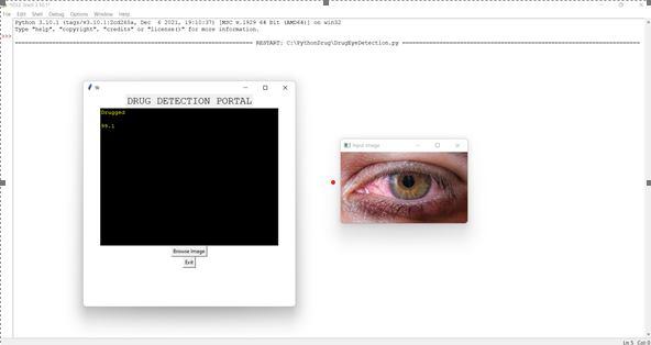
After applying the training set images, two base folders were used for identifying drugged eyes according to its accuracy. These files are called as two classes’ dataset. Another way is counting number of reddish places to identify drug consumed eye according to its stage. This method is called as alternative method.

Every training and testing time, rows of training files were shuffled randomly for increasing the accuracy of the model. Each training file was verified and tested in five times and accuracy was taken. Average of these accuracies is taken as accuracy of each model. Using this image dataset, two types of categories were found. Such as drugged eye and not drugged eye.
1) Accuracy is very high here.
2) Enhancing the values of drugged eye detection.
3) It takes only few seconds to provide exact result.
4) Result is provided with thehigh accuracy rate.
5) Applicable to both low and high pixel images.


