Synthesis and Characterization of Polyaniline Doped with Cu-Salts and Cu-Complexes
Madhab Upadhyaya1, Dilip K Kakati2
1Department of Chemistry, Chaiduar College, Gohpur, Assam, India
2Department of Chemistry, Gauhati University, Guwahati, Assam, India
In this work, we have synthesized polyaniline doped with Cu (II) salts and coordination complexes in presence of Aniline was polymerized in presence ammonium persulphate (APS). We varied the concentration of APS and also that of Cu (II) salts and complexes to see the effect of these on the properties of polyaniline. We investigated the effect of the dopant and ligand around Cu (II) ion on the morphology, crystallinity and conductivity of the resultant polyaniline. The products were characterized by UV-Vis, FT-IR spectroscopy, while the morphology and crystallinity were investigated by scanning electron microscopy, and X-ray diffraction studies respectively. Results show that the morphology, crystallinity and conductivity of the doped polyanilines arefound to beinfluenced by nature of ligand.
KEYWORDS: Conducting polymer, polyaniline, Cu-salts and Cucomplexes, dopant
How to cite this paper: Madhab Upadhyaya | Dilip K Kakati "Synthesis and Characterization of Polyaniline Doped with Cu-Salts and CuComplexes" Published in International Journal of Trend in Scientific Research and Development (ijtsrd), ISSN:24566470, Volume-6 | Issue-7, December 2022, pp.13231329, URL: www.ijtsrd.com/papers/ijtsrd52609.pdf
Copyright © 2022 by author (s) and International Journal of Trend in Scientific Research and Development Journal. This is an Open Access article distributed under the terms of the Creative Commons Attribution License (CC BY 4.0) (http://creativecommons.org/licenses/by/4.0)
1. INTRODUCTION
Among the family of intrinsically conducting polymers, polyaniline (PANI) has been most actively investigated due to its unique doping/dedoping behavior, facile synthesis and good environmental stability [1]. The beauty of this class of material is that the conductive properties could be tuned by doping. Polyaniline (PANI) is unique among the family of conjugated polymers since its doping level could be readily controlled through an acid doping and base dedoping process [2].
Besides the use of different protic acids as dopants [3], Lewis acids and transition metal ions [4-7] have also been used as dopants in polyaniline synthesis. Over the past years, there has been a growing interest in the research on polyaniline (PANI)/metal systems [8-10]. This is because of their potential practical applications, as redox-active catalyst [11], corrosion inhibitor [6] and organic electronic [12] and electroluminescent devices [13]. Transition metal ions can act as oxidizing agents as well. which can act as oxidizing agent, According to O.P Dimitriev [10]
doping occur through a two-step redox process. At first, the metal ions oxidize EB-PANI amine nitrogen atoms; afterwards, the reduced metal ions coordinate to imine nitrogen and finally, the reduced cations are oxidized by the imine groups, resulting in radical cation segments (ES-PANI) and the corresponding oxidized metal ions [10].O.P Dimitriev [10] reported that the formation of complex of transition metal and PANI are dependent on both the cation and anion of the inorganic salt used, as well as on the solvent. In some cases, formation of the complex occurred in the form of a precipitate directly in the solution. Change of anion with same metal cation produced different results. He observed that the addition of the LaNO3 to the dimethyl form amide (DMF) solution of EBPANI did not result in formation of any precipitate, whereas use of the LaCI3, yielded a dark-green precipitate in a few minutes. It has also been reported that the conductivity of films doped by nitrates of transition metals was usually 1 or 2 orders of magnitude smaller as compared to films doped by chlorides [4].
In the present work, our objective is to study the effect of the various ligands around the Cu+2 ion used as dopants on the properties of polyaniline and reported the effect of the size of ligands around the Cu+2 ions on the properties ofCu+2 doped polyaniline.
2. EXPERIMENTAL
2.1. Materials
Aniline was distilled under reduced pressure before use. Copper sulphate (CuSO4.5H2O), hydrochloric acid (HCl), ammonia, ethylene diamine, ammonium persulphate (APS), acetone and methanol were analytical grade chemicals ofMERCK-Indiaand they were used as received without further purification.
Table 1 Sample Coding and the Conductivity values
Dopant Name Dopant Conc. (M) Sample code Conductivity (Scm-1)
No Dopant Nil EBPANI 1.2x10⁻⁹ HCl 0.01 HClPANI 3.845 CuSO4.5H2O
2.3.
Synthesis of Polyaniline Hydrochloride (HClPANI)
Polyaniline Hydrochloride (HCl-PANI) was synthesized by the procedure of MacDiarmid et al.[17]. 1.82 mL (0.04 mol) of distilled aniline was dissolved in 100 mL 1M HCl solution and cooled to 0-5°C. To this, 20 mL cold ammoniumpersulphate (APS) solution containing 4.56 g (0.04 mol)APS was added very slowly in a dropwise manner. The reaction mixture was then agitated for 3 h. Polyaniline hydrochloride thus formed was filtered under suction, washed with dilute HCl, water and methanol and dried under vacuum. It was labeled as HCl-PANI
2.4. Preparation of Emeraldine base form of Polyaniline (EB-PANI)
The dried emeraldine salt form of polyaniline (ESPANI) was grounded to fine powder. The powdered HCl-PANI was then agitated with 0.1 M NH4OH solution (PH 9) for 6 h to form emeraldine base of polyaniline (EB-PANI). It was then filtered under vacuum, washed with water and dried. It was labeled EB-PANI.
0.002 0.006 0.02 0.04 0.08
0.002 0.006 0.02 0.04 0.08
[Cu(en)2]SO4
A B C D E
0.477 0.556 0.667 0.778 0.899 [Cu (NH3)4]SO4
A1 B1 C1 D1 E1
0.367 0.389 0.423 0.451 0.487
0.002 0.006 0.02 0.04 0.08
[Cu(en)3]SO4
002 0.006 0.02 0.04 0.08
A2 B2 C2 D2 E2
A3 B3 C3 D3 E3
0.321 0.345 0.365 0.388 0.422
0.167 0.189 0.246 0.271 0.295
2.2. Preparation of [Cu(en)2]SO4 and [Cu(en)3]SO4
Bis-ethylenediamine Copper (II) sulphate was prepared by the reaction of anhydrous copper (II) sulphate with ethylenediamine in a molar ratio of 1:1 in distilled water [15]. Dark blue prismatic crystals were obtained by slow evaporation of the solvent used. Tris-ethylenediamine copper (II) sulphate was prepared bymixing hydrated copper (II) sulphateand large excess of ethylenediamine and then recrystallising it from water [16].
2.5. Preparation of polyaniline doped with CuSO4.5H2O, [Cu(NH3)4]SO4 , [Cu(en)2]SO4 and [Cu(en)3]SO4 Aniline (1.82 ml 0.04 mol) was added to 100 mL water containing requisite amount of copper (II) compound or complex (table 1). The reaction mixture was stirred for 2 h and 4.56 g (0.04 mol) (NH4)2S2O8 dissolved in 20 mLwater was added drop wise over a period of 30 minutes. The reaction temperature was maintained at 0-5°C. It was kept undisturbed for 24 h for the reaction to complete. The resultant polyaniline was filtered, washed with rectified spirit and acetone and then vacuum dried at 60°C for 5 h.
3. CHARACTERIZATION
The FTIR spectra of all samples were recorded in a SHIMADZU IR Affinity-1 spectrophotometeras KBr pellet at room temperature in the range of 4000-400 cm -1. The UV-Vis absorption spectra were recorded in the 300-900 nm range in a SHIMADZU UV-1800 spectrophotometer using 1-methyl 2-pyrrolidone (NMP) as solvent. The phase identification of finely powdered samples were performed with an X’Pert Pro X-ray diffractometer, Phillips with nickel-filtered Cu kα radiation (wavelength = 1.5414 Å) in the 2θ range of 5-40˚. Thermo gravimetric analysis (TGA) of the samples was carried out in aPerkin ElmerPyris 1 TGA instrument at a heatingrateof10°C/min under N2 atmosphere over a temperaturerangeof30-800°C. SEM-micrographs were obtained in a JSM-6360 (JEOL) scanning electron microscope at an accelerating voltage of 20 kV. The electrical
conductivities of the samples were measured at room temperature with dry pressed pellets by standard four probe technique.
4. RESULTS AND DISCUSSION
4.1. UV-VISIBLE SPECTRAL STUDIES
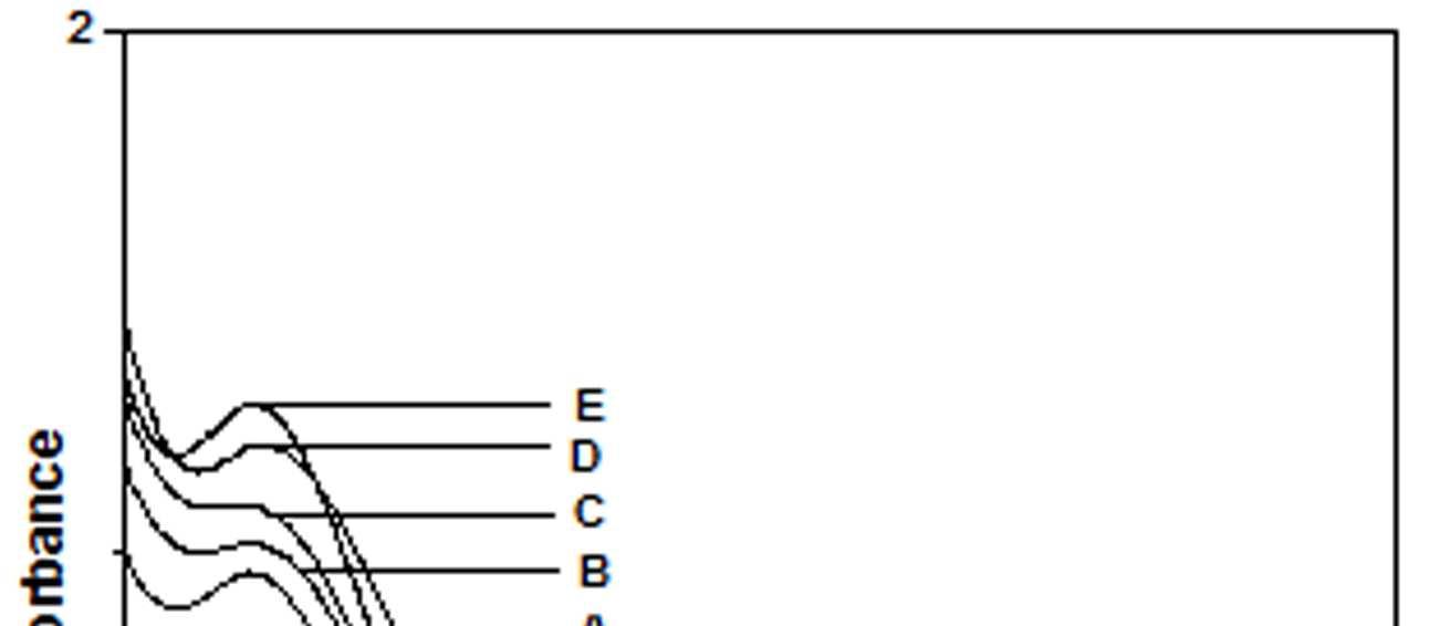
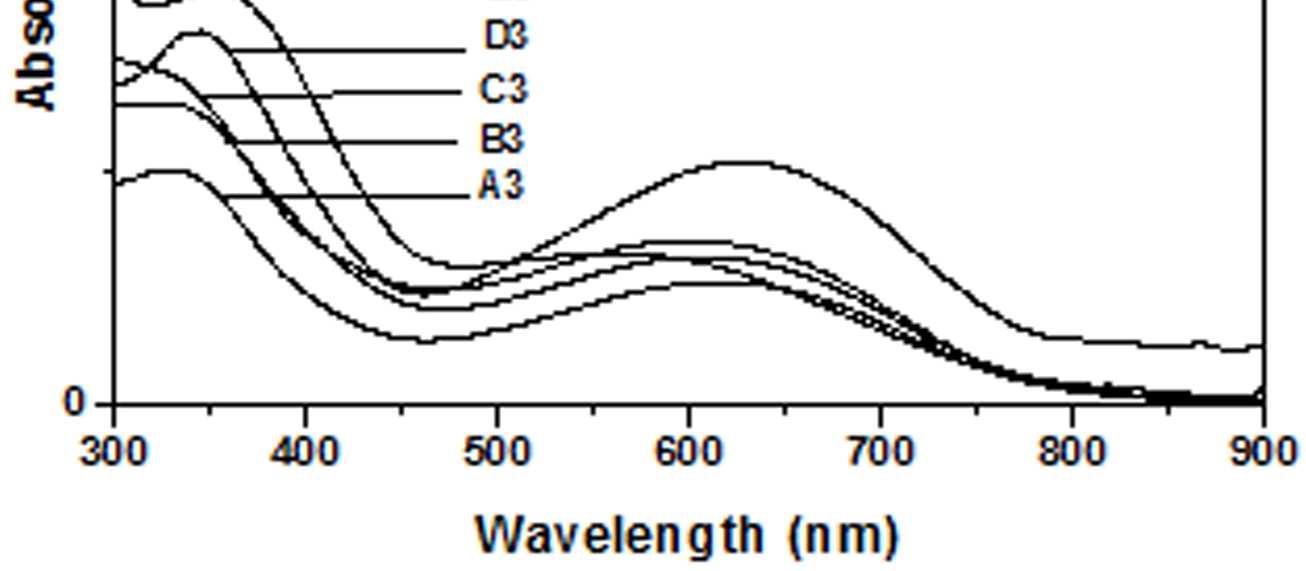
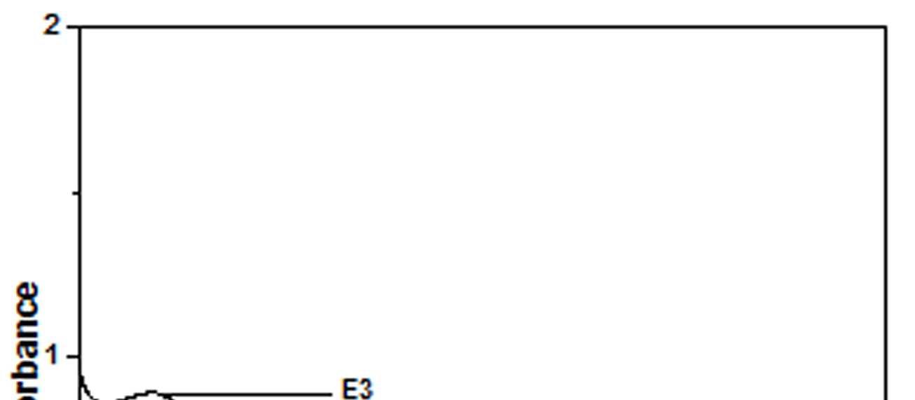
The UV-Vis spectra of EB-PANI, HCl-PANI and PANI doped with Cu+2 ion with different ligands are presented in figure 1. The benzenoid to quinoid excitation transition (BQET) was observed at 634 nm for the undoped PANI. For HCl doped PANI this peak disappeared. But on being doped by Cu+2 ion this peak undergoes a blue shift to the range of 533573 nm from 634 nm. Similar blue shift was also reported by Chen et al. [18]when they used a number of transition metal ions including Cu+2 to dopePANI. Hirao et al. [19] ascribed such a blue shift to the complexation of the Cu+2 ion with polyaniline and Chen et al. [18] also attributed this shift to the interaction of transition metal ions with polyaniline molecular chains. The observed blue shift maybedue to complexation of the Cu+2 ion with the polyaniline as well as due to partial oxidation of polymer backbone [20]. Further it was observed that this shift was dependent on the size or bulkiness of the ligands around Cu+2 ion. It is evident from figure 1 that larger the size of the ligands smaller is the blue shift. This is due to less efficient complexation of Cu+2 ion with polyaniline caused by the bulkiness of the ligands around the Cu+2 ion. Concentration of the dopant also has an influence on the extent of such a blue shift [21]. It was observed that for the same ligand higher the concentration of the dopant larger was the blue shift. This is evident from figure 2, figure 3, figure 4 and figure 5 where the concentration of the dopants was varied for the same ligand. When the dopant concentration was high, the extent of doping became more efficient showing a larger blue shift.
Figure 2 UV-Vis spectra of PANI doped with CuSO4.5H2O
Figure 1 UV-Vis spectra of EB-PANI, HCl-PANI C C1, C2 and C3
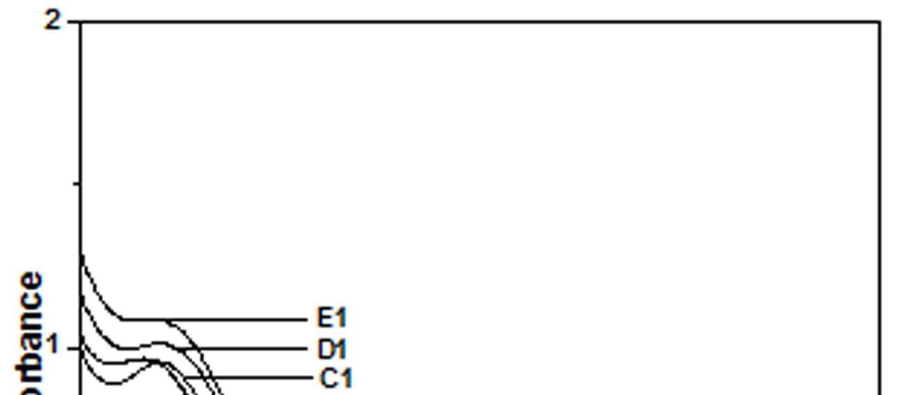
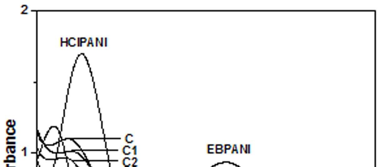
Figure 3 UV-Vis spectra of PANI doped with [Cu(NH3)4]SO4
Figure 4 UV-Vis spectra of PANI doped with [Cu(en)2]SO4
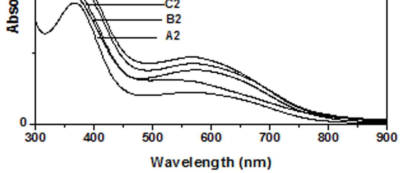
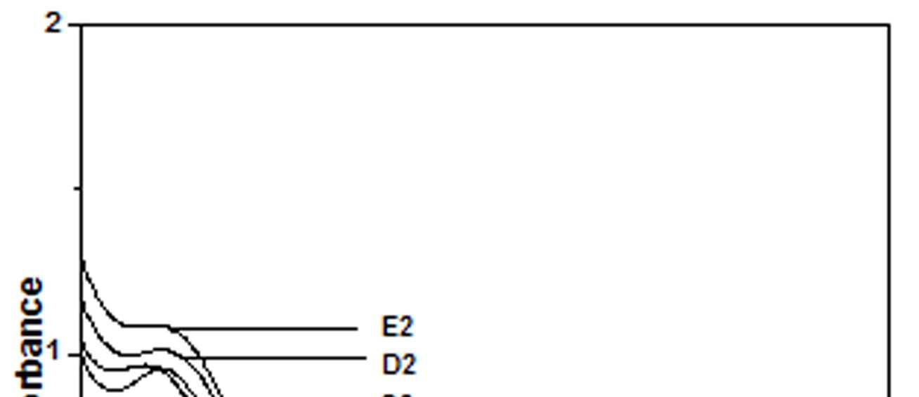
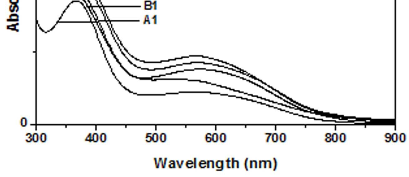
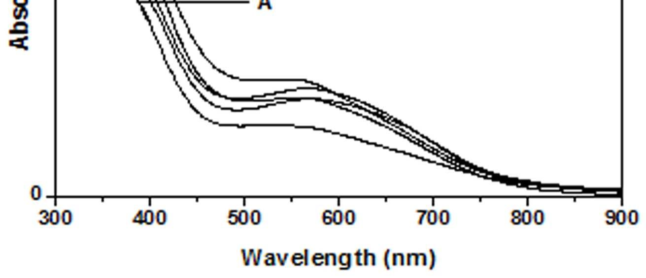
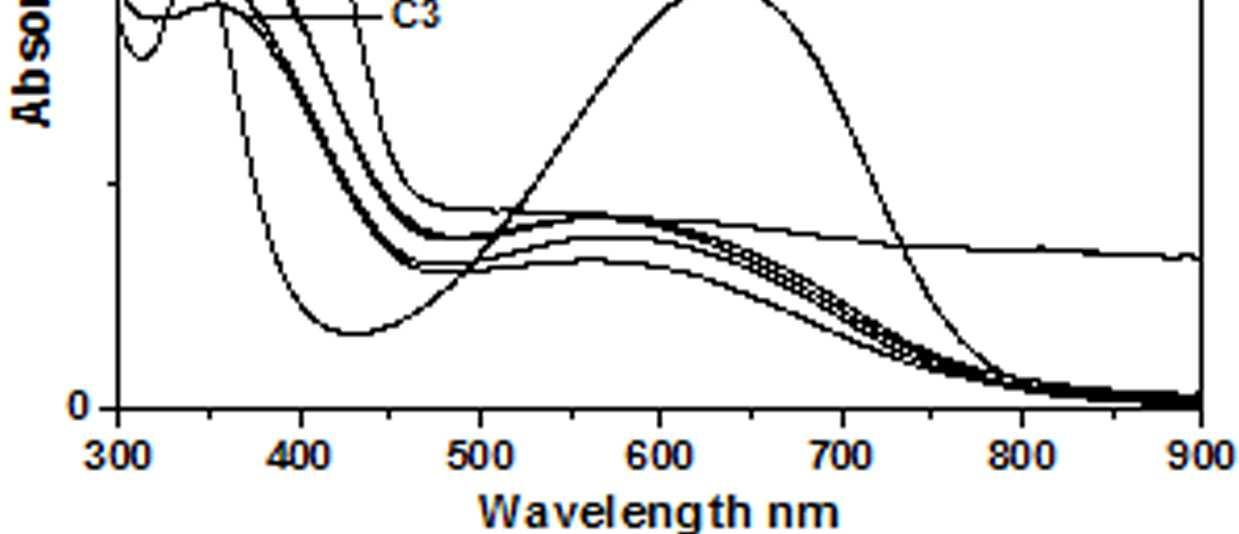
Figure 5 UV-Vis spectra of PANI doped with [Cu)en)3]SO4
The other absorption peak observed for undoped PANI was at 329 nm which is attributed to π-π* transition in the benzenoid rings [22-25]. On being doped, the π-π* peak undergoes a red shift to the polaronic range of 348-377 nm. This peak is related to the doping level and the formation of polaron [26]. For HCl-PANI, the intensity of this peak is maximum, which means a high degreeofdoping. The intensity of the polaronic peak decreases with increase in bulkiness of the ligands around Cu+2 ions. This is evident from plots C, C1, C2 and C3 of figure 1 with CuSO4.5H2O, [Cu(NH3)4]SO4, [Cu(en)2]SO4 and [Cu(en)3]SO4 as dopants respectively. Larger the ligand size, greater was the sterichindranceand lesser was the degree of doping. Increase in intensity of the polaronic peak afterdopingEB-PANIwith CuCl2 had also been observed by Nikolaidis et al. [25] from EPR studies. The polaronic peak for HCl-PANI is at 377 nm while for samples C, C1, C2 and C3 they are at 359 nm, 355 nm 354 nm and 348 nm respectively showing that larger the ligand size lesser was theshift towards polaronic range. That the peak in the range 348-377 nm was related to the doping level was further evident from figure 2, 3 & 4 where CuSO4.5H2O, [Cu(en)2]SO4 and [Cu(en)3]SO4 were used as dopants. It was observed that the intensity of this peak increased with the rise in concentration of the dopant. The increased concentration ofthedopant increased the doping level. The rise in the intensityof the polaron peak went parallel to the rise in the conductivity values.
4.2. FTIR SPECTROSCOPIC STUDIES
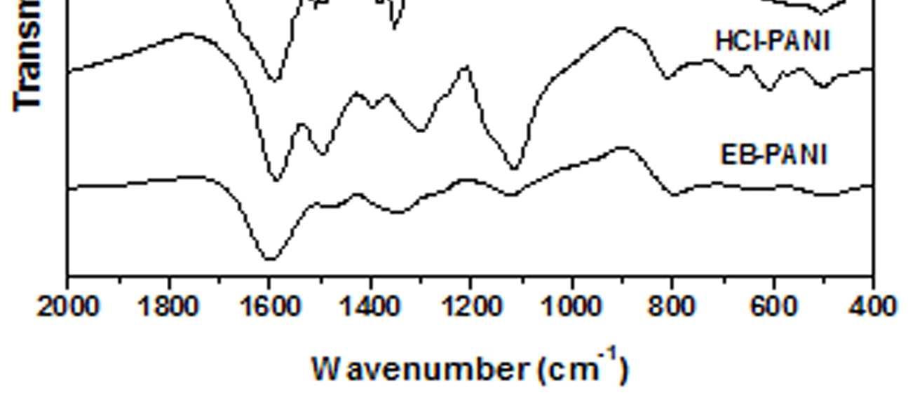
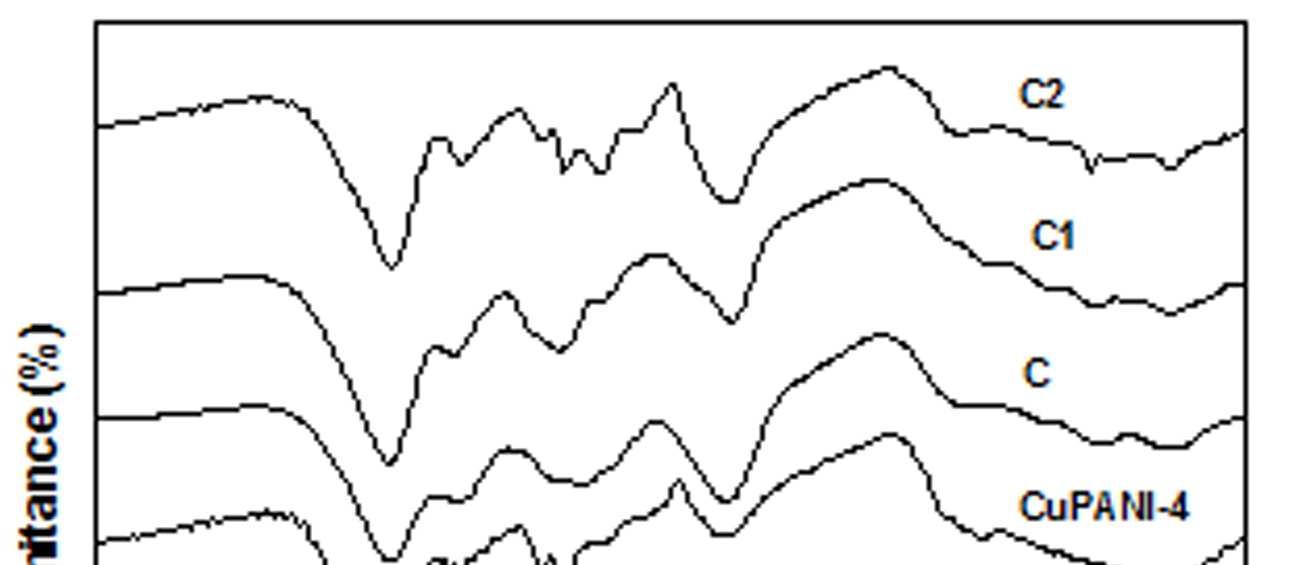
The FT-IR spectra of all the samples are presented in figure 6. The FTIR spectrum of EB-PANI shows characteristic peaks at 1597, 1487, 1346, 1149, and 810 cm-1. The peaks at 1597 cm-1 is attributed to C=C stretching mode of vibration for quinoid rings [4] and
the one at 1487 cm-1 is attributed to C=C stretching mode of vibration for benzenoid rings [4] and ring stretching due to C-C and C-H. The peak at 1346 cm-1 is due to C-N stretching, C=-N+ stretching and C-H bending mode benzenoid units. The band at 1149 cm1 is the characteristic mode of N=Q=N and the one at 810 cm-1 is due to C-H out of plane bending of 1,4 disubstituted benzene [19]. FTIR spectrum of HCl doped PANI shows vibration bands at 1584, 1502, 1352, 1114 and 804 cm-1. On being doped with HCl, there is a red shift of the quinoid peak while a blue shift is observed for the benzenoid peak. Similar behavior is observed on being doped with dopants based on Cu+2 ions viz. CuSO4.5H2O, [Cu(NH3)4]SO4, [Cu(en)2]SO4 and [Cu(en)3]SO4. It is seen that the band at 1597 cm-1 is red shifted to 1584 cm -1 while the band at 1487 cm-1 is blue shifted to 1502 cm-1 with much increase in intensityofthepeak. The characteristic mode of N=Q=N which appeared at 1149 cm-1 for EB-PANI is now red shifted to 1114 cm -1 for HCl-PANI again with a considerable increase in intensity of the peak. The C-H out of plane bending of 1,4 disubstituted benzene is slightly red shifted in HCl-PANI compared to EB-PANI. After doping with CuSO4.5H2O, [Cu(NH3)4]SO4, [Cu(en)2]SO4 and [Cu(en)3]SO4 the peak at 1597 cm-1 (EB-PANI) is red shifted to the range of 1585-1592 cm -1 with increase in peak intensity. The position of this red shift is found to be slightly dependent on the size of the ligands. Sample C3 with [Cu(en)3]SO4 as the dopant shows the peak at 1592 cm-1 and sample C2 with [Cu(en)2]SO4 as dopant shows it at 1589 cm-1 and while the dopant is CuSO4.5H2O the peak position is at 1586 cm-1 .
Figure 6 FTIR spectra of EB-PANI, HCl-PANI, C, C1, C2 and C3
The position of this band can be taken as indicative of the degree of oxidation of the polymer backbone. This red shifted band indicates the increase in the
relative number of quinoid groups in the polymer [8]. Another important change that observed with the Cu+2 ion doped PANI is that the weak peak at 1449 cm -1(EB-PANI) is blue shifted to 1502 cm-1 and becomes sharp. The sharpness is maximum in HClPANI and minimum in sample C3 with [Cu(en)3]SO4 as dopant indicating the influence of the ligand bulkiness. Similar red shift was observed with the peak at 1149 cm-1 after doping with Cu+2 salt and complexes. The peak at 1149 cm-1 which is ameasure of electron delocalization in PANI[26] was shifted to 1110-1114 cm-1 in CuSO4.5H2O, [Cu(NH3)4]SO4, [Cu(en)2]SO4 and [Cu(en)3]SO4 doped PANI and apparently becomes broader than in EB-PANI. Intensity of this peak increased with decrease in ligand size (figure 6). FTIR spectra indicates the complexation between the polyaniline and Cu+2 ion and the degree of complexation is influenced by the size of the ligands 4.3. MORPHOLOGY
STUDIES
Figure 7 shows the SEM images of EB-PANI and PANI doped with CuSO4.5H2O, [Cu(NH)3]SO4, [Cu(en)2]SO4 and [Cu(en)3]SO4. EB-PANI shows a regular scaly structure in its SEM micrograph as seen in the figure. When PANI is doped with Cu+2 ions the
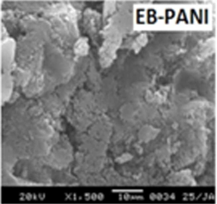
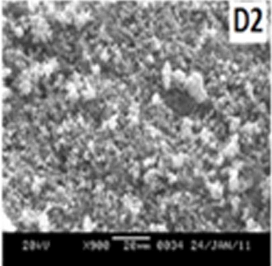
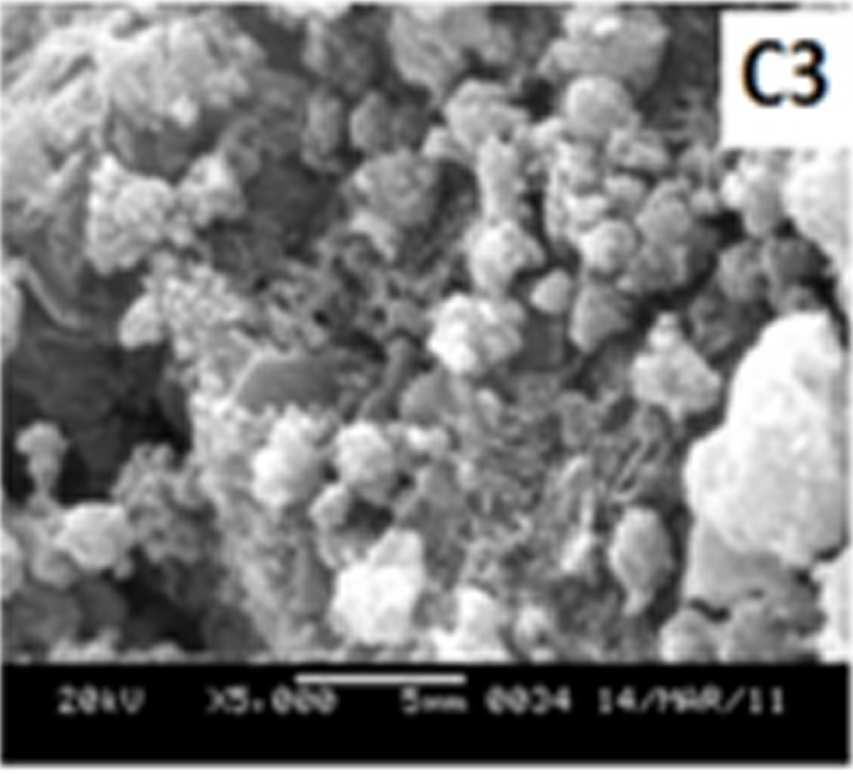
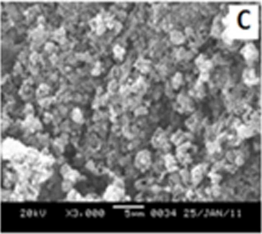
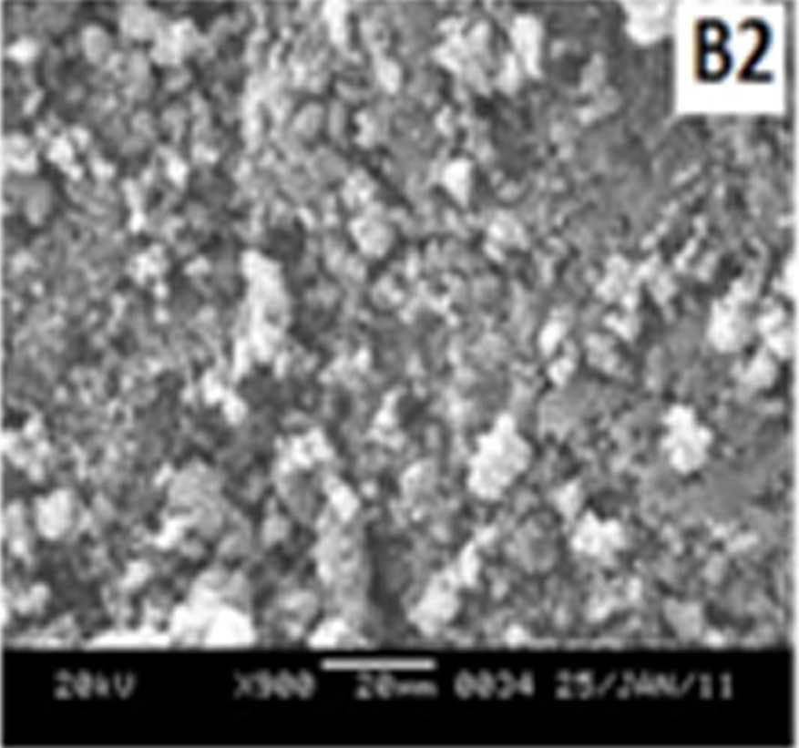
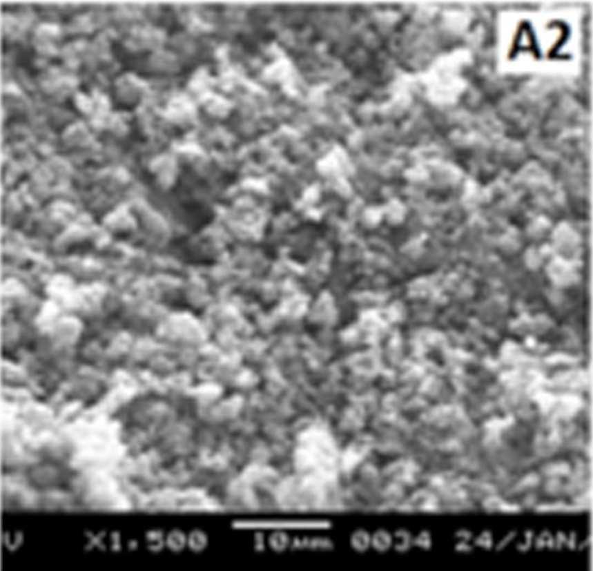
Figure 7 SEM images of EB-PANI and PANI doped with CuSO4.5H2O, [Cu(NH)3]SO4, [Cu(en)2]SO4 and [Cu(en)3]SO4
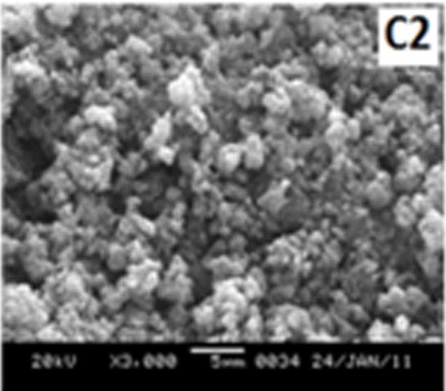
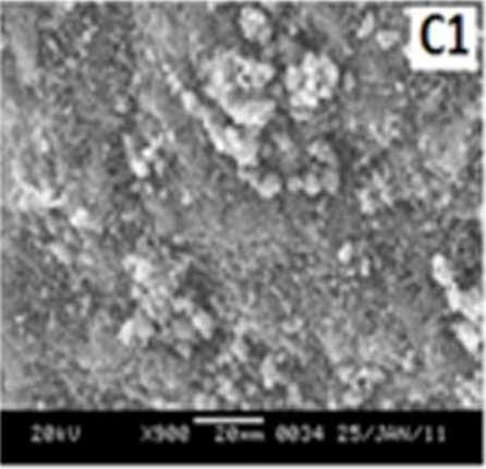
Morphology becomes irregular, granular and agglomerated. The particles are randomlyaggregated and rough surfaces appeared. Doping with Cu+2 ion results in an aggregation and networking of polyaniline molecular chains. This is because transition metal ions such as Cu+2 maybind to several nitrogen sites in the polyaniline chain or may form inter-chain linkages among several adjacent polyaniline chains by coordination [27-28]. Looking into the SEM images of C, C1, C2 and C3 it is observed that the size of the ligands around Cu+2 ion has an effect on the morphology. The size of the granules increases with increase in ligand size. Similar morphology of transition metal ion doped PANI was also observed by Chen et al. [18]
4.4.
XRD STUDIES
In addition to the polaronic species formed due to doping the electrical conductivity of doped PANI is also influenced by the crystalline domain formation [29]. Figure 8 shows the XRD patterns of EB-PANI, HCl-PANI, and PANI doped with CuSO4.5H2O (C), [Cu(NH3)4]SO4 (C1), [Cu(en)2]SO4 (C2), and [Cu(en)3]SO4 (C3). EB-PANI shows a very broad peak at 2θ = 12° and another one low intensity peak at 2θ =24°. HCl-PANIshow a broad peak at 2θ = 25°, another broad peak at around 2θ = 20° and also a sharp peak at around 2θ =6.4°. XRDplot ofEB-PANI shows its amorphous nature [30].
Figure 8 XRD Plots of EB-PANI, HCl-PANI, C, C1, C2 and C3
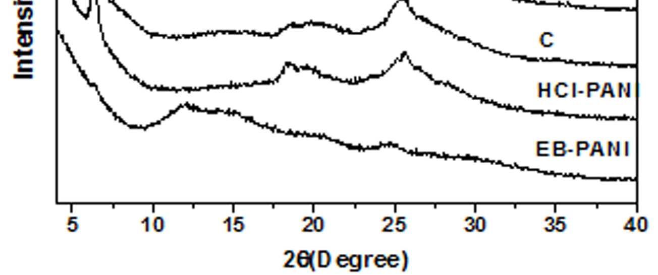
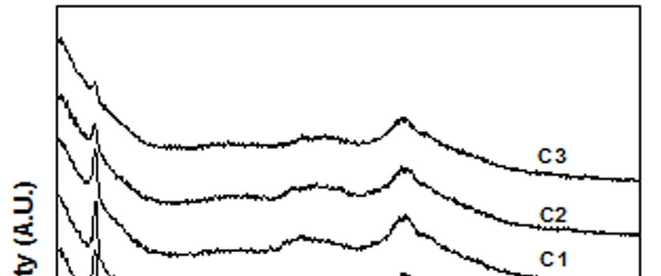
The presence of peak at 2θ = 6.4° for HCl-PANI implies the development of partial crystallinity in its structure.31 Similar peak is also observed for samples C, C1, C2 and C3 although with decreased intensity of the peak. This is indicative of the presence of low crystalline order in Cu+2 ion doped PANI in comparison to HCl doped PANI. Further the sharpness of the peak at 2θ = 6.4° decreases slightly with increase in ligand size which again reflects the effect of ligand size on the development ofcrystalline domains.
4.5. CONDUCTIVITY STUDIES
The conductivity measurements were carried out at room temperature in a four-probe electrode arrangement apparatus. The samples used were in pressed pellet forms. The results of conductivity studies are presented in table 1. The conductivity studies broadly revealed two facts: Firstly, the conductivity increased with the rise in concentration of Cu+2 ion with a particular ligand and secondly the conductivity decreased with the rise in the bulkiness of the ligand around the Cu+2 ion for the same concentration of Cu+2 ion.
We have observed that the conductivity is influenced by the nature of the groups or ligands around the dopant Cu+2 ion. The conductivity decreases with the rise in the steric crowding around the Cu+2 ion. (Table1). Steric crowding around Cu+2 ion hinders the chain alignment and causes the conductivity to decrease. Also, steric crowding reduces the accessibilityof Cu+2 ion to the polymerbackboneand this is responsible for the decreased conductivitywith increase in ligand size.
5. CONCLUSIONS
It is found that better the coordination higher the conductivity of the doped polyaniline [31] That is, by manipulating the extent of coordination between the Cu+2 and polyaniline it is possible to modulate the electrical conductivity of polyaniline. From our studies it was observed that by changing the nature and size of ligands around the Cu+2 ion, the electrical conductivity could be modulated. This was due to variation in the extent of interaction between theCu+2 ion and polyaniline as a result of interference from the ligands. Further, this also resulted in difference in morphology as well as crystallinity of the doped polyaniline.
6. ACKNOWLEDGEMENTS
The corresponding author acknowledges UGC–NERO(UniversityGrants Commission-North Eastern Regional Office) for awarding Minor Research Project vide letter No. F.5-1/2007-08 (MRP/NERO)/6096 dated 31/03/2008.
7. REFERENCES
[1] Ismail YA, Chang J, Shin SR, Mane RS, Han SH, Kim SJ, J Electrochem Soc 156: A313A317 (2009).
[2] Huang W, Humphrey BD, Macdiarmid AG, J Chem Soc Faraday Trans 1 82: 2385-2400 (1986).
[3] Li HL, Wang JX, Chu QX, Wang Z, Zhang FB, Wang SC, J Power Sources 190: 578-586 (2009).
[4] Dimitriev OP, Macromolecules 37: 3388-3395 (2004).
[5] Izumi CMS, Constantino VRL, Ferreira AMC, Temperini MLA, Synth Met 156: 654-663 (2006).
[6] Izumi CMS, Ferreira A, Constantino VRL, Temperini MLA, Macromolecules 40: 32043212 (2007).
[7] Izumi CMS, Brito HF, Ferreira A, Constantino VRL, Temperini MLA, Synth Met 159: 377384 (2009).
[8] Dimitriev OP, Synth Met 142: 299-303 (2004).
[9] Dimitriev OP, Kislyuk VV, Synth Met 132: 8792 (2002)
[10] Dimitriev OP, Polym Bull 50: 83-90 (2003).
[11] Palaniappan S, John A, Amarnath CA, Rao VJ J Mol Catal A: Chem 218: 47-53 (2004).
[12] Paloheimo J, Laakso K, Isotalo H, Stubb H, Synth Met 68: 249-257 (1995).
[13] Wang HL, MacDiarmid AG, Wang YZ, Wang YZ, Gebler DD, Epstein AJ, Synth Met 78: 3337 (1996).
[14] Kajnakova, M, Orendacova A, Orendac M, Feher A, Malarova M, Travnicek Z, Acta Physical Polonica A 113: 507-510 (2008).
[15] Inada Y, Ozutsumi K, Funahashi S, Soyama S, Kawashima T, Tanaka M, Inorg Chem 32: 3010-3014 (1993).
[16] MacDiarmid AG, Chiang JC, Richter AF, Somasiri NLD, Epstein AJ, In Aleacer (ed) Conducting Polymers, Reidel Publishing Company, 1987 p.105.
[17] Yang C, Chen C, Synth Met 153: 133-136 (2005).
[18] Higuchi M, ImodaD, Hirao T, Macromolecules 29: 8277-8279 (1996).
[19] Virji S, Fowler JD, Baker CO, Huang JX, Kaner RB, Weiller BH, Small 1 624-627 (2005).
[20] Genoud F, Kulszewicz-Bajer I, Bedel A, Odbou JL, Jeandey C, Pron A, Chem Mater 12: 744749 (2000).
[21] Amarnath CA, Kim J, Kim K, Choi J, Sohn D, Polymer 49: 432-437 (2008)
[22] Neelgund GM, Oki A, Polym Int 60: 12911295 (2011).
[23] Dhaoui W, Hasik M, Djurado D, Bernasik A, Pron A, Synth Met 160: 2546-2551 (2010).
[24] Gizdavic-Nikolaidis M, Travas-Sejdic J, Cooney RP, Bowmaker GA, Current Applied Physics 6: 457-461 (2006).
[25] Chiang JC, MacDiarmid AG, Synth Met 13: 193-205 (1086).
www.ijtsrd.com eISSN: 2456-6470
[26] Li J, Cui M, Lai Y, Zhang Z, Lu H, Fang J, Liu Y, Synth Met 160, 1228-1233 (2010).
[27] Tao S, Hong B, Kerong Z, Spectrochim Acta part A 66: 1364-1368 (2007).
[28] Kiattibutr P, Tarachiwin L, Ruangchuay L, Sirivat A, Schwank J, React and Funct Polym 53: 29-37 (2002).
[29] Dey A, De S, De A, De SK, Nanotechnology 15: 1277-1283 (2004).
[30] Kushwah BS, Upadhyaya SC, Shukla S, Singh Sikarwar A, Sengar RMS, Bhadauria S, Adv Mat Lett 2(1): 43-51 (2011).
[31] Ameen S, Lakshmi GBVS, Husain M, J Phys D Appl Phys 42, 105104 (2009).
