
14 minute read
Temporal-zygomatic lifting of the mandibular edge with high G’ fillers


Prof. Redaelli Alessio discusses his experience treating moderate ptosis of the mandibular edge with high G’ fillers and the results he witnessed in a single-centre study
REDAELLI ALESSIO, PHD Chairman and Scientific Director of IPAM International Congress, Private practice, Milan, Italy
email: mail@docredaelli.com
KEYWORDS hyaluronic acid, temporal lift, mandibular lift, cannula, filler, face medical lift ABSTRACT The orbital-zygomatic-mandibular ligament line divides the face in the medial and lateral midface. Settings and Design: a single centre study on case histories of temporo-zygomatic injections and their effects on the jawline. Materials and Methods: Fifteen patients were treated from March to May 2022 with upper lateral injections to the mandible on one side while on the other side, injections were placed at the jawline to the upper lateral face. Discussion: If we inject the upper and lateral face, the quantity of filler needed to correct the jaw line is smaller, and finally, the mandible is thinner and lighter. If we start from the mandible, the quantity of filler that we need to fill the mandibular edge is much higher, and result in a heavier and thicker mandible. Conclusions: injection of the upper and lateral face reduces the quantity of filler needed to improve the jawline.
THE CONCEPT THAT MODERN aesthetic medicine can correct specific defects caused by the ptosis of the face’s lateral tissues and that it is possible to obtain a lifting of the medialised tissues by injecting the face’s lateral and high tissues is rather new and, in my opinion, must be further confirmed through daily clinical practice. Many techniques have been suggested to obtain this result, such as suspension threads, fillers, and botulinum toxin–A. So-called temporo-zygomatic lifting to improve the mandibular edge has been amply confirmed in the scientific literature over the past 3–4 years. The fact this technique has, in recent years, become increasingly popular can be due to many factors, such as the simplicity, the near-total absence of complications, extremely short — I would even say non-existent — recovery time, and the notable duration of the results when using volumising high G’ hyaluronic acid.
For the reasons highlighted above, the demand for minimally invasive procedures continues to increase, to the detriment of surgical and invasive procedures. In some cases, aesthetic medicine can even be the Gold Standard in the field of tissue lifting when it comes to obtaining results that are very much appreciated by patients, which was the exclusive prerogative of the surgical approach until recently. Behind this technique, is the anatomical concept that the orbital-zygomatic-mandibular ligament line divides the face at the medial and lateral midface1,2 (Figure 1). Based on this anatomical concept, placing a temporal and zygomatic volumetric bolus makes it possible in certain selected cases to obtain excellent results on the mandibular edge3. The technique is very simple and the results are immediate. I have not encountered complications. The choice of the patient and the indication are still the fundamental parameters to obtain aesthetically valid results and avoid the complications described in the literature. I have personally treated 47 patients to date, all with good results, without encountering any significant complications. This study analyses a series of 15 consecutive female patients with good The fact this technique has, indications for lifting the in recent years, become increasingly mandibular edge popular can be due to many factors, such as the simplicity, the near total following standardised temporo-zygomatic filling. The purpose of this absence of complications, extremely article is to describe the short recovery time, and the notable personal technique and duration of the results when using the main practical steps so that the reader may volumising high G’ hyaluronic acid. execute it immediately.
Figure 1 Illustration of the anatomical concept that the orbital-zygomaticmandibular ligament line divides the face at the medial and lateral midface
LATERAL FACE

MEDIAL FACE
LIGAMENTS LINE
1 2

3
5 4
Figure 2 Injection points: 1. lateral temples, 2. medial temples, 3. lateral zygomatic, 4. medial zygomatic, 5. mandibular edge.
Materials and methods
The temporo-zygomatic lifting technique uses a single type of volumetric high G’ hyaluronic acid (Art Filler Volume by LABORATOIRES FILLMED, Paris, France4), a blunt cannula, usually 22G for 50 mm and 25G for 55 mm, and 27G needles supplied directly in the filler packaging.
Fifteen (15) consecutive patients were treated from March to May 2022; all were women, and their average age was 46 (37–58 years of age). The patients were all of normal weight and presented a modest relaxation of the mandibular rim with an initial temporo-zygomatic skeletonisation with moderate visibility of the nasojugal groove.
Four patients were treated on the left-hand side in the first session and on the right-hand side 7 days later. This was to make it possible to determine the exact quantity of filler required on each side for the mandibular line in the absence of oedema that appears immediately after the injections. When using Art Filler Volume, postinjection oedema was found to be very low, which is why all remaining patients were treated in the same session on both right- and left-hand sides, complying with the injection order described below.
Technique
No type of anaesthesia was used. The patients were treated while seated.
Injection points were standardised: lateral temples, medial temples, lateral zygomatic, medial zygomatic, and mandibular edge (Figure 2).
Injection point 1
The technique begins by making an entry point 1 cm in
The temporo-zygomatic lifting technique uses a single type of volumetric high G’ hyaluronic acid (Art Filler Volume by LABORATOIRES FILLMED, Paris, France), a blunt cannula, usually 22G for 50 mm and 25G for 55 mm, and 27G needles supplied directly in the filler packaging. 22 ❚ November/December 2022 | prime-journal.com

front of the tragus with a vertical upwards direction. For this initial opening, a 21G needle is used, which is only inserted up to the chamfer. We only insert through the epidermis and the dermis to reach the area’s subcutis. Next, a 22G cannula for 50 mm is inserted through this entry opening, strictly in the subcutaneous tissue. The direction is parallel to the ear to reach the subcutaneous tissue medial to the ear in the area of the temporal scalp (Figure 3). It is very important to be in the subcutaneous tissue to obtain a good projection of the surface tissue, which, trigonometrically, will result in lifting and elevation of the inferomedial tissue, especially of the mandibular edge.
The bolus injection is initiated once the 50 mm cannula has been inserted up to the temporal area, ensuring that we have remained in the subcutis and thus with total visibility of the subcutaneous cannula. It is typical to inject 0.6 ml in the subcutis. The injection is performed slowly to prevent the hyaluronic acid from spreading to the adjacent tissues instead of remaining localised. A finger of the non-injecting hand is placed 1 cm below the end of the cannula to avoid retrograde propagation, and the bolus is kept localised using lateral finger pressure. Too superficial an intradermal injection should be avoided since, in the author’s opinion, it could lead in particular cases to alopecia, due to the high pressure created on local hair bulbs; mid-subcutaneous tissue is the ideal target.
Injection point 2
The second area usually treated is the temples under the temporal crest to the zygomatic bone edge, laterally to the orbital bone margin.
The same cannula that was used for the posterior temple or a 25G cannula for 55 mm (Bio Nutri Lift Cannulas by FILLMED, Paris, France) can be used to inject this area. Personally, I still prefer the 22 G cannula, which is less traumatic and a lot safer, given the local presence of a very large number of subcutaneous veins and the superficial temporal artery and its branches in the superficial temporal fascia. Another entry opening is made at the edge of the scalp, immediately above the upper margin of the zygomatic bone. This entry opening is used to make a fanning injection to the entire temporal area up to the orbital edge. The channels must be very close together to prevent visible irregularities from the injections that can last for some time, despite being destined to disappear in any case. The usual practice for this area is to inject 0.6 ml of volumetric high G’ hyaluronic acid, the same as that used for the posterior temporal area. Lately, in most cases, I use a bolus of 0.6 ml for the posterior temporal area and a fanning injection of 0.6 ml for the subcrestal temporal area, using the entire first hyaluronic acid vial (1.2 ml). The malar zygomatic area is then injected during the same session.
Figure 3 An entry point is made 1 cm in front of the tragus with a vertical upwards direction. For this initial opening, a 21G needle is used. A 22G cannula is inserted for 50 mm through this entry opening, strictly in the subcutaneous tissue. The direction is parallel to the ear to reach the subcutaneous tissue medial to the ear in the area of the temporal scalp
Figure 4 A 25G x 55 mm cannula, 0.6 ml of high G’ hyaluronic acid is injected medially at the ligament line to even out the malar area and make it project
Figure 5 We refine the mandibular line with a blunt 22G x 70 mm cannula. The entry opening is performed with a 21G needle at the third medial of the mandibular edge. The cannula is inserted in the subcutaneous tissue above the facial artery and vein


Figure 1 Millilitres used on jawline
1.4
1.2
1
ml
0.8
0.6
0.4
0.2
0
1 2 3 4 5 6 7 8 9 10 11 12 13 14 15 Patient #
Temples first Jawline first
A B
C D


Injection point 3
The protocol envisages the injection with a deep bolus in the lateral zygomatic bone area at the ligament line with 0.4-0.6 ml of high G’ hyaluronic acid.
The injection is performed on the periosteum with two or three deep boluses following aspiration test.
Injection point 4
Having determined the malar eminence accurately with Powell’s method, an entry opening is made, and using a 25G x 55 mm cannula, 0.6 ml of high G’ hyaluronic acid is injected medially at the ligament line to even out the malar area and make it project. We normally use a second vial of volumetric filler for the malar-zygomatic area (Figure 4).
Injection point 5
At this point, we refine the mandibular line with a blunt 22G x 70 mm cannula. The entry opening is performed with a 21G needle at the third medial of the mandibular edge. The cannula is inserted in the subcutaneous tissue above the facial artery and vein (Figure 5). The material used after the temporo-zygomatic injection was consistently smaller in all patients compared to the contralateral side, where the mandibular line was injected first. The lateral part is filled first, followed by the medial. Not all patients required the medial injection.
Injection point 6
The contralateral side was injected in the opposite direction, from the mandibular edge to the temples. The quantity injected at the mandibular edge has always been significantly larger compared to the area previously treated with injections at the lateral temples to obtain symmetry of the bilateral mandibular edge. In 75% of cases, the patients were treated in the same session. The entry point is exactly the same as the previous side — at the third medial of the mandibular line, alongside the edge of the bone. A 22G x 70 mm cannula is introduced first laterally and then in the third medial to even out the mandibular edge. The injection must be made in the subcutaneous tissue, avoiding injecting under the SMAS.
Injection points 7–10
The injections at the zygomatic and temporal level are performed exactly as on the contralateral side, starting from the medial zygomatic area, then the lateral, to finally injecting the medial and lateral temple. The technique and filler quantity are exactly the same as those used for the previous side.
Discussion
The concept that the face can be divided into medial and lateral areas by the ligament line and that medial injections are volumising, whereas lateral injections displace tissue laterally and upwards, is fairly new; therefore, I have only recently fully accepted it in my clinical practice. It is certainly necessary to perform some treatments and see the practical results to truly espouse them and recommend them to patients3 .

In the cases I have presented in this article, I have confirmed that the quantity of filler required to improve the mandibular edge, if the face’s lateral and upper tissues are treated first, is a lot smaller than the contralateral side treated in the opposite way (see Table 1).
No significant collateral effects were encountered in any patient. The possibility of alopecia in the temporal area as described in the literature following the injection, was not witnessed in the author’s experience and was most likely caused by very superficial injections that can cause hyper compression of hair bulbs and, therefore, ischemia in local tissues. Additionally, intra-arterial injection to the superficial temporal artery is very unusual using a 22 G cannula even if theoretically possible.
The immediate effect is achieved primarily due to a trigonometric effect demonstrated in the literature5 , whereby the increase in lateral tissue thickness causes the superficial medial tissues to be lifted laterally and upwards by a few millimetres but sufficient enough to determine the lifting effect.
Furthermore, a neocollagenesis effect, which has been demonstrated with specific types of hyaluronic acid4 , surely plays a significant role in consolidating the results for some time following the injections. In my case, the patients injected at the start of my experience have certainly improved a few months down the line, and this must be proven in the future with additional practical clinical studies.
Moreover, we should not underestimate the fact that the smaller quantity of filler injected into the mandible makes it possible to finally obtain a thinner and lighter mandibular edge than the side on which the mandible is initially treated. On that side, the larger quantity of filler used makes the mandible a little bit heavier and thicker, even if this detail did not cause any problems for the patients.
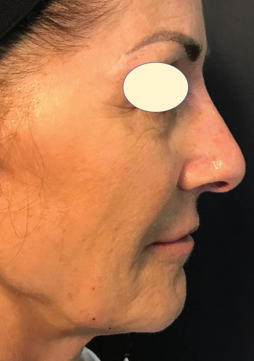
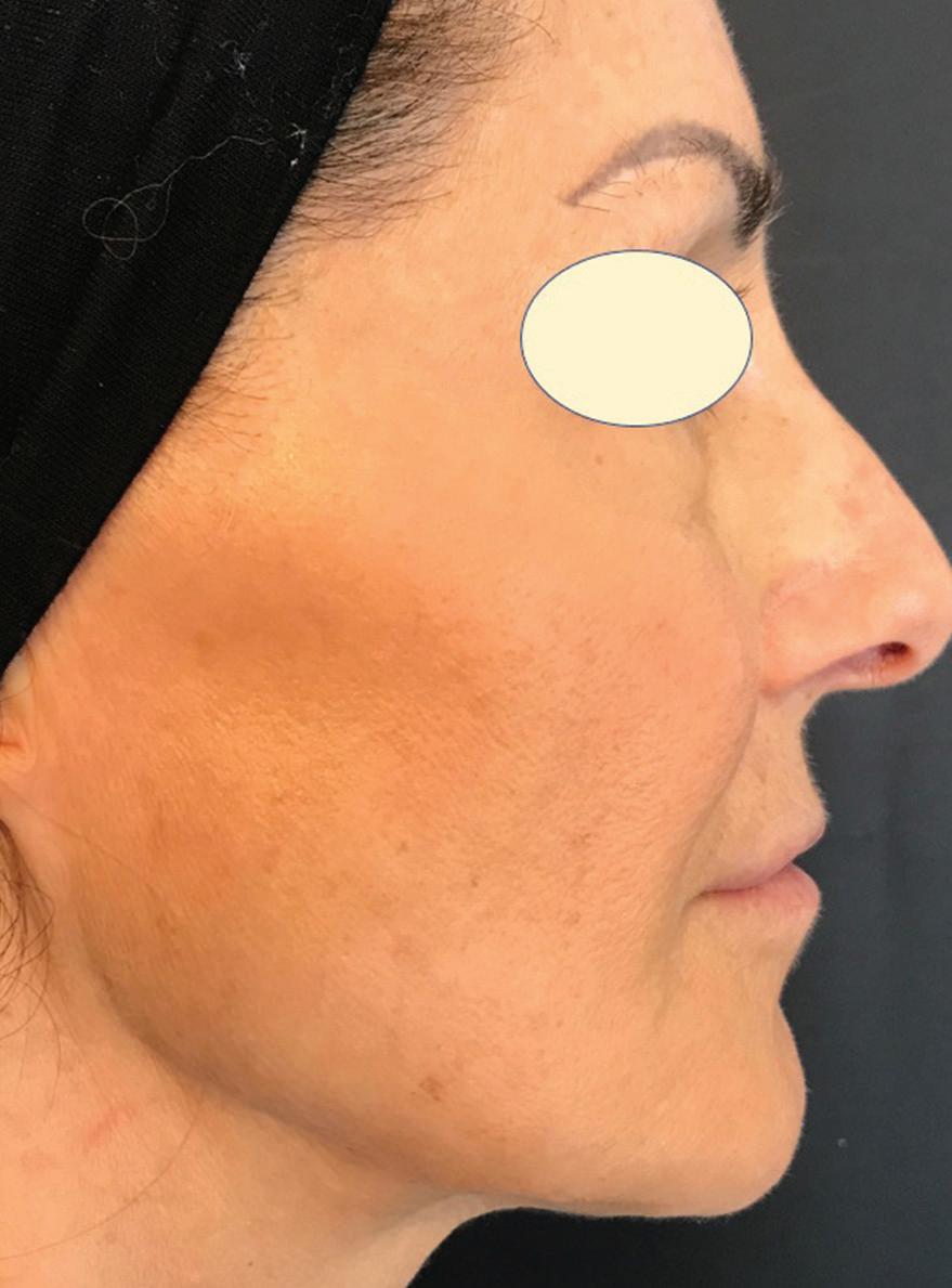
The final effect was very much appreciated by all patients who saw results immediately; this treatment, therefore, is suggested as the Gold Standard treatment in middle-aged female patients with moderate ptosis of the mandibular edge, with a thin or frankly skeletal face (Figure 6).
Key points
The face can be divided into upper, mid, and lower sections but also medial and lateral according to the line of ligaments
The concept that injection in one area of the face produces results in another area has now been confirmed (Domino effect).
Lateral and upper injection at the temples can lift medial tissues
Before injecting the mandibular edge and naso-labial folds it is better to inject the lateral and upper tissues (zygomas and temples)
Medical lifting of the face must be done with high G’ fillers to get an immediate but also long lasting result
The use of thick cannulas (at least 25G) is safer then thin cannulas or needles
Conclusions
Patient selection is the first essential element to obtain credible results. Ptosis that is too pronounced is not the right indication.
It is necessary to use a filler with specific rheological and volumetric characteristics to obtain visible and longlasting results.
The quantity of filler required for injection at the mandibular edge has been proven to be markedly smaller if the temples and cheekbones are filled first. However, the quantity of filler required for a full face result remains generally quite large, and the procedure is rather arduous for the patients.
The results are encouraging, but further studies are required to prove the difference in the quantity of filler injected and its effective duration over time.
Lastly, tolerability was excellent, and no significant side-effects were reported.
Declaration of interest Prof. Alessio is a KOL for Ipsen and Fillmed company.
Figures 3–6 © Redaelli Alessio, Table 1 © Redaelli Alessio
References
1. Hernandez C. et Al.: Clinical validation of the temporal lifting technique using soft tissue fillers. J Cosmet Dermatol 2020 Oct;19(10):25292535. doi: 10.1111/jocd.13621. Epub 2020 Jul 27. 2. Freytag L., Redaelli A. et Al.: Aging through Facial Biomechanics. A Clinically Applicable Guide for Improved Outcomes. Facial Plast Surg Clin N Am - (2022), https://doi.org/10.1016/j. fsc.2022.01.001 3. Casabona G. et Al: lifting vs volumizing. The difference in facial minimally invasive procedures when respecting the line of ligaments. JCD, 2019, Aug 12. doi: 10.1111/jocd. 13089. 4. Trevidick P. et Al: Prospective, Split-Face, Randomized, Long-Term Blinded Objective Comparison of the Performance and Tolerability of Two New Hyaluronic Acid Fillers. Dermatol Surg 2017 Dec;43(12):1448-1457. doi: 10.1097/DSS.0000000000001193 5. Suwanchinda A. et Al: The posterior temporal supraSMAS minimally invasive lifting technique using soft-tissue fillers. J Cosm Derm. 2018, Aug, 17(4): 617-24


