5 minute read
Mini hearts reducing heart disease risk

from In-SPHERE May 2023
by IN-SPHERE

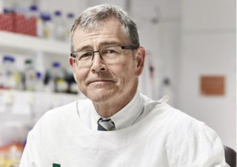
They may be tiny, but these mini hearts (pictured p14) are showing promise in helping reduce the risk of cardiovascular disease and death in women who have had gestational hypertension and pre-eclampsia in pregnancy
A team of researchers led by Associate Professor Lana McClements from University of Technology (UTS), in collaboration with St George Hospital and funded by SPHERE’s Cardiac and Vascular Health Clinical Academic Group, are seeking answers to explain why women who have had hypertensive disorders of pregnancy (HDP) are at an increased risk of developing and dying from cardiovascular disease (CVD).
And they’re doing it by creating mini hearts, composed of patients’ own blood cells, on which they can test biomarkers and treatments.
“Women who’ve had a hypertensive disorder of pregnancy have a 200300 per cent increased risk of developing CVD later in life than women who did not,” explains A/Prof McClements.
“Despite a well-established link between HDP and cardiovascular disease later in life, the mechanisms of this association are poorly understood. For this reason, we aim to re-create the complex CVD conditions associated with HDP by analysing our in-vitro bioengineered cardiac spheroid models of the human heart.
“If we can identify early signs of CVD at the molecular and cellular level, which can’t be picked up in the doctor’s office through regular tests like blood pressure monitoring, then we can potentially reduce the risk for these women by implementing correct therapy.”
Creating mini hearts
The first stage of the research involved developing 3D heart models specific to each patient. A/Prof McClements explains there are three main cell types in the heart: cardiac fibroblasts, cardiac endothelial cells and cardiomyocytes. Researchers extract stem cells from a patient’s blood with which they then create cardiomyocytes and cardiac endothelial cells. When these cells are combined with generic cardiac fibroblasts in a gel, researchers successfully create personalised cardiac spheroid models of the human heart.
“We then interrogate these models, at a molecular level, to see if there are any differences in the mini-hearts developed from women who had HDP five years prior compared with women who had normal healthy pregnancies.” The blood samples were collected at St George Hospital as part of the P4 study in collaboration with A/Prof Amanda Henry, A/Prof Greg Davis and Dr Lynne Roberts.
“Another way we did this is by incubating the generic mini hearts with blood plasma from women who had HDP or healthy pregnancies to see if it changed the way cells in the mini hearts contract which is a measure of heart function.
“What we found is that the hearts which had been incubated with plasma from women who had HDP contracted a lot faster which could be a sign of early heart dysfunction. We also observed that some of the inflammatory markers known to be linked to cardiac disease were also increased for women who had HDP,” explains A/Prof McClements.
“We plan to test some of the new and known preventative treatments on these models and see if we can inhibit these inflammatory markers.”
Having discovered these differences, researchers are now turning their attention to understanding what happens at the single cell level.
“What we’re doing now is RNA sequencing of individual cells to see if there are any differences in cells on a gene level.
“This is important as every organ including the heart is heterogenous with some sub-populations of cells showing certain patterns whilst others don’t. We want to understand in what specific types of cells are changes happening in women who had HDP compared to healthy controls, and how we can target these changes with treatments on a personalised basis.
What’s next?
This study is helping researchers not only understand the mechanism behind why women who had HDP have a higher incidence of future cardiovascular disease but also which biomarkers can be used to identify women at risk of developing CVD at an early stage.
“A blood sample can help us identify women who need closer monitoring, as well as identify therapeutic targets towards personalised treatment of women’s CVD,” says A/Prof McClements.
“Our unique approach combining expertise in cardiovascular and obstetric medicine, genomics and bioengineering will also allow us to fully understand, for the first time, HDP-induced cardiovascular disease and develop personalised monitoring platforms and treatment strategies to address the unmet needs of patients.”
Removing the need for animal research
The development of bioengineered hearts also removes the need for animal research.
“If we have patient-derived 3D models of hearts, we won’t have to use animals for research which is in line with the three R’s of animal research: Reduction, Replacement, Refinement. This is even more important considering the U.S. Food and Drug Administration no longer requires drugs to be tested on animals before human trials.”
The project is a collaboration between SPHERE’S Cardiac and Vascular Health Clinical Academic Group Maternal, Newborn and Women’s CAG, Diabetes, Obesity and Metabolic Disease CAG, Frontiers Technology CAG, SESLHD and UTS.

Read more: https://bsd.biomedcentral.com/ articles/10.1186/s13293-021-00376-1 https://www.mdpi.com/20734409/10/4/899