4 minute read
ESACROM Mandibular Bone Block Harvesting from the Retromolar Area with piezosurgery

from ImplantBook 2023
by Infodent srl

DDS Angelo Cardarelli Specialist in Oral Surgery
Member of Global Scientific Dental Alliance in Dubai
The reconstruction of alveolar defects after tooth loss is one of the biggest challenges in implant dentistry. In order to increase the bone thickness we can have several options alloplastic grafts, xenografts, allografts, and autografts. However, autogenous bone grafts are osteoinductive, osteogenic, and osteoconductive, with significant regenerative capacity in comparison to all other grafts. This is why autogenous bone remains the gold standard for augmentation. Extraoral donor sites for autogenous bone include the skull, the fibula, the ribs, and the iliac crest, all of which inevitably lead to additional patient morbility. Intraoral sources have the advantages of proximity of the donor and recipient sites, convenient surgical access, low morbility, and elimination of a hospital stay. The best anatomical area that allows to obtain a good cortical bone block grafts, suitable for two- or three- dimensional reconstructions of alveolar ridge defects is the retromolar and paramolar areas (external oblique ridge), or edentulous areas. The removal of large bone block grafts with drills or engraving or oscillating saws may be particularly dangerous in the anterior mandibular ramus. Piezosurgery is the state of the art bone cutting instrument in oral surgery and also in the harvesting of the ramus bone graft.The micro-oscillations, which are created at this frequency, cut only mineralized hard tissue while adjacent soft tissue, nerves and vessels remain unharmed. Using ultrasonic surgery, it is possible to cut mineralized tissue with greater precision and selectivity. Cavitation effect that is created by the irrigation/ cooling solution and oscillating tip of the device, provides blood- free surgical area, as a result greater visibility for the surgeon. With regard to bone formation and healing, it has been showed that ultrasound bone cutting is more favorable than it is with conventional bone cutting techniques.
Surgical Procedure
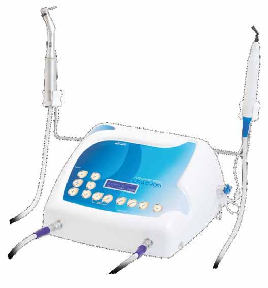
The procedure was performed under local anesthesia using Piezosurgery device (ESACROM Italy) with saw shape inserts. In the ramus zone, a midcrestal incision was performed, avoiding the lingual nerve trajectory. The donor area was exposed by extending a full thickness flap in the apical and distal aspect. Care was given to prevent any damage to the n. lingualis. For ramus bone harvesting, four osteotomies were made: one superior and 2 vertical as well as one osteotomy was made at the inferior border. The superior horizontal cut
By Dr. Andrea Tedesco
By Dr. Andrea Tedesco


was made 4 to 5 mm medial to the external oblique ridge with ES007lT Esacrom insert, cut was made from the edentulous area and continues posteriorly along the external oblique ridge to ascending ramus. The two vertical osteotomies, anterior and posterior, were also made with the ES007lT insert extending 10 to 12 mm in length in the supero inferior direction. Then, a cut connecting the inferior aspect of each vertical osteotomy was made with the angulated bone saw insert ES007LT. This special insert was exclusively produced for this inferior horizontal cut. After completed all the outline cuts of the graft, the harvest was usually pried out by gentle manipulation with a small flat chisel using hammer. fig 1-2 The donor site was primarily sutured back with 4-O SILK sutures. The patient received a single preoperative dose of oral antibiotics amoxicillin/ clavulanate sodium 2 gram, that same antibiotic regimen continued for 5 days postoperatively. Additionally, non steroidal anti-inflammatory agent naproxen sodium for pain and swelling, were prescribed as needed. Patient was also advised to rinse three times per day with 0.2% concentration chlorhexidine mouthwash for 7 days post-operatively. Extraoral application of a cold pack was recommended for 12 hours after the surgery. The harvested monocortical bone block was split in two pieces for horizontal augmentation in the maxilla and osteosynthesis screws were used to fix the plates bone to the recipient area. fig 3-4 The bone chips harvested from the bone block with the bone scrape were used to fill the gap around the blocks and the recipient bone. Any sharp edges or corners were rounded to avoid further soft tissue dehiscence.
Conclusions
Ultrasound surgery has certain advantages over traditional manual or high-speed motorized instruments in oral and maxillofacial surgery. Micro-oscillations of the tip of the device (ESACROM), operates in low frequency range allows for precise cutting and yields minimal wastage of bone. Low Frequencies causes minimal damage to soft tissues (nerves, vessels, mucosa). The unique phenomenon of cavitation effect gives operator a better visibility than using any conventional manual or rotary instrument. One of the important difficulties harvesting ramus block graft mentioned in literature is that managing the caudal horizontal cut due to close proximity of the IAN. With the specially angulated inserts of the device (ESACROM), ultrasound surgery has distinct advantage over conventional technique. Using this special tip for horizontal cut, surgeon does not need to reflect the flap extensively and making complete caudal cut is possible without damaging IAN.
References
1. Khoury F, Antoun A, Missika P. Bone Augmentation in Oral Implantology. Berlin, London: Quintessenz, 2007.
2. Zouhary K. Bone graft harvesting from distant sites: Concepts and techniques. Oral Maxillofac Surg Clin North Am 2010;22:301–316.
3. Nkenke E, Neukam FW. Autogenous bone harvesting and grafting in advanced jaw resorption: morbidity, resorption and implant survival. Eur J Oral Implantol 2014 Summer;7(suppl 2):S203–217.
4. Misch CM. Comparison of intraoral donor sites for onlay grafting to implant placement. Int J Oral Maxillofac Implants 1997;12:767–776.
6. Khoury F. Augmentation of the sinus floor with mandibular bone block and simultaneous implantation: A 6-year clinical investigation. Int J Oral Maxillofac Implants 1999;14:557–564.
I am passionate about new technology. exoplan enables me to use it effectively.
JEILMEDICAL Corporation provides a comprehensive line of high-quality products for Guided Bone Regeneration.
JEILMEDICAL has been developed in partnership with key opinion leaders to be innovative, technically advanced and lead to predictable patient outcomes.
GBR System consists of bone screws, bone-tac, mesh plate and instruments. Bone screws are made of a titanium alloy(ASTM F136), and the mesh is made of titanium(ASTM F67).

Bone Screws
Wide Head Screw (Ø1.4) – Fits better to GBR mesh and membrane thanks to the wider head.
Self-drilling Screw (Ø1.4 / Ø1.6) – Simplifies screwing process.
Tenting Screw (Ø1.6) – Creates and secures the space for bone graft
Mini Screw (Ø2.0) – Self-tapping screw
J-Tac – Tacking system for stabilizing a membrane on maxilla / mandible. Its dome-shaped head and barbed pin keep the membrane anchored on the bone after operation.
Meshes
Forms the space to protect the graft. Multiple pores on the mesh not only help blood supply but also minimize soft tissue exposures. Mesh can be cut to size depending on surgical needs.
Horizontal Smooth Bending Mesh
- Enables horizontal / vertical bone volume augmentation of implants site for guided bone regeneration. Its extra thin thickness and pre-cut design offer flexibility and shaping. Doctors can utilize this for multiple array of teeth cases, and also can fulfill bone formation and spatial retention for stable implant.
Fixation Hole Mesh
- Can be used as an alternative to a membrane, and also enables simultaneous GBR with dental Implant. Doctors can remove unnecessary edges.
www.jeilmed.co.kr global@jeilmed.co.kr // jamesjang@jeilmed.co.kr


