6 minute read
Sacroiliac Joint Pain
Sacroiliac Joint Pain
A common cause of low back pain, often under diagnosed, under treated for a problem that can be very debilitating.
by: Winston T. Capel, MD, MBA, FACS, FAANS
Low Back Pain (LBP) is a universal life experience for all of us. Most LBP is self-limiting and has a manageable baseline but commonly has exacerbations of varying severity and duration. Most LBP responds to observation, anti-inflammatories, heat, frequent changes in position and core conditioning. When LBP becomes intractable it is important to: exclude malignancy, infection or occult fracture.These sources of LBP make up to 5% of all LBP and need to be identified in order to preserve function, allow treatment when the delay of treatment can put patients in severe jeopardy of life and function. 95% of LBP is due to degenerative conditions and non-life threatening. Of these degenerative conditions, degenerative disc disease is the fundamental pathology and pain generator 75% of the time. Recent data identifies the Sacroiliac Joint (SIJ) as the responsible pain generator for the remaining 25%.
Given the frequency of LBP in the general population (2nd most common reason for primary care visits after the common cold) there are many people dealing with SIJ pain and dysfunction. Commonly there is significant delay in the accurate diagnosis of SIJ pain origin .
Sacroiliac Joint Pain
SIJ pain is more common in females; this is thought to be due to pregnancy and the pelvic adaptations of gestation and labor. SIJ is more common in patients that have had lumbar fusions. Pelvic trauma can also cause chronic SIJ pain.
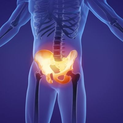
ANATOMY: The SIJ is the interface of the spine to the pelvis where major loads are transferred from the torso to the pelvis. This joint is broad with extensive ligament support. SIJ pain comes from arthritic changes in the joint, ligament or bone injury from trauma.The spine terminates at the sacrum the sacrum articulates with the pelvis at the important SIJ.

CLINICAL FEATURES: The back pain localizes low and off midline, this is indistinction to degenerative disc disease where patients localize their pain to more of a midline location. On exam patients will point to the SIJ as their pain source. It is always below L4-5 clinically.
• Pain is sensitive to different changes in pelvic loading like stairs, stooping, standing on one leg, ect.
• Leg pain: SIJ mediated leg pain can mimic radiculopathy from lumbar disc herniations. The key differences: SIJ leg pain starts in SIJ, is generally above the knee and without motor or sensory changes.
DIAGNOSIS: Patients will point to the SIJ on exam (Fortin’s Finger Test)
• SIJ pain is very sensitive to SIJ provocation on clinical exam where the joint is stressed.
• The diagnosis is a clinical diagnosis not by x-ray/MR imaging. When in doubt of the etiology of the leg pain an MRI lumbar should be obtained.
• The most definitive diagnostic method is a diagnostic block where the joint is blocked with a local anesthetic by an interventional pain specialist. Patients will experience an immediate > 80% or more reduction in their pain with the block which confirms the diagnosis
TREATMENT:
• Anti-inflammatories (NSAIDs) both oral and topical. This joint is relatively close to the surface and topical NSAIDS are safe adjunct to oral NSAIDs in most patients. Combined oral and topical Diclofenac is a good option unless medically contraindicated.
• Physical Therapy: exercise, manipulation and deep heat stimulation can help.
• Therapeutic Injection: at the time of the diagnostic block in the same syringe is corticosteroid which may relieve, control or give temporary benefit, its onset of effect is usually around 24 hours after the injection. There are 2 phases with the SIJ injection: diagnostic phase early and up to 8 hours after, then the diagnostic phase generally the next day.
• SIJ Belts: not commonly used due to limited effectiveness but some patients can have improvement of symptoms with the use of these belts that compress across the joint to reduce motion across the joint.
• Surgery: only for intractable pain that has been refractory to all non-surgical treatment. Many 3rd party payers require 2 positive diagnostic blocks and an MRI of the lumbar spine to exclude lumbar pathology.
Surgical Outcomes for SIJ Fusion
Prior to the development of highly effective minimally invasive SIJ fusion procedure, SIJ fusion was a large and open procedure with outcomes that rarely justified SIJ fusion except for pelvic trauma with overt instability.
The iFuse SIJ Fusion Technique was introduced in 2009 and was the first minimally invasive and the primary evidenced based technique today. Over 55,000 iFuse fusions have been done worldwide. It is one the best studied spinal surgical techniques in contemporary spine surgery with multiple randomized, prospectively controlled studies (Class I data) demonstrating efficacy and safety. The surgery is almost always performed on an outpatient basis with high level patient satisfaction. 82% of patients at 12, 24 and 60 months would “have the procedure again for the same result.” The technique is done with the patient in the prone position under fluoroscopic guidance (C arm) using 4 distinct views of the pelvis to navigate k-wires across the joint avoiding important structures like the sacral neuroforamina and sacral boundaries. Implants are passed across the joint over the k-wires after drilling and broaching. The implant immediately immobilizes the joint eliminating painful motion. The implant is designed for maximally osteogenesis leading to permanent bone growth across the joint. The risks of the procedure are low but include: bleeding, infection, nerve injury and failure of fusion.

Surgical Positioning of Implants Across the Joint
After surgery patients have restricted weight barring across the joint (touchdown only) with the use of crutches or a walker for 4 weeks. During the 5th week after surgery patients progressively add weight, if there is no pain, patients can bare full weight but avoid running, jumping and heavy lifting (>15-20 lbs.) until completely healed at approximately 3-6 months. There is generally slower healing in patients with osteoporosis and may require longer periods of restricted weight barring.

In summary, SIJ pain is common with effective tools for accurate diagnosis and effective treatment. These patients can have very incapacitating pain and if non-surgical treatments are ineffective in controlling pain and improving function the above described surgical procedure leads to highly satisfied patients with dramatically increased quality of life through a minimally invasive, well studied and effective procedure.
Alabama Bone & Joint Clinic
Winston T. Capel, MD, MBA, FACS, FAANS
205 621 3778


