CONTENTS International Microbiology (2016) 19:69-132 ISSN (print): 1139-6709. e-ISSN: 1618-1095 www.im.microbios.org
Volume 19 · Number 2 · June 2016 · pp. 69-132 RESEARCH REVIEWS
Núñez A, Amo de Paz G, Rastrojo A, García AM, Alcamí A, Gutiérrez-Bustillo AM, Moreno DA Monitoring of the airborne biological particles in outdoor atmosphere. Part 2: Metagenomics applied to urban environments
69
Romero D Unicellular but not asocial. Life in community of a bacterium
81
RESEARCH ARTICLES
Lasa A, Mira A, Camelo-Castillo A, Belda-Ferre P, Romalde JL Characterization of the microbiota associated to Pecten maximus gonads using 454 pyrosequencing
93
Thellin O, Zorzi W, Zorzi D, Delvenne P, Heinen E, ElMoualij B, Quatresooz P Lysozyme as a cotreatment during antibiotics use against vaginal infections: An in vitro study on Gardnerella vaginalis biofilm models
101
Keuter S, Rinkevich B Spatial homogeneity of bacterial and archaeal communities in the deep eastern Mediterranean Sea surface sediments
109
Thummeepak R, Kongthai P, Leungtongkam U, Sitthisak S Distribution of virulence genes involved in biofilm formation in multi-drug resistant Acinetobacter baumannii clinical isolates
121
BOOK REVIEWS PIONEERS IN MICROBIOLOGY: Paulina Beregoff (1902–1989), Colombia
131 A2
Journal Citations Reports 5-year Impact Factor of International Microbiology is 2,17. The journal is covered in several leading abstracting and indexing databases, including the following ones: Agricultural & Environmental Biotechnology Abstracts; ASFA/Aquatic Sciences & Fisheries Abstracts; BIOSIS; CAB Abstracts; Chemical Abstracts; SCOPUS; Current Contents/Agriculture, Biology & Environmental Sciences; EBSCO; EMBASE/Elsevier Bibliographic Databases; Food Science & Technology Abstracts; ICYT/CINDOC; IBECS/ BNCS; ISI Alerting Services; MEDLINE/Index Medicus; Latindex; MedBioWorld; PubMed; SciELO-Spain; Science Citation Index Expanded; SciSearch.
A1
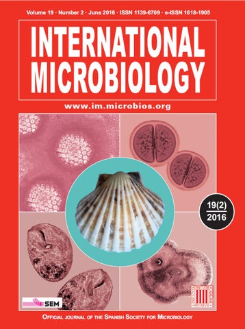
Front cover legends years, and become reproductively active at 3–5 years. The shell of great scallops commonly grow to a size of 120–210 mm width. [See article by Lasa et al., pp. 91-97 this issue.] Upper left. Papillomavirus, the causal agent of several human diseases, some of them developing as cancers. Several Spanish groups perform outstanding research on this virus and on the illnesses that it causes. The definitive link between the presence of the papillomavirus and cervix cancer in women was established by Colombian physician and researcher Nubia Muñoz, in Lyon, France. (Magnification, 600,000×)
Center. The great scallop (Pecten maximus) is a bivalve mollusc of important value in aquaculture. Great scallops are also known as St James Shell, due to the fact that in ancient times, pilgrims to the shrine of St James in Santiago de Compostela, Spain carried a scallop shell with them. They could expect to receive as much food as they could gather from one scoop of the shell at households along their journey. Great scallops can live for up to 20
being engulfed near the cytostome of the cell on the left. Photo by Rubén Duro, CIM, Barcelona. (Magnification, 3000×) Lower right. Macrophotograph of a growing colony of the mold Aspergillus sp. The colony is growing in a Petri dish. Note the whitish, button -like structure formed by a drop of liquid secreted by the sector on the left. Photo by Rubén Duro, CIM, Barcelona. (Magnification, 1.4×)
Upper right. Dark field micrograph of the cyanobacterium Chroococcus sp., isolated from a freshwater pond. Note the envelope surrounding the paired cells. Photo by Rubén Duro, Center for Microbiological Research (CIM), Barcelona. (Magnification, 1500×) Lower left. Dark field micrograph of the predator ciliate Pseudoprorodon sp., isolated from a freshwater lake. Note the pieces of food inside the large digestive vacuoles and the small ciliate
Back cover: Pioneers in Microbiology Paulina Beregoff (1902–1989), Colombia Paulina Beregoff was the first woman to obtain a degree in medicine in Colombia. She was born in 1902 in Kiev—by then a city of the Russian Empire—, in an aristocratic family of Jewish descent. Due to the political situation in her country, she was educated in the United States, where, in 1921, she graduated in Bacteriology and Parasitology and Pharmacy and Chemistry at the University of Pennsylvania. She started working at the laboratory of Pathology of that university and became a member of the Rivas Bacteriological Society of the University of Pennsylvania. In 1922, the Dean of the School of Medicine of the University of Cartagena, Colombia, asked the University of Pennsylvania for an expert in tropical diseases, including yellow fever. This disease was a great concern in Cartagena due to the high mortality rates it caused and because of the implications on the image of the city, which was a major commercial and harbor center. The University needed a qualified advisor that could also train local physicians, and the University of Pennsylvania chose Beregoff for that task. Once in Cartagena, she had to identify an epidemic outbreak that had been causing many fatalities, mostly among indigenous peoples living in the Magdalena River shores. Colombian phys icians were not familiar with symptoms and causal agents of diseases such as yellow fever, typhoid fever and malaria, but thought that the epidemic outbreak could be due to one of them. Beregoff sent samples of cultures
from corpses of people killed by the disease to be analyzed at the University of Pennsylvania. The disease turned out to be fiebre tifomalárica and not simply malaria, as they first had considered. Beregoff thought that the infection depended mostly on the deficiencies or resistance of the immune system and proposed that physicians should work to prevent the disease. Once she had achieved her task, she intended to go back to Philadelphia to study medicine at Temple University, but she was asked to remain in Cartagena, where she could also study medicine. In 1922 she enrolled at the University of Cartagena under special conditions. Due to her previous studies and qualification, she could be waived the first two years of the studies of medicine. She set up the first laboratories of bacteriology and parasitology in Cartagena, with microscopes and other equipment donated by the University of Pennsylvania. Her thesis director recognized her great contribution, she having been able to differentiate the various species of Laveran’s haematozoa, to observe the treponema causing yaws, to find the Piroplasma Donovani, the parasite of KalaAzar (visceral leishmaniasis) in the blood, and having been the first to isolate the “typhoid bacillus”, confirming thus the presence of typhoid fever in town. She could also to properly perform the Wassermann technique on syphilis. The fact that she was a foreign woman and the she had had some privileges in her medicine studies was criticized by some people. In 1933 she married bacteriologist Arthur Stanley Gillow and they moved to Canada. Since then she signed her publications as Pauline Beregoff-Gillow. After her husband’s death, in 1964, she returned to Colombia and dedicated his husband’s legacy to set up a foundation under his name that should work on preventive medicine. She died on September 20, 1989 and left her fortune to the foundation.
Front cover and back cover design by MBerlanga & RGuerrero
A2
RESEARCH REVIEW International Microbiology (2016) 19:69-80 doi:10.2436/20.1501.01.265. ISSN (print): 1139-6709. e-ISSN: 1618-1095
www.im.microbios.org
Monitoring of airborne biological particles in outdoor atmosphere. Part 2: Metagenomics applied to urban environments Andrés Núñez,1 Guillermo Amo de Paz,2 Alberto Rastrojo,3 Ana M. García,1 Antonio Alcamí,3 A. Montserrat Gutiérrez-Bustillo,2 Diego A. Moreno1* 1 Higher Technical School of Industrial Engineering, Technical University of Madrid, Madrid, Spain. 2Department of Plant Biology II, Faculty of Pharmacy, Complutense University of Madrid, Madrid, Spain. 3Center of Molecular Biology Severo Ochoa, CSIC-UAM, Madrid, Spain
Received 20 March 2016 · Accepted 20 April 2016 Summary. The air we breathe contains microscopic biological particles such as viruses, bacteria, fungi and pollen, some of them with relevant clinic importance. These organisms and/or their propagules have been traditionally studied by different disciplines and diverse methodologies like culture and microscopy. These techniques require time, expertise and also have some important biases. As a consequence, our knowledge on the total diversity and the relationships between the different biological entities present in the air is far from being complete. Currently, metagenomics and next-generation sequencing (NGS) may resolve this shortage of information and have been recently applied to metropolitan areas. Although the procedures and methods are not totally standardized yet, the first studies from urban air samples confirm the previous results obtained by culture and microscopy regarding abundance and variation of these biological particles. However, DNA-sequence analyses call into question some preceding ideas and also provide new interesting insights into diversity and their spatial distribution inside the cities. Here, we review the procedures, results and perspectives of the recent works that apply NGS to study the main biological particles present in the air of urban environments. [Int Microbiol 19(2): 69-80 (2016)] Keywords: airborne biological particles · metagenomics · next-generation sequencing (NGS) · air biomonitoring · urban aerobiology
Introduction Worldwide population is coarsely concentrated in urban environments where people are exposed to allergens and pathogens transported by the air like pollen, fungi, bacteria and viruses. Pollen and fungal spores may come from distant natural locations surrounding the metropolitan areas, while airborne active pathogenic bacteria and viruses come likely from closer sources inside the cities. We scarcely know the Correspondence: D.A. Moreno Escuela Técnica Superior de Ingenieros Industriales Universidad Politécnica de Madrid (ETSII-UPM) José Gutiérrez Abascal, 2 28006 Madrid, Spain Tel. +34-913363164. Fax +34-913363007 *
E-mail: diego.moreno@upm.es
biodiversity present in urban areas and, strikingly, a reliable automatic system has not been developed yet in order to monitor the bioaerosols present in the air, partly because of the low concentration of the biological particles and sample collection difficulties [23]. Moreover, the different airborne biological particles are usually studied by different disciplines independently and, hence, we have a lack of information concerning the existence of relationships about their relative abundance or fluctuations to each other. During the last years, metagenomics have increased significantly our knowledge on the biodiversity in every environment compared with conventional methods. Current technologies in sequencing are not restricted to bacteria, so any biological sample with a complex mixture of organisms can be analyzed. Thus, next-generation sequencing (NGS) offers an interesting alternative to study the metropolitan
70
Int. Microbiol. Vol. 19, 2016
NÚÑEZ ET AL.
atmosphere and uncover the real diversity present in the air. Metagenomics and the technologies related have simplified the steps required to characterize a particular environment like the urban air (Fig. 1). Here, from sample collection to taxa characterization, we review the current knowledge about airborne biodiversity in urban environments from DNA sequencing-based studies.
Traditional studies in microbiology for airborne bacteria and fungi are usually culture-based methods, whose bias concerning to biodiversity is widely known. Although several commercial devices are available, they are usually designed to fulfill air control legislation indoors, with fixed times and/ or volumes to sample. Sometimes it is the collection surface that determines these factors, like agar media, which tent to dehydration. Unfortunately, and according to our estimations, higher sampling volumes and times than those preselected in these devices may be required to study some biological particles present in the outdoor atmosphere by NextGeneration Sequencing (NGS) [55]. In addition to vacuum filtration used in outdoor urban and also non-urban areas [8,9,18,51,70], most metagenomic studies conducted in metropolitan environments employ Particulate Matter (PM) collectors, as those operating in air quality monitoring stations [7,10,14,25,56] (Table 1). The particles are harvested in fiber filters at flows >200 l/min and the DNA is subsequently extracted. Some samplers employed typically in aerobiological studies for visual fungi and pollen identification (Hirst-type spore trap, Fig. 2) have been also recently tested in NGS studies by some authors including us [27,43]. The particles are collected on an adhesive tape and the airflow rate is significantly lower than the formers (ca. 10 l/min), but closer to human inhalation rate. Thus, solid surfaces seem preferable to collect airborne biological particles when DNA-sequencing technologies are applied in urban environments. Only a few works have employed liquid collection [20,74], likely because water-based buffers tend to evaporate quickly, restricting the airflow rate and sampling time. However, new devices as the Spin-Cyclon used by Yooseph et al. [77] look an interesting option, with an airflow rate of 450 l/min. Fahlgren et al. [17] performed a study comparing the results obtained from three different devices (a modified impactor, a liquid impinger, and a Teflon membrane filter), concluding that there are no significant
Int Microbiol
Sampling procedures in NGS studies applied to urban atmosphere
Fig. 1. Workflow scheme for metagenomic studies applied to air samples designed for the Program AIRBIOTA-CM.
differences on the bacterial diversity and dominant species based on 16S rRNA sequences analyses. However, Hoisington et al. [33] conducted a similar survey indoors employing and comparing diverse air samplers, finding significant disparities in the OTUs of bacteria and fungi detected by each device. Thus, more studies analyzing the concordance between the results of different sampler-types for NGS analyses and also for different biological particles should be performed to confirm and standardize the sampling methodology.
BIOAEROSOLS METAGENOMICS OUTDOOR
DNA extraction In addition to the different methodology for sampling, the DNA extraction procedure is something to take into account. Quality and high concentration are desirable for NGS analysis but difficult to obtain when samples come from poorly inhabited environments like the air. Some studies comparing different methods suggest that there are remarkable differences regarding yield and purity, which could bias the further analysis [30], although it has been also exposed that DNA yield does not always correlate with differences in microbial diversity after the analysis [78]. The debate is even more controversial regarding DNA extraction
71
A
B
Int Microbiol
In addition to the variety in sampling devices, no consensus exists about the volume of air to analyze. Strikingly, works applying NGS to urban areas have employed air samples that range from 2.5 to 5000 m3 (see Table 1). Fahlgren et al. [18] for bacteria, and Fierer et al. [20] for both bacteria and fungi, were able to performed microbial identification by cloning the DNA collected in air samples of only ≤3 m3. Less than 10 m3 are necessary to conduct high-throughput sequencing according to the data provided by Kraaijeveld et al. [43] and Woo et al. [75], who employed different samplers and analyzed airborne eukaryotic organisms. In regards to viruses, Whon and colleagues [74] were able to carry out a metagenomic study with 54 m3 of air in the near-surface atmosphere. On the contrary, Yooseph et al. [77], despite the fact that they sampled large volumes of outdoor air (5900 m3 at the 22nd floor air, 101 m above street level in Midtown Manhattan, New York City) did not obtain enough DNA, highlighting the influence of the altitude on airborne biological particles abundance. Diverse sampling times have been also selected for each author, without any correlation with the final volume of air collected (see Table 1). Consequently, each work analyzes specimens from different devices, with different volume collected and different times sampled, what may lead the results between studies to differ, especially regarding the minor representatives of the airborne communities. As we previously reviewed [55], biological particles abundance and diversity vary significantly across the year and they are also susceptible of quick changes (in days or even hours). Therefore, sampling volume and time are two variables to consider in order to obtain a representative sample of the “airbiota� in a certain area, and it may be necessary to modify these parameters throughout the time and depending on the purposes of the study.
Int. Microbiol. Vol. 19, 2016
Fig. 2. (A) Volumetric Hirst-type spore trap for fungi and pollen collection employed in aerobiological studies placed on the roof of the Higher Technical School of Industrial Engineering, Technical University of Madrid (Madrid, Spain). (B) Agarose gel showing the results of the PCRs to detect the DNA from different groups of organisms obtained with such collector after sampling for 7 days (50 m3) in April 2015 (unpublished data from the Program AIRBIOTA-CM).
for viral metagenomic studies because of the difficulty of the procedures for purifying these organisms [5,72]. While some traditional methods (phenol:chloroform extraction or CTAB procedure) are still in use to study airborne microorganisms [18,51,63,74], the tendency is to incorporate commercial DNA purification kits when NGS technology is applied [4,7,9,10,68,70,76] (see Table 1). Nowadays, commercial kits provide similar quality than handling methods, saving time and preventing manipulation issues. A common factor is to include mechanical disruption such as a bead-beating step in addition to chemical disruption [78]. Accordingly, Jiang et al. [36] have recently described a protocol for purifying DNA from particulate matter to identify airborne microorganisms by metagenomics, which includes the use of a commercial kit for the DNA extraction with some modifications. Nonetheless, one remarkable conclusion from Hart et al.
72
Int. Microbiol. Vol. 19, 2016
[30] is that the method for DNA extraction must be amended for each case. This is particularly important when we require the characterization of different types of organisms (with their physical and chemical particularities) coming from a complex habitat like urban air.
Next-Generation Sequencing (NGS) Clinical and environmental samples containing a complex combination of organisms (viruses, archaea, bacteria, fungi, animals and/or plants) can be currently analyzed at once thanks to metagenomics, saving time and avoiding the previous bias of culturing. Traditional studies in aerobiology can also take advantage of this new branch of genomics to identify fungi and plants that are hardly assigned to low taxonomic levels (genus or species). Moreover, this novel methodology is particularly interesting for those unculturable organisms or obligate-intracellular parasites such as viruses or zoonotic pathogens from the genera Rickettsia, Coxiella or Chlamydia. Although some recent DNA-based identification studies still apply traditional molecular stages (cloning, Sanger sequencing of RFLPs, etc.), current high-throughput DNA sequencing technologies permit to skip these steps. 454 pyrosequencing (Life Science, Roche) was the pioneer platform for the so-called next-generation sequencing (NGS). While it is still in use, more recent platforms as Illumina (MiSeq/HiSeq), Ion Torrent/Proton (ThermoFisher Scientific), MinION (Oxford Nanopore Technologies) or PacBio RS system (Pacific Biosciences) have quickly replaced the former, improving the cost, timing and reliability [26]. Two main approaches exist in environmental NGS studies: • Targeted Amplicon Sequencing (TAS). This strategy implies the selection of a target region of the DNA to analyze. Any region can be chosen but it is usually one present in all the organisms to study, especially in biological diversity surveys (16S rRNA gene for bacteria or 18S rRNA gene/ITS for eukaryotic organisms). The technique requires a previous PCR amplification step by using specific primers for the preferred region. “Universal primers” have been described for each group of organisms (archaea and bacteria [2], fungi [67,73], eukarya [35], plants [13,34]), and the use of degenerated oligonucleotides reduces the bias for sequence poly morphisms. Unfortunately, there is always a fraction
NÚÑEZ ET AL.
of sequences that are amplified with less efficiency, so some groups of organisms can be underestimated or undetected while others are magnified, sloping the relative abundance. Furthermore, this methodology is limited for viral identification because these organisms do not share any common marker gene (such as 16S rRNA in bacteria or 18S rRNA in eukaryotes), so a shotgun approach is the only way to uncover viral communities [64,74]. Despite these limitations, targeted sequencing is currently widely adopted for biological diversity characterization. • Shotgun metagenomics (see Sharpton [69] for a full review). The entire DNA present in the sample is sequenced without specific-region enrichment, reducing the bias introduced in the other modality. Although bioinformatics analyses after sequencing are challenging, the results from this strategy include taxonomic biodiversity (mainly from ribosomal DNA fragments) and biological functions (from DNA coding sequences). This approach, frequently used for whole genome sequencing (WGS), is especially interesting for assembling genomes, functional metagenomics, viral identification and global biome characterization, providing also absolute abundance information. In regards to urban environments, far from exclusive, both approaches are complementary, offering critical knowledge for results interpretation. However, because of the cost, the difficulties to obtain enough DNA from some areas and the complexity of the bioinformatics analyses for shotgun analyses, TAS is the favored strategy to study microbial communities in urban airborne spaces (see Table 1).
Specific marker genes for sequencingbased analyses Although 16S rRNA gene (SSU) is widely accepted as a target for bacterial identification in metagenomic studies, one issue that remains controversial is the region to sequence to acquire the best representation. Ideally, the complete gene should be sequenced to obtain accurate taxonomic assignments. Since the diverse hypervariable regions present in SSU show different variability they provide different level of taxonomic discrimination [50]. Several studies involving direct cloning of the 16S rRNA gene from airborne microorganisms were published between 2008-2013 [17,18,20,58,60,61]. Universal primers 27F-1492R encompassing V1-V8 regions of the 16S rRNA [45] were very suitable for these analyses, providing
BIOAEROSOLS METAGENOMICS OUTDOOR
the first results extracting the microbial DNA directly from the collection surface. However, the current NGS tendency is to analyze the shortest length necessary to obtain good taxonomic identification, reducing the cost for sample and time for further bioinformatic analyses [26,42]. Accordingly, fragments comprising one or several regions within V1窶天5 hypervariable span have been successfully used for both environmental and clinical studies [42,47,50,52], discarding V6窶天9 regions for providing less resolution. Chakravorty et al. [12] and Salipante et al. [66] have demonstrated that the V1窶天3 regions are more suitable for clinical studies, distinguishing important pathogenic bacterial species from the genera Staphylococcus, Mycobacterium, Streptococcus or Haemophilus. In recent published works using NGS, the amplification of fragments covering total or partial V1窶天4 regions is favored to study airborne bacterial communities in urban environments (Table 1). Although the results from any of these regions may be acceptable within the same study, a global consensus about the specific hypervariable regions to analyze is required for comparing among different studies in order to extract correct conclusions. Similar to bacteria, ribosomal RNA is the favorite region for fungal taxa identification. Three regions: LSU (25S-28S), SSU (18S) and internal transcribed spacers (ITS1 and ITS2), are the most popular options for DNA sequence-based studies, being the latter (ITS2) proposed as universal barcode marker by the Fungal Barcoding Consortium [67]. Only a handful of studies performed in urban environments have analyzed fungal communities by NGS technologies (Table 1). Some authors have used LSU regions, domains D1 and/or D2 [56]; and some others the18S and/or ITS fragments [14,70,75,76]. Again, this dichotomy in the selected region makes more difficult to compare among different studies and extract significant conclusions, since the results obtained from LSU and ITS may not match completely [49]. Defining a DNA barcode for plants is a challenging task and the debate is still on. Starting with the genomic DNA region ITS2 [13,73], latest reports are focused on plastid genes such as rbcL, matK, trnH-pbsA, trnL or a combination of two for obtaining an undisputable taxonomic identification at species level [6,34]. Although affordable for individual analyses, current NGS platforms cannot integrate two distant markers at the same time. Sequencing-based identification is an attractive approach for pollen identification since morphological characters are not always sensitive enough to distinguish genus or species by microscopy (the plant families Poaceae or Chenopodiaceae, for instance). Just a few works
Int. Microbiol. Vol. 19, 2016
73
have been recently published evaluating NGS technologies for plant identification using trnL as gene marker [43], ITS2 [40], or rbcL [24,31], being the former the only one that analyzes urban airborne samples and generates promising results for using DNA sequence-based strategy as an alternative to traditional methods for pollen identification. As pointed above, viruses do not possess common sequences to use as a marker to design universal primers, so the shotgun approach must be adopted. In addition, the small genome size of viruses is another limiting factor for recovering enough DNA to carry out the analyses. Thus, previous random amplification procedures as Multiple Displacement Amplification (MDA) [46], or SequenceIndependent Single-Primer Amplification (SISPA) [38], are usually performed to increase the quantity of DNA or RNA, respectively. However, both techniques have some important biases [46,65]: MDA amplifies more efficiently circular DNA molecules than linear ones; and the SISPA method has a biased amplification that depends on the sequence of the primer used. Unfortunately, the best way to avoid such biases is, so far, to obtain sufficient viral biomass to sequence the sample directly. As a consequence, only Whon and colleagues [74] have confronted a complete airborne study of the viral communities in different locations in Korea using shotgun metagenomics, while two others surveys conducted in urban areas detected some viral sequences but not as a main goal [4,11].
Bioinformatics Open-source programs and web-based tools for bioinformatics processing have been strikingly promoted by NGS technology. A complete review about the analytical tools applied in DNA sequence-base studies was published by Kim and colleagues [41]. MEGAN, RDP Classifier, Mothur, QIIME or Muscle are some examples of software suites frequently used to process high-throughput sequencing data in addition to BLAST. Some of them require an open-access database to assign taxonomic affiliations to the sequences. Greengenes for bacteria, UNITE for fungi, and Silva for bacteria and eukarya, are the most popular curated databases for stand-alone workflow, all relying on the sequences and annotations submitted to GenBank and regularly updated. After the bioinformatics processing, DNA sequences are assigned to taxonomic classification, providing the information necessary to characterize the diversity of each biological community in our samples.
Portable Sampling Unit (BioWatch Program) High-volume aerosol samplerb High-volume aerosol samplerb High-volume aerosol samplerb Low volume gravimetric sampler High-volume aerosol samplerb Burkard volumetric spore trap High-volume aerosol samplerb 8-stage non-viable impactor
[4]
Two-stage bioaerosol cyclone sampler
[75]
3.5 l/min
19 l/min
24 l/min
28.3 l/min
30 l/min
10 l/min
200 - 500 l/min
38.33 l/min
1,130 l/min
1,130 l/min
250 l/min
ND
Flow rate
12 h (2.5 m3)
48 h (54 m3)
10 h (28.8 m3)
24 h x 10 days (360 m3) 3 days (5,000 m3) 72 h (4881 m3) 24 h (55 m3) 24 h (288 - 720 m3) 24 h (7.2 m3) 24 h (43 m3) 4 days (163 m3)
Time/Vol collecteda 24 h x 7 days UltraClean Soil DNA Isolation Kit1
Filter
PBS
0.45 μm cellulose ester filters
Chloroform, MiniElute Virus spin Kit3 DNeasy Plant Mini Kit3
FastDNA Spin for Soil Kit2
16S: V3-V4 18S: V1-V3 ITS: ITS1-2 ITS: 5.8S-ITS2
B E
NA
ITS: ITS2
F All
16S: V1-V3
16S: V3-V4
28S: D1-D2
plastid:TrnL
16S: V5-V6
16S: V3
ITS: ITS1-2
16S: V2
16S: V5-V6
Region sequenced NA
B
B
F
P
B
B
F
B
B
All
Extraction method Org.
Quartz fibre filter Kit FastDNA Spin for Soil2 Quartz fibre filter PowerSoil DNA isolation Kit1 Quartz fibre filter PowerSoil DNA isolation Kit1 PTFE filter FastDNA Spin for Soil Kit2 Quartz fibre filter FastDNA Spin for Soil Kit2 Adhesive tape QIAamp DNA Mini Kit3 0.2 μm Polyester UltraClean Soil membrane filters DNA Isolation Kit1 Uncoated PCTE phenol/chloroform filters protocol
Collection surface Filters
TAS
Shotgun
TAS
TAS
TAS
TAS
TAS
TAS
TAS
TAS
TAS
Shotgun
Approach
454
454
454
454
454
Ion
Illu
454
454
454
Illu
Illu
Seq. technol
Silva
RDP
CAMERA, NCBI
EzFungi, UNITE
Greengenes, RDP
RDP
NCBI
RDP
RDP
NCBI
RDP
RDP
NCBI
Database
Two-stage non28.3 l/min 4 weeks Glass fiber filter PowerMax Soil F TAS 454 NCBI viable Andersen (1,141 m3) DNA Isolation Kit1 sampler [77] SpinConHPAS 450450 l/min 7 days PBS MO BIO All NA Shotgun 454 NCBI 10A Wet Cyclone (5,900 m3) PowerWater Kit1 Portable Air B 16S: V3-V5 TAS Sampler 1 : MoBio Laboratories, 2: MP Biomedicals, 3: Qiagen; NA: Not applicable; ND: Not described; TAS: Targeted amplicon-sequencing; NCBI: National Center for Biotechnology Information; RDP: Ribosomal Database Project .Organisms: B (Bacteria); F (Fungi); P (Plants); E (Eukaryotes). Sequencing Technology: Illu (Illumina); 454 (454-Roche); Ion (ion Torrent). a: Estimated when not specified; b: Particulate matter collector
Impinger
[74]
[76]
Vacuum filtration
[70]
[63]
[56]
[43]
[25]
[21]
[14]
[10]
[7]
Sampler
Ref.
Table 1. Studies of airborne organisms in urban environments implying direct purification of the DNA and NGS analysis (studies about particular events such as dust storms have been omitted)
74 Int. Microbiol. Vol. 19, 2016 NÚÑEZ ET AL.
BIOAEROSOLS METAGENOMICS OUTDOOR
Lessons from DNA-sequencing studies in urban spaces Abundance of microorganisms in the air. It is well characterized that some biological entities present in the air suffer important seasonal fluctuations as the noticeable variation in abundance and composition of the pollen grains throughout the year [28]. Likewise, culture-dependent studies performed in urban areas suggest that fungi and bacteria are, in general, more abundant in summer than in winter [19,37,54]. Recent culture-independent studies are in concordance with these results. Woo et al. [75] described that there is a peak of DNA concentration in the air in urban environments during summer season (June to September) independent on rural or urban environments. Consistently, some other authors have used different approaches as DAPI [10], shotgun sequencing [4] or qPCR [7] to show that airborne bacteria abundance is higher in summer, only outnumbered in spring by the group Plant-Fungi [4]. Moreover, both Dannemiller et al. [14] and Frรถhlich-Nowoisky et al. [22] also agree on the fact that fungal richness is higher in spring, being reduced in winter. However, Fierer et al. [20] showed that some airborne organisms are submitted to more rapid variations in metropolitan areas. In this study, they observed that the abundance of bacteria and fungi changes significantly within a period of 10 days, based on cloned sequences. Similar results were obtained by Shin et al. [70] and Oh et al. [56] using NGS. Furthermore, some culture-based studies have reported that daily precipitations can affect fungal abundance during the following days [32], and even diurnal oscillations for bacteria have been described [19,48,79]. Thus, some divergences between studies may be obtained depending on environmental factors as the weather or the time of day when the sampling is performed. Relative abundance of different taxa. Contrary to culture-based studies in which spore-forming Gram-positive bacteria (e.g. Bacillus, Micrococcus) are usually the most abundant group identified outdoors [44,48,79], NGS has proved this is not necessarily real, showing an unexpected diversity of Gram-negative bacteria [18,25,75,77]. A general conclusion (independent on shotgun or TAS approaches) is that the phyla Actinobacteria and Proteobacteria are the most abundant in urban outdoor environments, followed by Firmicutes and Bacteroidetes [7,18,21,25,70,75]. Interestingly, controversial results using NGS have been found within the fungal kingdom. It is unclear which group is
Int. Microbiol. Vol. 19, 2016
75
more abundant in metropolitan spaces. Several authors have reported that the abundance of Ascomycetes in metropolitan spaces is greater than Basidiomycetes, and they are at all seasons [14,56,75]. The opposite is shown by other authors like Shin et al. [70] and Pashley et al. [57], supported by morphological identification, and these contradictory results are independent on methodology (cloning, TAS, shotgun) or the DNA region considered (LSU, ITS or 18S). However, as underlined above, some works suggest that significant changes in fungal abundance may occur within short periods of time (days or even hours). Since most of NGS analyses only register a few isolated days, both conclusions may be plausible. To shed some light on the matter, some morphology and culture-based surveys performed systematically along the year concluded independently that Ascomycetes spores are the main group in urban areas, being Cladosporium the most abundant fungal spore [15,16]. Additionally, it is widely accepted that Dothideomycetes (in which Cladosporium is included) is the most represented taxa in Ascomycetes, as Agaricomycetes is within the Basidiomycetes group [14,70,76]. Moreover, a positive correlation has been observed between relative humidity and Basidiomycetes spores. Analyzing the LSU region of the fungi present in urban atmosphere, Pashley and colleagues [57] found an increment of these microorganisms during wet days, what partly could explain the differences among studies. Regarding airborne viruses, Cao et al. [11] and Be et al. [4] have been able to detect viral sequences from urban airborne samples, mainly bacteriophages and some human-related viruses as herpesviruses and Adenovirus C. Additionally, most of the viruses identified by Whon and coworkers [74] in the city of Korea were related to geminivirus, circovirus, microvirus, nanovirus and bacteriophages (Caudovirales). The influence of meteorological factors is a challenge to address in urban environments employing NGS technologies. While traditional studies agree temperature and wind positively correlate with an increment of bacterial abundance according to counting and culture-based results [29,53], some NGS studies found no correlation with meteorological parameters. Shin et al. [70] did not find a direct correspondence between the abundance of microbial taxa and temperature or relative humidity during the study in childcare facilities (indoors and outdoors), neither did Bowers et al. [9] in bacterial composition from different land-use sites. Seasonal differences in microbial communities have been also detected [10,18,25,74], however, several parameters change altogether throughout the year so it is difficult to analyze the influence of each one separately. The attempts to detect pathogenic organisms and
76
Int. Microbiol. Vol. 19, 2016
allergens using high-throughput sequencing technologies deserve a special remark. During the study conducted by Woo et al. [75] using TAS approach, they were capable of identifying members of the genera Legionella, Salmonella, Staphylococcus (potentially dangerous), in addition to Clostridium perfringens and Escherichia coli O157:H7 sequences. The same tactic was followed by Shin et al. [70] to detect fungal allergens such as Cladosporium and Alternaria both indoors and outdoors. However, the best strategy may be shotgun metagenomics, employed by Be et al. [4] and Cao et al. [11]. Thus, sequences from Streptococcus pneumoniae, Klebsiella penumoniae, Staphylococcus epidermidis and fungi Alternaria, Cladosporium, and Aspergillus fumigatus can be detected in the same analysis and additional information about absolute abundance is provided. Differential distribution of microorganisms in urban spaces. One interesting subject is whether particular places in the city have singular microbial communities and its correlation with anthropologic activities. Prussin et al. [62] studied virus-like and bacteria-like particles (VLPs and BLPs) concentration indoors and outdoors at different facilities, showing that the air composition outdoors (with higher VLPs counts) is the main source influencing the concentration of the indoor particles, both VLPs and BLPs. Similarly, Amend and coworkers [1] studied the fungal presence at different sites and countries, analyzing the ITS sequences extracted from dust samples. Besides a latitude connection, they concluded that fungal communities indoors are highly influenced by the outdoor environment, not finding differences among nearby buildings with diverse human activities. Some comparative studies performed at schools and childcare facilities support the idea that fungal populations are similar indoors and outdoors, while bacterial diversity differs due to human occupancy [63,70]. Thus, amplicon-sequencing results have shown common bacterial communities in both environments (indoors and outdoors) with species from Rhodobacteria and the genera Sphingomonas and Pseudomonas, but an increase in the relative abundance of human skin-associated bacteria indoors: Staphylococcus, Corynebacteria or Propionibacteria [18,63,70]. It has been also proposed that areas with high traffic density or sewage pollution have much higher concentrations of airborne bacteria [19,29], suggesting a strong influence of the antropogenic activities as a source of particular airborne biological communities. Accordingly, a metagenomic study conducted by Yooseph and colleagues [77] confirmed a common bacterial population in urban atmosphere with
NÚÑEZ ET AL.
remarked abundance of some genera depending on the use of the building: Klebsiella and Bordetella in hospitals or marine-related Plantomyces, Pirellula and Synechococcus in piers. Alike, viral communities in the air of Korea can be distinguished based on the land use according to the study of Whon et al. [74]. BarberĂĄn and colleagues [3] performed a large-scale study collecting samples over a thousand houses in USA and evaluated dust-associated microorganisms by 16S and ITS analysis. Although they found that microbial communities compositions were highly variable across the United States (explained by climatic and soil variables), their results pointed out that microbial communities in urban air tend to homogenization, with less variability compared with rural areas (in accordance with Bowers et al. [9]). Additionally, Prussin et al. [62] observed that the concentration of VLPs and BLPs was similar between different city locations, supporting that the air is, in fact, a homogeneous fluid distributing microorganisms through all the spaces in the city and providing a common base of microbial diversity. Nonetheless, despite its singularities as a microbial biome, urban environments are susceptible to external incomes from sporadic events like dust storms. Several studies have analyzed the biological diversity associated to these particles coming from long-distance locations as those conducted by Katra et al. [39] and Maki et al. [51]. Though performed in different regions, they agree the richness of DNA sequences in the air belong to prokaryotes and eukaryotes during these events is significantly increased with uncommon taxa that usually correlates with the soil and vegetation of the dust origin. Taken all these studies as a whole, they clearly highlight the complexity of the urban airborne dynamics and the requirement to normalize the study procedures to reach a better comprehension of microbial communities in metropolitan environments. In perspective, these results remark the critical importance of the sampling organization (time, volume, synchronized sampling, etc.) and additional annotation regarding meteorological factors, human activity, microbial sources, etc. in order to obtain conclusive and comparable results.
Conclusions and final remarks Recent studies have shown unexpected roles of the biological particles in the air, proving how important is to get a better knowledge of this habitat for environmental and clinical
BIOAEROSOLS METAGENOMICS OUTDOOR
reasons. The first challenge to face during the characterization of these airborne particles using NGS technologies is the low abundance of them and, as a result, low amount of DNA. Two approaches can be adopted to solve this problem: (i) to collect large volumes of air in a short time by using high volume cyclone samplers-type; (ii) to sample at lower flows but longer times. Assuming that the abundance and diversity of bioaerosols can shift very fast, the second option seems more appropriate to obtain a more representative sample. Even so, the first approach can be suitable for detecting specific pathogens or allergens in a particular site. Additionally, sampling at different weeks in a season can provide significant information to get a better characterization of the airborne biological diversity in a particular location. Either way, sampling methodology and devices should be adapted to the aims of the study. Until shotgun metagenomics becomes economically more affordable, TAS is still a good proxy despite the loss of functional genes information and absolute abundance. V1-V4 regions within the 16S rRNA gene for bacteria and ITS2 for fungal propagules and spores are the most favored regions for identification and taxonomic assignation in airborne studies (see Table 1). In regards to pollen/plant identification, the discussion about the DNA barcode is still open. Up to now, Kraaijeveld and colleagues [43], using trnL as gene marker, have published encouraging results from metropolitan environments comparing with morphological determination. We have also tested the 18S rRNA gene to perform similar assays [27], although poor resolution at genus or species level can be obtained from this marker. More recently, we have evaluated the resolution of ITS2, confirming that this region is more suitable as a genomic marker and overcome the trouble with 18S gene (unpublished data). Currently, Illumina is the preferred platform for highthroughput DNA sequencing in most researches. However, technical procedures are evolving and advancing strikingly fast. Ion Torrent, PacBio RS system or MinION are very promising platforms but they still need to prove their value in the environmental field. The studies under reviewed suggest that microbial diversity of urban environments holds singular features, as a particular biome. Fungal, viral and majorly bacteria communities are under the influence of human presence and the building use [63,70,74,77]. Both culture-dependent and -independent studies indicate that the spring and summer seasons correlate with higher abundance and richness of microorganisms in the air and, as a result, an increase in DNA concentration. However, some apparent contradictions
Int. Microbiol. Vol. 19, 2016
77
can be found, especially when the abundance of organisms from different taxa are examined. The abundance of some biological entities in the air like bacteria or fungi can shift quickly [20,48,79]. Since most NGS studies are usually restricted to a few discrete days or even a few hours, some disagreements may be expected. Consequently, the application of combined techniques (sequence-based and traditional) to solve the discrepancies is the best strategy until sequencing becomes complete trustworthy, and standard procedures are established. In accordance, NGS yields an overwhelming amount of data, which is processed essentially by computers. Reviewing the results to confirm they are plausible is essential and a multidisciplinary collaboration among microbiologists, botanists and bioinformaticians is highly recommended to curate the NGS outcomes. On the other hand, culture-independent studies have proved to be useful to detect allergens and airborne pathogens [4,11,70,75]. However, one intrinsic weakness of DNA sequencing methods is that it cannot be distinguished between alive and dead, or complete and fragmented biological particles. For pollen and fungal allergens, even fragments can induce clinical symptoms when a threshold is overpassed [71]. In contrast, pathogenic fungi, viruses, bacteria or resistant spores usually need to be metabolically active to induce any disease. In both cases, any approach for quantification may be quite helpful. The results obtained from TAS permit to calculate relative abundance but we must take into consideration that it is based on several prior PCR amplifications. Although the number of cycles of these reactions is kept at minimum to keep the proportional ratio, the conclusions on relative abundance must be carefully considered. Moreover, the values of abundance can be biased for the number of copies of the selected DNA region. Several fungal genera (e.g. Alternaria, Leptosphaeria, Stemphylium, Pleospora) produce multicellular spores, so they contain several copies of genomic DNA, magnifying their representation. A similar effect exists in pollen grains from polyploid species when sequences from genomic DNA are chosen (SSU, LSU or ITS). Likewise, plastid sequences are controversial because of the number of chloroplasts that can be found in pollen grains or if they are even present in the pollen of all species [6,24,43]. As a result, DNA-based scores compared with morphology surveys might differ, so novel and unexpected results must be supported by other methods. Some attempts to infer absolute abundance from the sequencing outcome have been proposed by some authors [14,59], and recently the RDP Classifier have implemented an algorithm with a gene-copy-number adjustment for bacteria
78
Int. Microbiol. Vol. 19, 2016
and fungi to make the analyses more quantitative. Yet, more studies evaluating the effectiveness of these approaches are needed. Overall, high-throughput sequencing is an outstanding technology with an extraordinary potential to come. Although some adjustments are needed to apply this methodology to metropolitan environments, its qualities for easy identification of any type of organism at once, flexibility to adapt to specific goals and its potential for pathogens and allergens identification make NGS a promising tool for real-time bioaerosols monitoring and revealing the air genome. Acknowledgements. This study was funded by the Community of Madrid, Spain, under the AIRBIOTA-CM Program (S2013/MAE-2874). Competing interests. None declared.
References 1. Amend AS, Seifert KA, Samson R, Bruns TD (2010) Indoor fungal composition is geographically patterned and more diverse in temperate zones than in the tropics. Proc Natl Acad Sci USA 107:13748-13753 doi:10.1073/pnas.1000454107 2. Baker GC, Smith JJ, Cowan DA (2003) Review and re-analysis of domainspecific 16S primers. J Microbiol Methods 55:541-555 doi:10.1016/j. mimet.2003.08.009 3. Barberán A, Ladau J, Leff JW, Pollard KS, Menninger HL, Dunn RR, Fierer N (2015) Continental-scale distributions of dust-associated bacteria and fungi. Proc Natl Acad Sci USA 112:5756-5761 doi:10.1073/ pnas.1420815112 4. Be NA, Thissen JB, Fofanov VY, Allen JE, Rojas M, Golovko G, Fofanov Y, Koshinsky H, Jain CJ (2015) Metagenomic analysis of the airborne environment in urban spaces. Microb Ecol 69:346-355 doi:10.1007/ s00248-014-0517-z 5. Behzad H, Gojobori T, Mineta K (2015) Challenges and opportunities of airborne metagenomics. Genome Biol Evol 7:1216-1226 doi:10.1093/ gbe/evv064 6. Bell KL, Burgess KS, Okamoto KC, Aranda R, Brosi BJ (2016) Review and future prospects for DNA barcoding methods in forensic palynology. Forensic Sci Int: Genet 21:110-116 doi:10.1016/j.fsigen.2015.12.010 7. Bertolini V, Gandolfi I, Ambrosini R, Bestetti G, Innocente E, Rampazzo G, Franzetti A (2013) Temporal variability and effect of environmental variables on airborne bacterial communities in an urban area of Northen Italy. Appl Microbiol Biotechnol 97:6561-6570 doi:10.1007/s00253012-4450-0 8. Bowers RM, McCubbin IB, Hallar AG, Fierer N (2012) Seasonal variability in airborne bacterial communities at a high-elevation site. Atmos Environ 50:41-49 doi:10.1016/j.atmosenv.2012.01.005 9. Bowers RM, McLetchie S, Knight R, Fierer N (2011) Spatial variability in airborne bacterial communities across land-use types and their relationship to the bacterial communities of potential source environments. ISME J 5:601-612 doi:10.1038/ismej.2010.167 10. Bowers RM, Sullivan AP, Costello EK, Collett JL, Knight R, Fierer N (2011) Sources of bacteria in outdoor air across cities in the midwestern United States. Appl Environ Microbiol 77:6350-6356 doi:10.1128/ AEM.05498-11 11. Cao C, Jiang WJ, Wang BY, Fang JH, Lang JD, Tian G, Jiang JK, Zhu
NÚÑEZ ET AL.
TF (2014) Inhalable microorganisms in Beijing’s PM2.5 and PM10 pollutants during a severe smog event. Environ Sci Technol 48:14991507 doi:10.1021/es4048472 12. Chakravorty S, Helb D, Burday M, Connell N, Alland D (2007) A detailed analysis of 16S ribosomal RNA gene segments for the diagnosis of pathogenic bacteria. J Microbiol Methods 69:330-339 doi:10.1016/j. mimet.2007.02.005 13. Chen S, Yao H, Han J, Liu C, Song J, Shi L, Zhu Y, Ma X, et al. (2010) Validation of the ITS2 region as a novel DNA barcode for identifying medicinal plant species. PLoS One 5:e8613 doi:10.1371/journal. pone.0008613 14. Dannemiller KC, Lang-Yona N, Yamamoto N, Rudich Y, Peccia J (2014) Combining real-time PCR and next-generation DNA sequencing to provide quantitative comparisons of fungal aerosol populations. Atmos Environ 84:113-121 doi:10.1016/j.atmosenv.2013.11.036 15. De Antoni Zoppas BC, Valencia-Barrera RM, Vergamini Duso SM, Fernández-González D (2006) Fungal spores prevalent in the aerosol of the city of Caxias do Sul, Rio Grande do Sul, Brazil, over a 2-year period (2001–2002). Aerobiologia 22:119-126 doi:10.1007/s10453-006-9022-2 16. Díez Herrero A, Sabariego Ruiz S, Gutiérrez Bustillo M, Cervigón Morales P (2006) Study of airborne fungal spores in Madrid, Spain. Aerobiologia 22:135-142 doi:10.1007/s10453-006-9025-z 17. Fahlgren C, Bratbak G, Sandaa RA, Thyrhaug R, Zweifel UL (2011) Diversity of airborne bacteria in samples collected using different devices for aerosol collection. Aerobiologia 27:107-120 doi:10.1007/ s10453-010-9181-z 18. Fahlgren C, Hagström A, Nilsson D, Zweifel UL (2010) Annual variations in the diversity, viability, and origin of airborne bacteria. Appl Environ Microbiol 76:3015-3025 doi:10.1128/AEM.02092-09 19. Fang Z, Ouyang Z, Zheng H, Wang X, Hu L (2007) Culturable airborne bacteria in outdoor environments in Beijing, China. Microb Ecol 54:487496 doi:10.1007/s00248-007-9216-3 20. Fierer N, Liu ZZ, Rodriguez-Hernandez M, Knight R, Henn M, Hernandez MT (2008) Short-term temporal variability in airborne bacterial and fungal populations. Appl Environ Microbiol 74:200-207 doi:10.1128/ AEM.01467-07 21. Franzetti A, Gandolfi I, Gaspari E, Ambrosini R, Bestetti G (2011) Seasonal variability of bacteria in fine and coarse urban air particulate matter. Appl Microbiol Biotechnol 90:745-753 doi:10.1007/s00253-0103048-7 22. Fröhlich-Nowoisky J, Pickersgill DA, Després VR, Pöschl U (2009) High diversity of fungi in air particulate matter. Proc Natl Acad Sci USA 106:12814-12819 doi:10.1073/pnas.0811003106 23. Fronczek CF, Yoon JY (2015) Biosensors for monitoring airborne pathogens. J Lab Automat 20:390-410 doi:10.1177/2211068215580935 24. Galimberti A, De Mattia F, Bruni I, Scaccabarozzi D, Sandionigi A, Barbuto M, Casiraghi M, Labra M (2014) A DNA barcoding approach to characterize pollen collected by honeybees. PLoS One 9:e109363 doi:10.1371/journal.pone.0109363 25. Gandolfi I, Bertolini V, Bestetti G, Ambrosini R, Innocente E, Rampazzo G, Papacchini M, Franzetti A (2015) Spatio-temporal variability of airborne bacterial communities and their correlation with particulate matter chemical composition across two urban areas. Appl Microbiol Biotechnol 99:4867-4877 doi:10.1007/s00253-014-6348-5 26. Glenn TC (2011) Field guide to next-generation DNA sequencers. Mol Ecol Resour 11:759-769 doi:10.1111/j.1755-0998.2011.03024.x 27. Gutiérrez-Bustillo AM, Ferencova Z, Núñez A, Alcamí A, Campoy P, Guantes R, Moreno DA (2016) Análisis por técnicas morfológicas y secuenciación de ADN del polen atmosférico de la Comunidad de Madrid: estudios preliminares. Rev Salud Ambient 16:71-77 http://ojs. diffundit.com/index.php/rsa/article/view/804 [In Spanish] 28. Gutiérrez-Bustillo AM, Sáenz C, Aránguez E, Ordóñez JM (2001) Polen
BIOAEROSOLS METAGENOMICS OUTDOOR
atmosférico en la Comunidad de Madrid. Documento Técnico de Salud Pública No. 70. Consejería de Sanidad, Comunidad de Madrid, 204 pp. [In Spanish] 29. Harrison RM, Jones AM, Biggins PDE, Pomeroy N, Cox CS, Kidd SP, Hobman JL, Brown NL, Beswick A (2005) Climate factors influencing bacterial count in background air samples. Int J Biometeorol 49:167-178 doi:10.1007/s00484-004-0225-3 30. Hart ML, Meyer A, Johnson PJ, Ericsson AC (2015) Comparative evaluation of DNA extraction methods from feces of multiple host species for downstream next-generation sequencing. PLoS One 10:e0143334 doi:10.1371/journal.pone.0143334 31. Hawkins J, de Vere N, Griffith A, Ford CR, Allainguillaume J, Hegarty MJ, Baillie L, Adams-Groom B (2015) Using DNA metabarcoding to identify the floral composition of honey: A new tool for investigating honey bee foraging preferences. PLoS One 10:e0134735 doi:10.1371/ journal.pone.0134735 32. Hjelmroos M (1993) Relationship between airborne fungal spore presence and weather variables: Cladosporium and Alternaria. Grana 32:40-47 doi:10.1080/00173139309436418 33. Hoisington AJ, Maestre JP, King MD, Siegel JA, Kinney KA (2014) Impact of sampler selection on the characterization of the indoor microbiome via high-throughput sequencing. Build Environ 80:274-282 doi:10.1016/j.buildenv.2014.04.021 34. Hollingsworth PM, Forrest LL, Spouge JL, Hajibabaei M, Ratnasingham S, van der Bank M, Chase MW, Cowan RS, et al. (2009) A DNA barcode for land plants. Proc Natl Acad Sci USA 106:12794-12797 doi:10.1073/ pnas.0905845106 35. Hugerth LW, Muller EEL, Hu YOO, Lebrun LAM, Roume H, Lundin D, Wilmes P, Andersson AF (2014) Systematic design of 18S rRNA gene primers for determining eukaryotic diversity in microbial consortia. PLoS One 9:e95567 doi:10.1371/journal.pone.0095567 36. Jiang WJ, Liang P, Wang BY, Fang JH, Lang JD, Tian G, Jiang JK, Zhu TF (2015) Optimized DNA extraction and metagenomic sequencing of airborne microbial communities. Nat Protoc 10:768-779 doi:10.1038/ nprot.2015.046 37. Kaarakainen P, Meklin T, Rintala H, Hyvärinen A, Kärkkäinen P, Vepsäläinen A, Hirvonen MR, Nevalainen A (2008) Seasonal variation in airborne microbial concentrations and diversity at landfill, urban and rural sites. Clean-Soil, Air, Water 36:556-563 doi:10.1002/clen.200700179 38. Karlsson OE, Belák S, Granberg F (2013) The effect of preprocessing by sequence-independent, single-primer amplification (SISPA) on meta genomic detection of viruses. Biosecur Bioterror 11(Suppl 1): S227-S234 http://online.liebertpub.com/doi/pdf/10.1089/bsp.2013.0008 39. Katra I, Arotsker L, Krasnov H, Zaritsky A, Kushmaro A, Ben-Dov E (2014). Richness and diversity in dust stormborne biomes at the southeast Mediterranean. Sci Rep 4:5265 doi:10.1038/srep05265 40. Keller A, Danner N, Grimmer G, Ankenbrand M, von der Ohe K, von der Ohe W, Rost S, Härtel S, Steffan-Dewenter I (2015) Evaluating multiplexed next-generation sequencing as a method in palynology for mixed pollen samples. Plant Biol 17:558-566 doi:10.1111/plb.12251 41. Kim M, Lee KH, Yoon SW, Kim BS, Chun J, Yi H (2013) Analytical tools and databases for metagenomics in the next-generation sequencing era. Genom Inform 11:102-113 doi:10.5808/GI.2013.11.3.102 42. Kim M, Morrison M, Yu Z (2011) Evaluation of different partial 16S rRNA gene sequence regions for phylogenetic analysis of microbiomes. J Microbiol Methods 84:81-87 doi:10.1016/j.mimet.2010.10.020 43. Kraaijeveld K, de Weger LA, Ventayol García M, Buermans H, Frank J, Hiemstra PS, den Dunnen JT (2015) Efficient and sensitive identification and quantification of airborne pollen using next-generation DNA sequencing. Mol Ecol Resour 15:8-16 doi:10.1111/1755-0998.12288 44. Kumar B, Gupta GP, Singh S, Kulshrestha UC (2013) Study of abundance and characterization of culturable bioaerosol at Delhi, India. Int J
Int. Microbiol. Vol. 19, 2016
79
Environ Res Manag 4:219-226 http://www.ripublication.com/ijeem_spl/ ijeemv4n3_10.pdf 45. Lane DJ (1991) 16S/23S rRNA sequencing. In: Stackebrandt E, Goodfellow M (eds) Nucleic acid techniques in bacterial systematics, pp 115-147 46. Lasken RS (2009) Genomic DNA amplification by the multiple displacement amplification (MDA) method. Biochem Soc Trans 37:450453 doi:10.1042/BST0370450 47. Li H, Zhang Y, Li DS, Xu H, Chen GX, Zhang CG (2009) Comparisons of different hypervariable regions of rrs genes for fingerprinting of microbial communities in paddy soils. Soil Biol Biochem 41:954-968 doi:10.1016/j.soilbio.2008.10.030 48. Lighthart B (1997) The ecology of bacteria in the alfresco atmosphere. FEMS Microbiol Ecol 23:263-274 doi:10.1111/j.1574-6941.1997. tb00408.x 49. Liu J, Yu Y, Cai Z, Bartlam M, Wang Y (2015) Comparison of ITS and 18S rDNA for estimating fungal diversity using PCR-DGGE. World J Microbiol Biotechnol 31:1387-1395 doi:10.1007/s11274-015-1890-6 50. Liu Z, DeSantis TZ, Andersen GL, Knight R (2008) Accurate taxonomy assignments from 16S rRNA sequences produced by highly parallel pyrosequencers. Nucleic Acids Res 36:e120 doi:10.1093/nar/gkn491 51. Maki T, Puspitasari F, Hara K, Yamada M, Kobayashi F, Hasegawa H, Iwasaka Y (2014) Variations in the structure of airborne bacterial communities in a downwind area during an Asian dust (Kosa) event. Sci Total Environ 488-489:75-84 doi:10.1016/j.scitotenv.2014.04.044 52. Methé BA, Nelson KE, Pop M, Creasy HH, Giglio MG, Huttenhower C, Gevers D, Petrosino JF, et al. (2012) A framework for human microbiome research. Nature 486:215-221 doi:10.1038/nature11209 53. Mouli PC, Mohan SV, Reddy SJ (2005) Assessment of microbial (bacteria) concentrations of ambient air at semi-arid urban region: Influence of meteorological factors. Appl Ecol Environ Res 3:139-149 54. Naruka K, Gaur J (2014) Distribution pattern of airborne bacteria and fungi at market area. Am-Eurasian J Sci Res 9:186-194 doi:10.5829/ idosi.aejsr.2014.9.6.86254 55. Núñez A, Amo de Paz G, Rastrojo A, García AM, Alcamí A, GutiérrezBustillo AM, Moreno DA (2016) Monitoring of the airborne biological particles in outdoor atmosphere. Part 1: Importance, variability and ratios. Int Microbiol 19:1-13 doi:10.2436/20.1501.01.258 56. Oh SY, Fong JJ, Park MS, Chang L, Lim YM (2014) Identifying airborne fungi in Seoul, Korea using metagenomics. J Microbiol 52:465-472 doi:10.1007/s12275-014-3550-1 57. Pashley CH, Fairs A, Free RC, Wardlaw AJ (2012) DNA analysis of outdoor air reveals a high degree of fungal diversity, temporal variability, and genera not seen by spore morphology. Fungal Biol 116:214-224 doi:10.1016/j.funbio.2011.11.004 58. Pearce DA, Hughes KA, Lachlan-Cope T, Harangozo SA, Jones AE (2010) Biodiversity of air-borne microorganisms at Halley station, Antarctica. Extremophiles 14:145-159 doi:10.1007/s00792-009-0293-8 59. Pitkäranta M, Meklin T, Hyvärinen A, Paulin L, Auvinen P, Nevalainen A, Rintala H (2008) Analysis of fungal flora in indoor dust by ribosomal DNA sequence analysis, quantitative PCR, and culture. Appl Environ Microbiol 74:233-244 doi:10.1128/AEM.00692-07 60. Polymenakou PN, Mandalakis, M (2013) Assessing the short-term variability of bacterial composition in background aerosols of the eastern Mediterranean during a rapid change of meteorological conditions. Aerobiologia 29:429-441 doi:10.1007/s10453-013-9295-1 61. Polymenakou PN, Mandalakis M, Stephanou EG, Tselepides A (2008) Particle size distribution of airborne microorganisms and pathogens during an intense African dust event in the eastern Mediterranean. Environ Health Persp 116:292-296 doi:10.1289/ehp.10684 62. Prussin AJ, Garcia EB, Marr LC (2015) Total concentrations of virus and bacteria in indoor and outdoor air. Environ Sci Technol Lett 2:84-88 doi:10.1021/acs.estlett.5b00050
80
Int. Microbiol. Vol. 19, 2016
63. Qian J, Hospodsky D, Yamamoto N, Nazaroff WW, Peccia J (2012) Size-resolved emission rates of airborne bacteria and fungi in an occupied classroom. Indoor Air 22:339-351 doi:10.1111/j.16000668.2012.00769.x 64. Rosario K, Nilsson C, Lim YW, Ruan Y, Breitbart M (2009) Metagenomic analysis of viruses in reclaimed water. Environ Microbiol 11:2806-2820 doi:10.1111/j.1462-2920.2009.01964.x 65. Rosseel T, Van Borm S, Vandenbussche F, Hoffmann B, van den Berg T, Beer M, Höper D (2013) The origin of biased sequence depth in sequence-independent nucleic acid amplification and optimization for efficient massive parallel sequencing. PLoS One 8:e76144 doi:10.1371/ journal.pone.0076144 66. Salipante SJ, Sengupta DJ, Rosenthal C, Costa G, Spangler J, Sims EH, Jacobs MA, Miller SI, et al. (2013) Rapid 16S rRNA next-generation sequencing of polymicrobial clinical samples for diagnosis of complex bacterial infections. PLoS One 8:e65226 doi:10.1371/journal. pone.0065226 67. Schoch CL, Seifert KA, Huhndorf S, Robert V, Spouge JL, Levesque CA, Chen W; Fungal Barcoding Consortium Author List (2012) Nuclear ribosomal internal transcribed spacer (ITS) region as a universal DNA barcode marker for Fungi. Proc Natl Acad Sci USA 109:6241-6246 doi:10.1073/pnas.1117018109 68. Seifried JS, Wichels A, Gerdts G (2015) Spatial distribution of marine airborne bacterial communities. MicrobiologyOpen 4:475-490 doi:10.1002/mbo3.253 69. Sharpton TJ (2014) An introduction to the analysis of shotgun metagenomic data. Front Plant Sci 5:209 doi:10.3389/fpls.2014.00209 70. Shin SK, Kim J, Ha SM, Oh HS, Chun J, Sohn J, Yi H (2015) Metagenomic insights into the bioaerosols in the indoor and outdoor environments of childcare facilities. PLoS One 10:e0126960 doi:10.1371/journal. pone.0126960 71. Taylor PE, Flagan RC, Miguel AG, Valenta R, Glovsky MM (2004) Birch pollen rupture and the release of aerosols of respirable allergens. Clin Exp Allergy 34:1591-1596 doi:10.1111/j.1365-2222.2004.02078.x
NÚÑEZ ET AL.
72. Thurber RV, Haynes M, Breitbart M, Wegley L, Rohwer F (2009) Laboratory procedures to generate viral metagenomes. Nat Protoc 4:470483 doi:10.1038/nprot.2009.10 73. White TJ, Bruns T, Lee S, Taylor J (1990) Amplification and direct sequencing of fungal ribosomal rna genes for phylogenetics. In: Innis MA, Gelfand DH, Sninsky JJ, White TJ (eds). PCR protocols: a guide to methods and applications. Academic Press, New York, pp. 315-322 74. Whon TW, Kim MS, Roh SW, Shin NR, Lee HW, Bae JW (2012) Metagenomic characterization of airborne viral DNA diversity in the near-surface atmosphere. J Virol 86:8221-8231 doi:10.1128/JVI.0029312 75. Woo AC, Brar MS, Chan YK, Lau MCY, Leung FCC, Scott, JA, Vrijmoed LLP, Zawar-Reza P, Pointing SB (2013) Temporal variation in airborne microbial populations and microbially-derived allergens in a tropical urban landscape. Atmos Environ 74:291-300 doi:10.1016/j. atmosenv.2013.03.047 76. Yamamoto N, Bibby K, Qian J, Hospodsky D, Rismani-Yazdi H, Nazaroff WW, Peccia J (2012) Particle-size distributions and seasonal diversity of allergenic and pathogenic fungi in outdoor air. ISME J 6:1801-1811 doi:10.1038/ismej.2012.30 77. Yooseph S, Andrews-Pfannkoch C, Tenney A, McQuaid J, Williamson S, Thiagarajan M, Brami D, Zeigler-Allen L, Hoffman J, Goll JB, Fadrosh D, Glass J, Adams MD, Friedman R, Venter JC (2013) A metagenomic framework for the study of airborne microbial communities. PLoS One 8:e81862 doi:10.1371/journal.pone.0081862 78. Yuan S, Cohen DB, Ravel J, Abdo Z, Forney LJ (2012) Evaluation of methods for the extraction and purification of DNA from the human microbiome. PLoS One 7:e33865 doi:10.1371/journal.pone.0033865 79. Zhu H, Phelan PE, Duan T, Raupp GB, Fernando HJS, Che F (2003) Experimental study of indoor and outdoor airborne bacterial concentrations in Tempe, Arizona, USA. Aerobiologia 19:201-211 doi:10.1023/B:AERO.0000006571.23160.8a
RESEARCH REVIEW International Microbiology (2016) 19:81-90 doi:10.2436/20.1501.01.266 ISSN (print): 1139-6709. e-ISSN: 1618-1095
www.im.microbios.org
Unicellular but not asocial. Life in community of a bacterium Diego Romero Institute of Subtropical and Mediterranean Horticulture “La Mayora”, University of Malaga, CSIC, Department of Microbiology, Faculty of Sciences, Malaga, Spain. Received 20 March 2016 · Accepted 20 April 2016
Summary. All living organisms have acquired the outstanding ability to overcome the limitations imposed by changeable environments through the gain of genetic traits over years of evolution and the tendency of individuals to associate in communities. The complementation of a singular weakness, the deployment of reinforcement for the good of the community, the better use of resources, or effective defense against external aggression are advantages gained by this communal behavior. Communication has been the cohesive element prompting the global responses that promote efficiency in two features of any community: specialization in differentiated labor and the spatio-temporal organization of the environment. These principles illustrate that what we call human ecology also applies to the cellular world and is exemplified in eukaryotic organisms, where sophisticated cell-to-cell communication networks coordinate cell differentiation and the specialization of multiple tissues consisting of numerous cells embedded in a multifunctional extracellular matrix. This sophisticated molecular machinery appears, however, to be invented by the “simple” but still fascinating bacteria. What I will try to expand in the following sections are notions of how “single prokaryotic cells” organize a multicellular community. [Int Microbiol 19(2):81-90 (2016)] Keywords: evolution · molecular machinery · multicellular community · prokaryotic cells · global responses
From “animalcules” to multicellular bacterial behavior The first idea of the complexity of the microbial world should likely be attributed to Antonie van Leeuwenhoek (18th century), whose observations established an important principle in microbial ecology: the lives of microbes in multispecies communities embedded in a sort of regenerative Correspondence: D. Romero Departamento de Microbiología, Universidad de Málaga Centro de Supercomputación y Bioinnovación Parque Tecnológico de Andalucía C/Severo Ochoa, 34 29590, Málaga, Spain *
Tel. +34-951953057; E-mail: diego_romero@uma.es
shield [66], a description of what is now known as the multispecies biofilm that constitutes dental plaques [39]. It is now believed that any bacterial species is capable of organizing biofilms on any given surface, biotic or abiotic, and research has been devoted to understanding basic aspects of why bacterial cells decide to assemble these multicellular communities and how this process is accomplished. Prof. Claude ZoBell was one of the first scientists who took an interest in this bacterial behavior, specifically in the bacteria that form “biofouling” on the submerged side of surfaces. In his description of this biofouling, “…our observations show that bacteria […] form a mucilaginous surface to which the fouling organisms adhere. It is entirely possible that bacterial film forms a protecting coating”, a fundamental concept of bacterial biofilms emerges: the cells are embedded in a
This article is based on the Closing Lecture of the 25th National Congress of the Spanish Society for Microbiology (SEM), given by the author in Logroño, Spain, on 10 July, 2015. The author was awarded the SEM’s 2015 Jaime Ferrán Prize. See list in p. 98 of this issue.
82
Int. Microbiol. Vol. 19, 2016
multifunctional extracellular matrix [98]. In his study on the relevance of surfaces to the metabolic activity of bacteria, ZoBell concluded that most marine bacterial species must live in association with solid surfaces as a result of the active attachment of bacterial cells [97]. While ZoBell and other authors began suggesting the presence of stalks from some adhered cells to the surfaces, how bacterial cells reach this level of organization and how they respond globally to certain environmental triggers remained to be discovered. By the mid-20th century, diverse scientists were amazed by the fascinating intrinsic abilities of some organisms to produce light [36,53]. Among these scientists, Prof. Osamu Shimomura was awarded with the 2008 Nobel Prize in Chemistry for the discovery and further development of green fluorescent protein [80]. This phenomenon, called bioluminescence, also occurs in bacteria and was extensively investigated by Prof. J. Woodland Hastings [35]. Prof. Hastings and collaborators elegantly demonstrated that the spent medium of a Vibrio fischeri culture accumulated something they intuitively termed autoinducer, a molecule that triggers the production of bioluminescence. This observation established the basis for the further elucidation of the structure of the first bacterial cell-to-cell chemical communication molecule, N-(3oxohexanoyl)-3-aminodihydro-2(3H)-furanone [25,57]. Further investigation by Prof. Peter Greenberg and Prof. Bonnie Bassler, among others, proved that the accumulation of the autoinducer triggers a global response in the bacterial population, leading to antibiotic production, the expression of virulence factors or the formation of biofilms [58,76]. Continuing research in the field is taking us into a fascinating and unpredictable world of chemical communication networks among cells of the same species, different species and even members of other kingdoms [61,71,89,90]. The concepts introduced above, including bacterial communication, global population response and multicellular behavior, were connected to biofilms by the work of Prof. JW Costerton: “…in all aquatic systems with adequate concentration of nutrients, bacteria form glycocalyx-enclosed biofilms adherent to available surfaces and these sessile populations usually attain numerical and physiological predominance in medical, natural, and industrial aquatic ecosystems.” [23,33]. As in human communities, the transition of individual bacterial cells to multicellular communities is attainable due to the combination of the following factors: i) the existence of an inducible communication system ii) the specialization of individuals for different functions, and iii) their spatio-temporal organization through a multifunctional structure called the extracellular matrix.
ROMERO
Lessons in multicellularity from harm less bacteria The predicted complexity of microbial communities in nature has motivated the use of reductionist approaches to really understand the mechanisms of the developmental program that orchestrates the assembly of single-species biofilms. Pathogenic bacteria have received special attention, since their biofilms are involved in the contamination of medical devices, serving as a reservoir of pathogens that can cause future host infections. They are also involved in the contamination of instruments in the food industry and are difficult to eradicate due to their resilience to many antimicrobials [21,22,34,44,74,95]. Furthermore, studies of the soil-dwelling, non-pathogenic Bacillus subtilis have contributed enormously to our understanding of the basis of biofilm formation. At the cellular level, Bacillus forms spores that are extremely resistant to environmental offenses [17,54,84]. To form a spore, B. subtilis deploys an intricate machinery consisting of receptors, signals and genetic cascades to determine the moment when sporulation is initiated [28,43,88]. The sporulation developmental program and the multifaceted properties of the spores have definitively contributed to making B. subtilis a model in studies of gene regulation [3,86].
The complex communication network that coordinates cell differentiation The joint work of the laboratories of Prof. Roberto Kolter and Prof. Richard Losick, among many other outstanding scientists, has contributed to our understanding of the developmental program that allows B. subtilis to form biofilms. In a chemically defined medium, B. subtilis assembles either colonies or pellicles with wrinkles as the most visible morphological feature, a simple but extremely effective experimental setup for the screening of genes dedicated to biofilm formation (Fig. 1A-B) [9,10]. Sporulation and biofilm formation are connected by Spo0A, a master regulator that is phosphorylated (Spo0A-P) following a cascade of signals. The intracellular levels of Spo0A determine the fate of the cell, which will become a biofilm producer at an intermediate level of Spo0A or sporulate if the level is high. Readers are highly encouraged to consult other reviews in which this topic is extensively treated [46,93]. The pheromone ComX, which reaches maximum levels during the stationary phase, triggers the development of
Int. Microbiol. Vol. 19, 2016
83
Int Microbiol
BUILDING A BACTERIAL COMMUNITY
Fig. 1. The experimental set up for the in vitro study of Bacillus biofilms. (A) The strain Bacillus subtilis subsp. subtilis NCIB 3610 forms pellicles in the biofilminducing medium MSgg with no agitation at 30ยบC. Left image: side view of the thickness of the pellicle. Right image: Top view of the pellicle with visible wrinkles. (B) These wrinkles are also morphological features characterizing B. subtilis colonies grown for at least 72 h in MSgg agar at 30ยบC. C) Bacillus species closely related phylogenetically to Bacillus subtilis subsp. subtilis NCIB 3610 form morphologically different colonies in the biofilm-inducing medium MSgg agar. Bsp, Bacillus subtilis subsp. spizizenii; Bamp, Bacillus amyloliquefaciens subsp. plantarum; Bam, Bacillus amyloliquefaciens DSM7; Bli, Bacillus licheniformis.
competence, allowing certain cells to take up DNA from their surroundings. ComX also allows the expression of the operon involved in the synthesis of surfactin, a fascinating molecule known for decades but with hidden features still waiting to be discovered. It is the amphiphilic structure of surfactin, a peptide ring fused to a fatty acid tail, that makes it a multivalent molecule in bacterial multicellular behavior: i) the reduction of water surface tension, a physical phenomenon closely linked to the flagella-dependent bacterial social movement called swarming, and ii) insertion in biological membranes, which can lead to cytoplasmic imbalance and cell death, thus providing protection against competitors [79]. This tendency to target membranes appears to be behind the role of surfactin as a self-produced trigger of biofilm formation in B. subtilis. Surfactin provokes a leakage of K+ in a certain subpopulation of cells, which is in turn sensed by the histidine kinase KinC, which then, through Spo0A, activates the expression of genes dedicated to the synthesis of the major components of the extracellular matrix: the adhesive protein TasA and the exopolysaccharide EPS [45,47]. In the end, the coordination of these and other signals and their corresponding genetic cascades orchestrates an effective response to a variety of signals and promote divergent cell fates and their coexistence
within the same extracellular matrix [12,64,93]. Despite the universality of these genetic factors in the Bacillus genus, variations of this developmental program result in variable biofilm architectures. Indeed, bacteria closely related to B. subtilis develop visually different biofilms, suggesting the involvement of additional factors (Fig.1C) [49,56]. The integrity and functionality of multicellular organisms are achieved by the processes of cell differentiation and spatial organization of different cell types, and the same is true in bacterial communities [40]. A feature of Bacillus biofilms is the spongy appearance of the outermost layers, which, at the microscopic level, reveal the presence of structures reminiscent of the fruiting bodies of Myxococcus xanthus, where sporulation preferentially occurs [9,41]. The connection between cell differentiation and spatial localization was further expanded by Hera Vlamakis, Claudio Aguilar and their collaborators in their beautiful anatomical study of biofilms [92]. Using reporter cells for diverse cell fates, they demonstrated that motile cells occupied the bottom of the biofilms, sporulating cells were present in the outer layers, and the matrix producers were embedded in between. This arrangement is characteristic of the wild-type strain; however, a mutant disrupted in the assembly of the extracellular matrix,
Int. Microbiol. Vol. 19, 2016
ROMERO
Int Microbiol
84
Fig. 2. The extracellular matrix is responsible for the final architecture of Bacillus subtilis biofilms. (A) The colony morphology of B. subtilis changes with time and reaches maturity, characterized by visual wrinkles, after 72 h of growth in MSgg agar at 30ºC. B) The environmental electron micrograph of a 72-h-old B. subtilis biofilm reveals the cells and extracellular matrix to be gathered in a tissue-like structure that delimits channels in which nutrients, water, signals and gases flow freely. (C) Mutants in any of the main components of the B. subtilis extracellular matrix are defective in biofilm formation: (i) the TasA amyloid-like fibers (sipW encodes the signal peptidase that processes TapA and TasA; tapA encodes the auxiliary protein TapA; and tasA encodes the major component of the amyloid-like fibers); (ii) the hydrophobin protein BslA (bslA), which forms a hydrophobic coat; and (iii) an exopolysaccharide (eps operon). Bar equals 20 μm in B.
and thus lacking the morphological features of the biofilm, failed to achieve cell differentiation and spatial organization. These findings indicate the relevance of the extracellular matrix in maintaining not only the structural integrity of the biofilm but also the regulation of the cell-to-cell communication network. What, then, is the extracellular matrix made of?
The multifunctional extracellular matrix The biofilm is a dynamic biostructure in which water, nutrients and hazardous compounds flow freely. To reach this goal, cells engineer the assembly of a tissue-like structure, the extracellular matrix [29,94]. The extracellular matrix is responsible for the final architecture of the biofilm: examining B. subtilis biofilms by electron microscopy reveals a system of channels delimited by masses of spatially organized cells (Fig. 2A-B), an architecture absent from mutants lacking this tissue-like structure [68,94]. Although not reported directly, this spatial organization of cells prompted by an extracellular matrix should be credited to Ferdinand Cohn. He and another
two of the finest scholars in the field, Louis Pasteur and Robert Koch (Koch won the 1905 Nobel Prize in Physiology or Medicine), investigated B. anthracis, the etiological agent of a devastating disease called anthrax. In his work, Prof. Cohn described, with great precision, and without the help of potent electron microscopes (not yet available), all the cell types and morphologies of B. anthracis. Among them, we note tubular structures consisting of cells within a shield (Fig. 1 reproduces the original color plate in [1]), which, astonishingly, resembles the most recent electron microscopic observations of the channels of cells that characterize the biofilms of B. subtilis (Fig. 2B) [68]. The remarkable hydrophobicity of the extracellular matrix promotes the tight adhesion of the colony to surfaces and the enhanced resistance of cells encased in biofilms to antibiotics and other antimicrobials. These two attributes additionally contribute to the difficulty of eradicating biofilms [12,64]. In B. subtilis, at least two proteins, TasA and BslA, and an exopolysaccharide, are among the most relevant elements that define the chemical and biological features of the extracellular matrix (Fig. 2C) [8,10,62], although the involvement of
BUILDING A BACTERIAL COMMUNITY
Int. Microbiol. Vol. 19, 2016
85
additional molecules should not be excluded [12]. New studies on the extracellular matrices of diverse related bacterial species will contribute exponentially to our understanding of the functionality of each of these components.
Within a biofilm, cells of B. subtilis are interconnected by a network of long fibers consisting of TasA (Fig. 3). A deep analysis demonstrated that TasA possesses the intrinsic ability to polymerize to form of fibers that are morphologically and biochemically similar to amyloid proteins [69]. A similar protein called curli was previously reported in E. coli, and diverse studies have demonstrated the wide distribution of these proteins in the microbial world [7,16,20,26,30,32,50, 51,60,70,75,77,87,91]. Prof. Virchow was the first to use the term amyloid to refer to the corpora amylacea of the nervous system, based on the similar appearance of the plaques to starch after staining with iodine. Other authors, however, questioned Virchow’s observations and thus the hypothetical starchy nature of the plaques: “…It has been stated by Virchow that, by a dexterous adjustment of sulfuric acid and iodine, a blue tint may be given to the “amyloid” deposit, but, like many other observers, I have never succeeded in obtaining any color but reddish brown, merging into shades of dirty black. This color, due to the precipitation of the iodine by the acid, would probably never have been looked upon as blue except by a person whose impartiality of observation had been warped by a desire to connect the morbid change with the production of starch” [25]. After a long controversy regarding the chemical composition of amyloid plaques, it was demonstrated that amyloid fibers, as observed by electron microscopy, consisted of proteins. However, despite the rejection of the starchy hypothetical composition of the fibers, the pathological term amyloid has prevailed to the present [81,82]. Amyloid proteins do not share similarity at the amino-acid level, but all of them assemble into fibers enriched in β-sheets capable of binding the specific dyes Congo Red and thioflavin T, which resist proteolysis and detergent denaturalization [20,30]. In B. subtilis, the TasA amyloid-like fibers form a resistant network that spatially organizes the biofilm, but other amyloids hide cells from the host immune system, protect the cells from the environment or even scavenge toxic monomers, resulting in the term “functional amyloids” in an attempt to distinguish them from pathogenic amyloids [2,5,32,75,85]. Further studies have highlighted interesting
Int Microbiol
The exoskeleton consisting of func tional amyloids
Fig. 3. The extracellular matrix of Bacillus subtilis biofilms contains functional amyloids. Transmission electron micrographs of uranyl acetatecontrasted samples show the tendency of TasA to form fibers. (A) Anti-TasA immunogold-labeled samples shows TasA fibers emerging from the surfaces of cells. (B) Double anti-TasA and anti-TapA immunogold-labeled B. subtilis biofilm samples reveal the presence of TasA (10 nm gold particles) and TapA (15 nm gold particles) in the TasA amyloid-like fibers. t, TasA fibers; f, flagella. (C) TasA protein purified to homogeneity from B. subtilis cells retains the ability to form fibers. Bars equal 500 nm in A and C, 100 nm in B.
differences between functional and pathogenic amyloids, such as the way they polymerize. In general, it can be said that amyloidogenesis in bacteria is the result of an efficient and highly regulated process that defines how and when the fibers are produced [75]. Beyond the intrinsic aggregative nature of amyloid proteins, additional factors assist the monomers in
86
Int. Microbiol. Vol. 19, 2016
the development of fibers. In B. subtilis, the accessory protein TapA seems to play two roles: accelerating the polymerization of the TasA fibers and anchoring the growing fibers to the cell envelope [72,73]. These functions, however, are not universal among the Bacillus genus. Members of the B. cereus group possess three orthologs of TasA but lack orthologs of TapA. Furthermore, TasA in B. cereus is still functional and assembles into fibers that resemble the ones in B. subtilis. More interestingly, the genomic region dedicated to the synthesis of the fibers in B. cereus can be transferred to a B. subtilis mutant lacking any known amyloid-related proteins and still promotes the formation of fibers in the cell surfaces [13]. In addition to these proteins, it is thought that the hydrophobicity that characterizes the bacterial cell surfaces and the extracellular matrix also influences the polymerization of the fibers. This idea is based on the fact that aggregates of TasA purified from B. subtilis evolve to form fibers when deposited on hydrophobic surfaces but not on hydrophilic ones [15].
The highly hydrophobic members of the extracellular matrix The exopolysaccharides, and especially the protein BslA, are responsible for the hydrophobicity of the extracellular matrix [37,42]. Exopolysaccharides (EPS) are polymers with hydrophobic or repellent features, two distinctive traits of the extracellular matrix [59]. EPS from different bacteria differ in their chemical composition, which defines their morphological and staining features as well as their biological relevance. Pseudomonas putida is known to possess up to four different gene clusters dedicated to the production of EPS [52]. In contrast, B. subtilis synthesizes at least one EPS, which is non-cellulosic given the failure of staining with Congo Red [69]. Rheological studies on Pseudomonas aeruginosa biofilms, which also contain more than one EPS, have shown the modulatory use of diverse exopolysaccharides as an efficient way for bacterial populations to modify and adapt to the changeable microenvironment[19]. In B. subtilis, the osmotic pressure gradients associated with the EPS appear to serve as a driving force to facilitate colony spreading [78]. BslA is a protein that forms a layer covering the entire biofilm of B. subtilis and thus contributes greatly, even more than the EPS, to the repellent nature of the extracellular matrix. BslA is similar in size to TasA but polymerizes in the form of regular, markedly hydrophobic aggregates, similarly to fungal hydrophobins [11]. TasA, EPSs, BlsA and other components
ROMERO
yet to be found coordinately contribute to the construction of the extracellular matrix, the infrastructure that permits the assembly of this remarkable bacterial community.
From the laboratory to the field The reductionist approaches have delved into the mechanisms of the sophisticated program by which bacteria form biofilms in the laboratory: external signals, receptors, genetic cascades and structural elements. All this knowledge can now be applied to the study of bacterial biofilms in more complex scenarios and how these factors integrate with many other external/ environmental signals that might interfere with the biofilm developmental program. As mentioned above, B. subtilis lives in association with plants, a compelling and useful niche for testing all our accumulated knowledge on biofilms. Plants are truly fascinating living organisms that, due to their static lifestyle, have evolved the ability to handle and respond efficiently to the multitude of changeable abiotic (desiccation, light, UV radiation, drought) and biotic factors (animals, other plants, microbes), thus becoming able to colonize any environment found in the world [65]. Plants live in association with a large variety of microbes, some of them pathogenic and thus responsible for deleterious metabolic imbalances, and others beneficial that may contribute positively to the health of the plants [55]. B. subtilis is one of these beneficial microbes: it lacks any virulence factors, such as toxins, and contributes to plant health in a multifaceted way (Fig. 4A-B) [63]. To maintain this mutualistic interaction, B. subtilis and plants must use a mutually understandable language (Fig. 4C). Diverse organic acids and polysaccharides secreted by plant roots are sensed by Bacillus cells, which activate the formation of biofilms via the histidine kinase KinD. KinD is one of the receptors that triggers the phosphorelay leading to the formation of Spo0A-P, which ultimately activates the expression of the extracellular matrixrelated genes [4,18]. Reciprocally, Bacillus cells colonizing the roots produce surfactin, which contributes to this beneficial interaction in at least two roles. First, it prompts biofilm formation, probably via KinC, as demonstrated in vitro. In this way, Bacillus cells efficiently colonize the plant surfaces and secrete antimicrobials, which coordinately and locally repress the spread of pathogens [96]. Second, surfactin activates the immune system of the plants, which thus become better able to locally and systemically defeat pathogens in other parts of the plant. This process of “immunization” is called “priming” and is mediated by the activation of diverse plant hormone
Int. Microbiol. Vol. 19, 2016
87
Int Microbiol
BUILDING A BACTERIAL COMMUNITY
Fig. 4. Biofilms in beneficial Bacillus-plant interactions. Diverse Bacillus species contribute to plant health in a multifaceted way. (A) Cell suspensions of beneficial Bacillus strains spread on melon leaves protect the plant against the fungal disease powdery mildew caused by Podosphaera fusca and the bacterial soft-rot disease caused by Pectobacterium carotovorum subsp. carotovorum. (B) Scanning electron micrograph of a bacterial biofilm on melon leaves 21 days after the application of the Bacillus cell suspension. A section of the biofilm shows multiple layers of cells connected by fibrillar material (arrows). (C) The beneficial Bacillus-plant interaction is the result of a complex chemical communication network. Aboveground, antimicrobials (e.g., iturins, fengycin) and surfactin, a trigger of biofilm formation self-produced by Bacillus cells, contribute to the efficient targeting of pathogens, to protection from other possible competitors and environmental conditions and also to long-term persistence. Belowground, the plants produce diverse organic acids or polysaccharides that trigger the formation of the Bacillus biofilm. In parallel, Bacillus cells produce surfactin, which reinforces the development of biofilms and also induces systemic resistance of the plant (ISR), providing protection against pathogens in the aerial part of the plant. Bar equals 5 Îźm in B. Figure 4(B) courtesy of Maria Luisa Antequera.
88
Int. Microbiol. Vol. 19, 2016
signaling pathways [14,24,31]. In addition to plants, there are other organisms might affect the fitness of Bacillus cells. All the knowledge accumulated over the years on single-species biofilms has prompted the study of multispecies biofilms. Recent research demonstrates that different Bacillus species share the same niche, and some molecules of one player can trigger the expression of the biofilm developing program in another, which could benefit the entire community [6]. This interaction, however, does not occur between Bacillus and Pseudomonas or between Bacillus and Streptomyces, which prefer to exclude each other, or at least do not cooperate in the formation of a mixed biofilm [38,67]. These examples of interspecies communication validate the sophisticated developmental programs studied in our laboratories and demonstrate the variability of outcomes depending on the repertoire of chemical signals and receptors implicated [48,83]. Bacterial cells have built a sophisticated platform consisting of signals, receptors, and structural components that are finely interconnected to respond efficiently to variations in the environment. One of the most fascinating adaptive responses is the arrangement into perfectly organized communities called biofilms. The chemical communication among bacterial cells promotes a global and therefore more efficient response, and macrostructures made of exopolysaccharides and proteins, among others, constitute the infrastructure that organizes the space. In this way, cells obtain a number of benefits: they are better protected from external aggression and can efficiently manage nutrient limitations or modify the environment. The research on microbial biofilms persuasively argues that bacterial cells may be unicellular but are definitively not asocial. Acknowledgements. I thank especially to my PhD mentors Prof. A. de Vicente and Prof. A. Pérez-García (University of Málaga, Spain), and my postdoc mentor Prof. R. Kolter (Harvard Medical School, Boston, MA) for teaching me and guiding me in the different stages of my training. I also thank to members of the Plant Pathology group at Malaga University and members of Kolter or Losick lab at Harvard University, for all their suggestions and help during my stays in their labs. I feel grateful to Marta Martínez-Gil for critical reading and multiple suggestions and improvement of the manuscript. This work has been partially founded by grant AGL-2012-31968 from the Plan Nacional de I+D+I from Ministerio de Economía y Competitividad (Spain) and co-financed by FEDER funds, and Starting Grant BacBio ERC637971, both from the European Union.
References 1. Aguilar C, Vlamakis H, Losick R, Kolter R (2007) Thinking about Bacillus subtilis as a multicellular organism. Curr Opin Microbiol 10:638-643 2. Badtke MP, Hammer ND, Chapman MR (2009) Functional amyloids signal their arrival. Sci Signal 2:pe43
ROMERO
3. Bate AR, Bonneau R, Eichenberger P (2014) Bacillus subtilis systems biology: applications of -omics techniques to the study of endospore formation. Microbiol Spectr 2 4. Beauregard PB, Chai Y, Vlamakis H, Losick R, Kolter R (2013) Bacillus subtilis biofilm induction by plant polysaccharides. Proc Natl Acad Sci USA 110:e1621-30 5. Blanco LP, Evans Ml, Smith DR, Badtke MP, Chapman MR (2012) Diversity, biogenesis and function of microbial amyloids. Trends Microbiol 20:66-73 6. Bleich R, Watrous JD, Dorrestein PC, Bowers AA, Shank EA (2015) Thiopeptide antibiotics stimulate biofilm formation in Bacillus subtilis. Proc Natl Acad Sci USA 112:3086-3091 7. Bokhove M, Claessen D, De Jong W, Dijkhuizen L, Boekema EJ, Oostergetel GT (2013) Chaplins of Streptomyces coelicolor self-assemble into two distinct functional amyloids. J Struct Biol 184:301-309 8. Branda SS, Chu F, Kearns DB, Losick R, Kolter R (2006) A major protein component of the Bacillus subtilis biofilm matrix. Mol Microbiol 59:1229-1238 9. Branda SS, Gonzalez-Pastor JE, Ben-Yehuda S, Losick R, Kolter R (2001) Fruiting body formation by Bacillus subtilis. Proc Natl Acad Sci USA 98:11621-11626 10. Branda SS, Gonzalez-Pastor JE, Dervyn E, Ehrlich SD, Losick R, Kolter R (2004) Genes involved in formation of structured multicellular communities by Bacillus subtilis. J Bacteriol 186:3970-3979 11. Bromley KM, Morris RJ, Hobley L, Brandani G, Gillespie Rm, Mccluskey M, Zachariae U, Marenduzzo D, Stanley-Wall NR, MacPhee CE (2015) Interfacial self-assembly of a bacterial hydrophobin. Proc Natl Acad Sci USA 112:5419-5424 12. Cairns LS, Hobley L, Stanley-Wall NR (2014) Biofilm formation by Bacillus subtilis: new insights into regulatory strategies and assembly mechanisms. Mol Microbiol 93:587-598 13. Caro-Astorga J, Perez-Garcia A, De Vicente A, Romero D (2014) A genomic region involved in the formation of adhesin fibers in Bacillus cereus biofilms. Front Microbiol 5:745 14. Cawoy H, Mariutto M, Henry G, Fisher C, Vasilyeva N, Thonart P, Dommes J, Ongena M (2014) Plant defense stimulation by natural isolates of Bacillus depends on efficient surfactin production. Mol Plant Microbe Interact 27:87-100 15. Chai L, Romero D, Kayatekin C, Akabayov B, Vlamakis H, Losick R, Kolter R (2013) Isolation, characterization, and aggregation of a structured bacterial matrix precursor. J Biol Chem 288:17559-17568 16. Chapman MR, Robinson LS, Pinkner JS, Roth R, Heuser J, Hammar M, Normark S, Hultgren SJ (2002) Role of Escherichia coli curli operons in directing amyloid fiber formation. Science 295:851-855 17. Checinska A, Paszczynski A, Burbank M (2015) Bacillus and other spore-forming genera: variations in responses and mechanisms for survival. Annu Rev Food Sci Technol 6:351-369 18. Chen Y, Cao S, Chai Y, Clardy J, Kolter R, Guo JH, Losick R (2012) A Bacillus subtilis sensor kinase involved in triggering biofilm formation on the roots of tomato plants. Mol Microbiol 85:418-430 19. Chew SC, Kundukad B, Seviour T, van Der Maarel Jr, Yang L, Rice SA, Doyle P, Kjelleberg S (2015) Erratum for “dynamic remodeling of microbial biofilms by functionally distinct exopolysaccharides”. Mbio 6:e00688 20. Chiti F, Dobson CM (2006) Protein misfolding, functional amyloid, and human disease. Annu Rev Biochem 75:333-366 21. Chua SL, Yam JK, Hao P, Adav SS, Salido MM, Liu Y, Givskov M, Sze SK, Tolker-Nielsen T, Yang L (2016) Selective labelling and eradication of antibiotic-tolerant bacterial populations in Pseudomonas aeruginosa biofilms. Nat Commun 7:10750 22. Clinton A, Carter T (2015) Chronic wound biofilms: pathogenesis and potential therapies. Lab Med 46:277-284
BUILDING A BACTERIAL COMMUNITY
23. Costerton JW, Geesey GG, Cheng KJ (1978) How bacteria stick. Sci Am 238:86-95 24. Debois D, Jourdan E, Smargiasso N, Thonart P, De Pauw E, Ongena M (2014) Spatiotemporal monitoring of the antibiome secreted by Bacillus biofilms on plant roots using maldi mass spectrometry imaging. Anal Chem 86:4431-4438 25. Dickinson WH (1867) On the nature of the waxy, lardaceous, or amyloid deposit. Med Chir Trans 50:39-56.3 26. Dueholm MS, Petersen SV, Sonderkaer M, Larsen P, Christiansen G, Hein KL, Enghild JJ, Nielsen JL, Nielsen KL, Nielsen PH, Otzen DE (2010) Functional amyloid in Pseudomonas. Mol Microbiol 77:1009-1020 27. Eberhard A, Burlingame Al, Eberhard C, Kenyon GL, Nealson KH, Oppenheimer NJ (1981) Structural identification of autoinducer of Photobacterium fischeri luciferase. Biochemistry 20:2444-2449 28. Errington J (2003) Regulation of endospore formation in Bacillus subtilis. Nat Rev Microbiol 1:117-126 29. Flemming HC, Wingender J (2010) The biofilm matrix. Nat Rev Microbiol 8:623-633 30. Fowler DM, Koulov AV, Balch WE, Kelly JW (2007) Functional amyloid-from bacteria to humans. Trends Biochem Sci 32:217-224 31. Garcia-Gutierrez L, Zeriouh H, Romero D, Cubero J, De Vicente A, Perez-Garcia A (2013) The antagonistic strain Bacillus subtilis UMAF6639 also confers protection to melon plants against cucurbit powdery mildew by activation of jasmonate- and salicylic acid-dependent defence responses. Microbial Biotechnology 6:264-274 32. Gebbink MF, Claessen D, Bouma B, Dijkhuizen L, Wosten HA (2005) Amyloids-a functional coat for microorganisms. Nat Rev Microbiol 3:333-341 33. Geesey GG, Richardson WT, Yeomans HG, Irvin RT, Costerton JW (1977) Microscopic examination of natural sessile bacterial populations from an alpine stream. Can J Microbiol 23:1733-1736 34. Gopal N, Hill C, Ross PR, Beresford TP, Fenelon MA, Cotter PD (2015) The prevalence and control of Bacillus and related spore-forming bacteria in the dairy industry. Front Microbiol 6:1418 35. Greenberg EP, Nealson KH, Johnson CH (2014) Woody Hastings: 65 years of fun. Proc Natl Acad Sci USA 111:14964-14965 36. Harvey EN (1914) On the chemical nature of the luminous material of the firefly. Science 40:33-34 37. Hobley L, Ostrowski A, Rao FV, Bromley KM, Porter M, Prescott AR, Macphee CE, van Aalten DM, Stanlet-Wall NR (2013) Bsla is a self-assembling bacterial hydrophobin that coats the Bacillus subtilis biofilm. Proc Natl Acad Sci USA 110:13600-13605 38. Hoefler BC, Gorzelnik KV, Yang JY, Hendricks N, Dorrestein PC, Straight PD (2012) Enzymatic resistance to the lipopeptide surfactin as identified through imaging mass spectrometry of bacterial competition. Proc Natl Acad Sci USA 109:13082-13087 39. Jakubovics NS (2015) Intermicrobial interactions as a driver for community composition and stratification of oral biofilms. J Mol Biol 427:36623675 40. Julien B, Kaiser AD, Garza A (2000) Spatial control of cell differentiation in Myxococcus xanthus. Proc Natl Acad Sci USA 97:9098-9103 41. Kaiser D (2004) Signaling in myxobacteria. Annu Rev Microbiol 58:7598 42. Kobayashi K, Iwano M (2012) Bsla (Yuab) forms a hydrophobic layer on the surface of Bacillus subtilis biofilms. Mol Microbiol 85:51-66 43. Kroos L (2007) The Bacillus and Myxococcus developmental networks and their transcriptional regulators. Annu Rev Genet 41:13-39 44. Lister JL, Horswill AR (2014) Staphylococcus aureus biofilms: recent developments in biofilm dispersal. Front Cell Infect Microbiol 4:178 45. Lopez D, Fischbach MA, Chu F, Losick R, Kolter R (2009) Structurally diverse natural products that cause potassium leakage trigger multicel-
Int. Microbiol. Vol. 19, 2016
89
lularity in Bacillus subtilis. Proc Natl Acad Sci USA 106:280-285 46. Lopez D, Vlamakis H, Kolter R. 2009. Generation of multiple cell types in Bacillus subtilis. FEMS Microbiol Rev 33:152-163 47. López D, Vlamakis H, Losick R, Kolter R (2009) Paracrine signaling in a bacterium. Genes Dev 23:1631-1638 48. Lyons NA, Kraigher B, Stefanic P, Mandic-Mulec I, Kolter R (2016) A combinatorial kin discrimination system in Bacillus subtilis. Curr Biol 26:733-742 49. Magno-Perez-Bryan MC, Martinez-Garcia PM, Hierrezuelo J, RodriguezPalenzuela P, Arrebola E, Ramos C, De Vicente A, Pérez-García A, Romero D (2015) Comparative genomics within the Bacillus genus reveal the singularities of two robust Bacillus amyloliquefaciens biocontrol strains. Mol Plant Microbe Interact 28:1102-1116 50. Maji SK, Perrin MH, Sawaya MR, Jessberger S, Vadodaria K, Rissman RA, Singru PS, Nilsson KP, Simon R, Schubert D, Eisenberg D, Rivier J, Sawchenko P, Vale W, Riek R (2009) Functional amyloids as natural storage of peptide hormones in pituitary secretory granules. Science 325: 328-332 51. Marcoleta A, Marin M, Mercado G, Valpuesta JM, Monasterio O, Lagos R (2013) Microcin e492 amyloid formation is retarded by posttranslational modification. J Bacteriol 195:3995-4004 52. Martinez-Gil M, Quesada JM, Ramos-Gonzalez MI, Soriano MI, de Cristobal RE, Espinosa-Urgel M (2013) Interplay between extracellular matrix components of Pseudomonas putida biofilms. Res Microbiol 164:382-389 53. Mcelroy WD, Ballentine R (1944) The mechanism of bioluminescence. Proc Natl Acad Sci USA 30:377-382 54. Mckenney PT, Driks A, Eichenberger P (2013) The Bacillus subtilis endospore: assembly and functions of the multilayered coat. Nat Rev Microbiol 11:33-44 55. Mendes R, Garbeva P, Raaijmakers JM (2013) The rhizosphere microbiome: significance of plant beneficial, plant pathogenic, and human pathogenic microorganisms. FEMS Microbiol Rev 37:634-663 56. Mielich-Suss B, Lopez D (2015) Molecular mechanisms involved in Bacillus subtilis biofilm formation. Environ Microbiol 17:555-565 57. Nealson KH, Platt T, Hastings JW (1970) Cellular control of the synthesis and activity of the bacterial luminescent system. J Bacteriol 104:313-322 58. Ng WL, Bassler Bl (2009) Bacterial quorum-sensing network architectures. Annu Rev Genet 43:197-222 59. Nwodo UU, Green E, Okoh AI (2012) Bacterial exopolysaccharides: functionality and prospects. Int J Mol Sci 13:14002-14015 60. Oli MW, Otoo HN, Crowley PJ, Heim KP, Nascimento MM, Ramsook CB, Lipke PN, Brady LJ (2012) Functional amyloid formation by Streptococcus mutans. Microbiology 158:2903-2916 61. Olive AJ, Sassetti CM (2016) Metabolic crosstalk between host and pathogen: sensing, adapting and competing. Nat Rev Microbiol 14:221234 62. Ostrowski A, Mehert A, Prescott A, Kiley TB, Stanley-Wall NR (2011) Yuab functions synergistically with the exopolysaccharide and TasA amyloid fibers to allow biofilm formation by Bacillus subtilis. J Bacteriol 193:4821-4831 63. Perez-Garcia A, Romero D, De Vicente A (2011) Plant protection and growth stimulation by microorganisms: biotechnological applications of Bacilli in agriculture. Curr Opin Biotechnol 22:187-193 64. Peterson Bw, He Y, Ren Y, Zerdoum A, Libera MR, Sharma PK, Van Winkelhoff Aj, Neut D, Stoodley P, van Der Mei HC, Busscher HJ (2015) Viscoelasticity of biofilms and their recalcitrance to mechanical and chemical challenges. FEMS Microbiol Rev 39:234-245 65. Pineda A, Zheng SJ, Van Loon JJ, Pieterse CM, Dicke M (2010) Helping plants to deal with insects: the role of beneficial soil-borne microbes. Trends Plant Sci 15:507-514
90
Int. Microbiol. Vol. 19, 2016
66. Porter JR (1976) Antony van Leeuwenhoek: Tercentenary of his discovery of bacteria. Bacteriol Rev 40:260-269 67. Powers MJ, Sanabria-Valentin E, Bowers AA, Shank EA (2015) Inhibition of cell differentiation in Bacillus subtilis by Pseudomonas protegens. J Bacteriol 197:2129-2138 68. Romero D (2013) Bacterial determinants of the social behavior of Bacillus subtilis. Res Microbiol 164:788-798 69. Romero D, Aguilar C, Losick R, Kolter R (2010) Amyloid fibers provide structural integrity to Bacillus subtilis biofilms. Proc Natl Acad Sci USA 107:2230-2234 70. Romero D, Kolter R (2014) Functional amyloids in bacteria. Int Microbiol 17:65-73 71. Romero D, Traxler MF, Lopez D, Kolter R (2011) Antibiotics as signal molecules. Chem Rev 111:5492-5505 72. Romero D, Vlamakis H, Losick R, Kolter R (2011) An accessory protein required for anchoring and assembly of amyloid fibres in B. subtilis biofilms. Mol Microbiol 80:1155-1168 73. Romero D, Vlamakis H, Losick R, Kolter R (2014) Functional analysis of the accessory protein tapa in Bacillus subtilis amyloid fiber assembly. J Bacteriol 196:1505-1513 74. Sarkar S, Vagenas D, Schembri MA, Totsika M (2016) Biofilm formation by multidrug resistant Escherichia coli ST131 is dependent on type 1 fimbriae and assay conditions. Pathog Dis 74(3) 75. Sawyer EB, Claessen D, Gras SL, Perrett S (2012) Exploiting amyloid: how and why bacteria use cross-beta fibrils. Biochem Soc Trans 40:728-734 76. Schuster M, Sexton DJ, Diggle SP, Greenberg EP (2013) Acyl-homoserine lactone quorum sensing: from evolution to application. Annu Rev Microbiol 67:43-63 77. Schwartz K, Syed AK, Stephenson RE, Rickard AH, Boles BR (2012) Functional amyloids composed of phenol soluble modulins stabilize Staphylococcus aureus biofilms. Plos Pathog 8:e1002744 78. Seminara A, Angelini TE, Wilking JN, Vlamakis H, Ebrahim S, Kolter R, Weitz DA, Brenner MP (2012) osmotic spreading of Bacillus subtilis biofilms driven by an extracellular matrix. Proc Natl Acad Sci USA 109:1116-1121 79. Sen R (2010) Surfactin: biosynthesis, genetics and potential applications. Adv Exp Med Biol 672:316-323 80. Shimomura O (2009) Discovery of green fluorescent protein (GFP) (Nobel Lecture). Angew Chem Int Ed Engl 48:5590-5602 81. Shirahama T, Cohen AS (1967) High-resolution electron microscopic analysis of the amyloid fibril. J Cell Biol 33:679-708 82. Sipe JD, Cohen AS (2000) Review: history of the amyloid fibril. J Struct Biol 130:88-98 83. Stefanic P, Kraigher B, Lyons NA, Kolter R, Mandic-Mulec I (2015) Kin discrimination between sympatric Bacillus subtilis isolates. Proc Natl Acad Sci USA 112:14042-14047
ROMERO
84. Stenfors Arnesen LP, Fagerlund A, Granum PE (2008) From soil to gut: Bacillus cereus and its food poisoning toxins. FEMS Microbiol Rev 32:579-606 85. Stevens FJ (2004) Amyloid formation: an emulation of matrix protein assembly? Amyloid 11:232-244 86. Stragier P, Losick R (1996) Molecular genetics of sporulation in Bacillus subtilis. Annu Rev Genet 30:297-241 87. Taglialegna A, Navarro S, Ventura S, Garnett JA, Matthews S, Penades JR, Lasa I, Valle J (2016) Staphylococcal bap proteins build amyloid scaffold biofilm matrices in response to environmental signals. PLoS Pathogens 12:e1005711 88. Tan IS, Ramamurthi KS (2014) Spore formation in Bacillus subtilis. Environ Microbiol Rep 6:212-225 89. Traxler MF, Seyedsayamdost MR, Clardy J, Kolter R (2012) Interspecies modulation of bacterial development through iron competition and siderophore piracy. Mol Microbiol 86:628-644 90. Vannini C, Carpentieri A, Salvioli A, Novero M, Marsoni M, Testa L, De Pinto Mc, Amoresano A, Ortolani F, Bracale M, Bonfante P (2016) An interdomain network: the endobacterium of a mycorrhizal fungus promotes antioxidative responses in both fungal and plant hosts. New Phytol 211:265-275 91. Villar-Pique A, Espargaro A, Sabate R, De Groot NS, Ventura S (2012) Using bacterial inclusion bodies to screen for amyloid aggregation inhibitors. Microbial Cell Factories 11:55 92. Vlamakis H, Aguilar C, Losick R, Kolter R (2008) Control of cell fate by the formation of an architecturally complex bacterial community. Genes Dev 22:945-53 93. Vlamakis H, Chai Y, Beauregard P, Losick R, Kolter R (2013) Sticking together: building a biofilm the Bacillus subtilis way. Nat Rev Microbiol 11:157-68 94. Wilking JN, Zaburdaev V, de Volder M, Losick R, Brenner MP, Weitz DA (2013) Liquid transport facilitated by channels in Bacillus subtilis biofilms. Proc Natl Acad Sci USA 110:848-852 95. Winstanley C, O’Brien S, Brockhurst MA (2016) Pseudomonas aeruginosa evolutionary adaptation and diversification in cystic fibrosis chronic lung infections. Trends Microbiol 24:327-337 96. Zeriouh H, de Vicente A, Perez-Garcia A, Romero D (2014) Surfactin triggers biofilm formation of Bacillus subtilis in melon phylloplane and contributes to the biocontrol activity. Environ Microbiol 16:2196-2211 97. Zobell CE (1943) The effect of solid surfaces upon bacterial activity. J Bacteriol 46:39-56 98. Zobell CE, Allen EC (1935) The significance of marine bacteria in the fouling of submerged surfaces. J Bacteriol 29:239-251
BUILDING A BACTERIAL COMMUNITY
Int. Microbiol. Vol. 19, 2016
91
SEM Biennial Prize The Spanish Society for Microbiology (SEM) Biennial Prize dates back to 1983, when the SEM decided that a lecture should be given by a young researcher at each SEM National Congress. The nominees are selected from among the SEM membership; they must be under 40 years of age, and carrying out research of excellence in a field of microbiology. The following researchers have been awarded the SEM Biennial Prize (the centers indicated are those where the scientists worked when they received the prize). • First: Juan Ortín. Center for Molecular Biology (CBM), CSIC-Autonomous University of Madrid (10th SEM National Congress, Valencia, 1985) • Second: Enrique Herrero. Department of Microbiology, University of Valencia (11th SEM National Congress, Gijón, 1987) • Third: Ernesto García López. Biological Research Center (CIB), CSIC, Madrid (12th SEM National Congress, Pamplona, 1989) • Fourth: Antonio Ventosa. Department of Microbiology, University of Sevilla (13th SEM National Congress, Salamanca, 1991) • Fifth: Alicia Estévez Toranzo. Department of Microbiology, University of Santiago de Compostela (14th SEM National Congress, Zaragoza, 1993) • Sixth: Sergio Moreno, Department of Microbiology, University of Salamanca (15th SEM National Congress, Madrid, 1995) • Seventh: Daniel Ramón Vidal. Department of Biotechnology, Institute for Agrochemistry and Food Technology (IATA), CSIC, Valencia (16th SEM National Congress, Barcelona, 1997). Published in Microbiología SEM 1997; 13(4):405-412 • Eighth: José Antonio Vázquez Boland. Department of Animal Pathology, Complutense University of Madrid (17th SEM National Congress, Granada, 1999). Published in the special issue on Microbial Patogenesis, Int Microbiol 1999; 2(3):131-198 • Ninth: Jesús L. Romalde. Departament of Microbiology and Parasitology, University of Santiago de Compostela (18th SEM National Congress, Alicante, 2001). Published in Int Microbiol 2002; 5(1):3-9
• Tenth: Eduardo Díaz. Biological Research Center (CIB), CSIC, Madrid (19th SEM National Congress, Santiago de Compostela, 2003). Published in Int Microbiol 2004; 7(3):171-178 • Eleventh: Iñigo Lasa. Institute of Agrobiotechnology and Department of Agrarian Production, Public University of Navarra-CSIC, Pamplona (20th SEM National Congress, Cáceres, 2005). Pubished in Int Microbiol 2006; 9(1):21-28 • Twelveth: Luis Á. Fernández Herrero. National Center for Biotechnology, CSIC-Autonomous University of Madrid (21st SEM National Congress, Sevilla, 2007) • Thirteenth: Alejandro Mira. Center for Advanced Research in Public Health (CSISP), Valencia (22nd SEM National Congress, Almería, 2009). Published in Int Microbiol 2010; 13(2):45-57 • Fourteenth: Bruno González-Zorn. Department of Aniumal Health, Faculty of Veterinary, Complutense University of Madrid (23rd SEM National Congress, Salamanca, 2011). Published in Int Microbiol 2012; 15(3):101-109 • Fifteenth: David Rodríguez Lázaro. Institute of Agricultural Technology of Castilla y León, Valladolid (24th SEM National Congress, L’Hospitalet-Barcelona, 2013). • Sixteenth: Diego Romero. Institute of Subtropical and Mediterranean Horticulture “La Mayora”, CSIC-University of Malaga. (25th SEM National Congress, Logroño, 2015). See this issue, pp. 81-90
RESEARCH ARTICLE International Microbiology (2016) 19:93-99 doi:10.2436/20.1501.01.267 ISSN (print): 1139-6709. e-ISSN: 1618-1095
www.im.microbios.org
Characterization of the microbiota associated to Pecten maximus gonads using 454-pyrosequencing Aide Lasa, 1Alex Mira,2 Anny Camelo-Castillo,2 Pedro Belda-Ferre,2 Jesús L. Romalde1* Department of Microbiology and Parasitology, CIBUS, Universidade de Santiago de Compostela, Santiago de Compostela, Spain. 2Department of Genomics and Health, Foundation FISABIO, High Research Center of Public Health, Valencia, Spain
1
Received 9 March 2016 · Accepted 3 June
Summary. A next-generation sequencing (NGS) approach was used to study the microbiota associated to Pecten maximus broodstock, applying pyrosequencing of PCR-amplified V1-V4 16S rRNA gene regions. We analysed the resident bacterial communities in female and male scallop gonads before and after spawning. DNA samples were amplified and quality-filtered reads were assigned to family and genus taxonomic levels using the Ribosomal Database Project classifier. A total of 18,520 sequences were detected, belonging to 13 phyla, including Proteobacteria (55%), Bacteroidetes (11,7%), Firmicutes (3%), Actinobacteria (2%) and Spirochaetes (1,2%), and 110 genera. The major fraction of the sequences detected corresponded to Proteobacteria, Beta- and Gammaprotebacteria being the most abundant classes. The microbiota of P. maximus gonad harbour a wide diversity, however differences on male and female samples were observed, where female gonad samples show a larger number of genera and families. The dominant bacterial genera appeared to be Delftia, Acinetobacter, Hydrotalea, Aquabac terium, Bacillus, Sediminibacterium, Sphingomonas, and Pseudomonas that were present among the four analysed samples. This next generation sequencing technique, applied for the first time in P. maximus (great scallop) gonads was useful for the study of the bacterial communities in this mollusc, unravelling the great bacterial diversity in its microbiota. [Int Microbiol 19(2): 93-99(2016)]
Keywords: Pecten maximus · gonads microbiota · next-generation sequencing (NGS) · molluscs pathogens · aquaculture
Introduction Great scallop (Pecten maximus) is a bivalve mollusc species of great value in aquaculture due to its high market price. The main producers of this bivalve mollusc are France and United Kingdom [7]. However, the production of cultured scallops is still low since scallops at early life stages are susceptible
Correspondence: J.L. Romalde E-mail: jesus.romalde@usc.es *
to high mortalities. Due to their filter-feeding mechanism, bivalve molluscs have an abundant associated microbiota that can play an important role in their nutrition [5,6,25,29]. It has been proposed that the microbiota of shellfish is associated with the aquatic habitat and varies with factors such as salinity, bacterial load in the water, temperature, diet and rearing conditions [12,21]. Considering that scallops live in an environment with high concentration of microorganisms, opportunistic pathogens might be present [13,19], although their virulence would depend on the environment and bivalve conditions.
94
Int. Microbiol. Vol. 19, 2016
Most studies of bacterial communities associated with bivalves have been based on cultured strains, and in a study made in Chile with P. maximus, the main isolated bacteria were specific pathogens or probionts [24]. However, recently molecular methods have been applied to bacterial communities associated to molluscs. Sandaa et al. studied the bacterial communities associated with great scallop larvae using denaturing gradient gel electrophoresis (DGGE) and subsequent sequencing [30]. They found that more than 50% of the 16S rRNA gene sequences among the different samples (algal cultures, hatchery systems and scallop larvae) belonged to Gammaproteobacteria class. Sequences from DGGE bands were assigned to uncultured CytophagaFlexibacter group, Pseudoalteromonas sp., Vibrio sp. and Alteromonas/Pseudoalteromonas group. In a study focused on the microbiota associated to oysters (C. gigas and C. corteziensis) in three different stages, temperature gradient gel electrophoresis (TGGE) bands were sequenced to determine the bacterial community revealing low bacterial diversity, with the main groups belonging to the Proteobacteria and Firmicutes phyla [33]. In the last few years, high-throughput pyrosequencing has been developed and applied to 16S rRNA gene analysis to enhance the knowledge about bacterial communities present on different natural and host-associated samples. The study of bacterial diversity associated to three oyster species [34], analysing V3-V5 regions of the 16S rRNA gene, generated sequences belonging to 13 phyla. Proteobacteria was the most abundant phylum, and within this phylum Alpha- and Gammaproteobacteria comprised the dominant classes. The results presented here, to the best of our knowledge, mean the first data on the analysis of the microbiota associated to the gonads of cultured great scallop, before and after spawning, applying pyrosequencing of the 16S rRNA gene. The process of the spawning induction represents an important stressful factor, which may cause considerable changes in the resident microbiota of the scallop, increasing the risk of disease. Within the main routes of bacterial contamination to larval culture, such as incoming seawater, broodstock, and microalgal food, breeders could be one important route of pathogens input. Additionally, there are some evidences on other oyster species of bacterial transfer from parents to larval stages after the spawning [23], which may contain potential pathogens to larvae. In this sense, the current work aims to determine the effect of the spawning induction on the microbiota associated to scallop gonads. Our results will lead to a better understanding of the interactions between the host and its associated microbiota. In addition, it might
LASA ET AL.
constitute the optimization of a suitable model for future indepth studies of the overall bacterial composition associated to the great scallop.
Material and methods Sample collection. Four gonad samples of scallop broodstock were collected in a Norwegian hatchery located at Bergen (60º 30′ 53.77′′ N, 4º 54′ 14.75′′ W) on January 2011 and used for the analysis. Four samples, including two female (F) and two male (M) gonad samples were collected before (bS) and after (aS) spawning. The spawning was induced by thermal shock by the method described by Gruffyd and Beamont [10]. Before and after spawning, the external surface was washed by scrubbing under running water, washed with 70% ethanol and allowed to dry. The scallops were then opened aseptically by cutting the adductor muscle with a sterile scalpel. One gram of the gonad tissue was homogenized in 1 ml of artificial sterile seawater (ASW).The samples were stored at –20ºC until DNA extraction. DNA extraction. DNA was extracted using the MasterPure Complete DNA and RNA Purification kit (Epicentre Biotechnologies) following the manufacturer´s instructions with a previous step of lysozyme (SIGMA) treatment (1 mg/ml, 37 ºC for 30 min). The DNA concentration and quality was determined by agarose gel electrophoresis (1% wt/vol agarose in Trisacetate-EDTA buffer) and using NanoDrop ND-1000 spectrophotometer (Thermo Scientific). DNA extracted was stored at –20 ºC until use for PCR amplification. PCR and pyrosequencing. A fragment of the 16S rRNA gene was amplified using the universal primers 27F and 785R with annealing temperature of 52 ºC and 20 cycles to minimize PCR biases [32]. The number of cycles was increased with equal conditions when the DNA concentration was insufficient. This approach has been used to reduce the frequency of nonspecific amplification [20]. The 27F universal primer was modified to contain an 8-bp “tag sequence” specific to each sample [16]. Barcodes were different in at least two nucleotides from each other to minimize mistakes in sample assignments. PCR products were purified using Nucleofast plates (Macherey-Nagel) following the manufacturer’s instructions. The final concentration of DNA per sample was measured by picogreen fluorescence in a Modulus 9200 fluorimeter from Turner Biosystems. The pyrosequencing was performed unidirectionally from the forward primer at the Center for Advanced Research in Public Health (CSISP; Valencia, Spain) using 454FLX sequencer (Roche) with Titanium Plus chemistry. Quality assessment. Sequence end-trimming was performed to remove 10-bp windows with average quality values <20, as well as sequences of <250 bp and sequences with more than one ambiguous base call were removed using Ribosomal Database Project pyrosequencing pipeline (RDP) [35]. The program was used to separate samples according to the barcode sequences tagged to each forward primer. The possible chimeric sequences were detected with the UCHIME program [6], and an average of 2% of sequences for each sample were removed as possible chimeras. Taxonomic analyses of sequence reads. The taxonomic assignment of the sequences was made using the RDP multiclassifier [35] using an 80% bootstrap confidence cutoff. For unidentified sequences a Blastn search was performed in order to lighten this issue. Many chloroplast sequences were detected (1550 sequences) that could be amplified with universal primers of the 16S rRNA gene [2], and removed before the
MICROBIOTA OF P. MAXIMUS
Int. Microbiol. Vol. 19, 2016
Results The high-throughput sequencing approach was successfully applied to the microbiota associated to scallops. The number of reads filtered and the reads assigned at the genus level were higher on samples after spawning (Table 1). Despite the fact that differences on sequencing efficiency were observed, they being lower in sample M-bS, the numbers of genera and families were similar in the four samples. Evenness of samples appeared to be at the same level (Shannon–Wiener indexes) among samples, ranging from 5.56 to 6.20 values, indicating similar microbial diversity. However, Chao1 indexes showed differences among samples, and F-aS sample appeared to be the sample with highest richness (Table 1). The rarefaction analysis showed that the microbiota from female gonad samples was more diverse than the microbiota from male gonad samples (Fig. 1). A total of 18,520 sequences were detected among the samples and sets of unclassified bacteria (data not shown), due to the 97% cutoff level of assignment, were manually assigned using a Blastn search, they having been classified as unculturable bacteria from other pyrosequencing studies.
Int Microbiol
diversity analyses. For bacterial diversity estimation in the samples, the number of operational taxonomic units (OTUs) at 97% sequence identity was determined, and rarefaction analysis was carried out. Rarefaction curves were obtained by plotting the number of observed OTUs against the number of sequences. Equal number of sequences were used to minimize the biases caused by differences in the sequencing effort [31], using the minimum sample size. The diversity and richness of the samples were studied by calculating the Shannon and Chao1 indexes using the RDP Pipeline [35]. The overall composition of the microbial communities was compared using principal coordinate analysis (PCoA) performed by Fast Unifrac [11,15] using the weighted algorithm. This tool measures the similarity between bacterial communities based on phylogenetic distances. The number of shared OTUs between communities/samples was visualized using the Venn function in gplots [cran.r-project.org/package=gplots]. Significance tests based on the phylogenetic UniFrac distances [11] were performed. The P-values reported for multiple comparisons were adjusted by Bonferroni correction [25].
Fig. 1. Comparison of rarefaction curves among the four gonad samples, showing the number of OTUs (at 97% 16S rRNA gene sequence similarity) as a function of the number of sequences analysed. F-aS: female gonad sample after spawning; M-aS: male gonad sample after spawning; F-bS: female gonad sample before spawning; M-bS: male gonad sample before spawning.
However, the taxonomic assignment of the sequences pointed out a great diversity, with a total of 13 phyla (including Bacteroidetes, Proteobacteria, Spirochaetes, Actinobacteria and Firmicutes) and showed that the main bacterial classes were Betaproteobacteria, Gammaproteobacteria, Alpha proteobacteria, Sphingobacteria, Actinobacteria and Bacilli. These groups represented more than 80% of the bacterial classes in samples with the exception of M-aS sample (Fig. 2A). Dominant families were the same among the four samples, namely Chitinophagaceae, Moraxellaceae, Comamonadaceae, Pseudomonadaceae and Sphingomonadaceae, they showing differences in their relative frequencies. Note that some families were more abundant in some pair of samples, such as Xanthomonadaceae and Rhodocyclaceae with higher frequencies in female gonad samples compared to male gonad samples, or the Enterobacteriaceae family that was not present in samples before spawning.
Table 1. Summary of the characteristics of the scallop samples, sequences analysed and diversity/richness indexes. F-aS: female gonad sample after spawning; M-aS: male gonad sample after spawning; F-bS: female gonad sample before spawning; M-bS: male gonad sample before spawning Reads filtered
OTUs
No of genera
No of families
Shannon–Wiener Index
Chao1 index
F-bS
4395
1399
67
43
5.85
777.73
M-bS
2240
1162
41
35
5.56
526.50
F-aS
6742
2990
66
44
6.20
1482.12
M-aS
5143
2859
45
32
5.67
622.59
Samples
95
96
Int. Microbiol. Vol. 19, 2016
LASA ET AL.
A
Int Microbiol
B
Fig. 2. Relative abundances of the bacterial classes (A) and genera (B) in the microbiota associated to scallop gonads. The graphs show the percentage (>1%) of 16S pyrosequencing reads assigned to different bacterial taxa. F-aS: female gonad sample after spawning; M-aS: male gonad sample after spawning; F-bS: female gonad sample before spawning; M-bS: male gonad sample before spawning.
Genera with the largest number of sequences detected, namely Delftia, Acinetobacter, Hydrotalea, Aquabacterium, Bacillus, Sediminibacterium, Sphingomonas, and Pseudo monas, appear in every sample with abundances over 1% of
the total sequences assigned (Fig. 2B). The assignment at the genus level revealed the presence of more than 110 genera. However, approximately half of the sequences could not be assigned to any bacterial genus.
MICROBIOTA OF P. MAXIMUS
97
Int Microbiol
Int Microbiol
Int. Microbiol. Vol. 19, 2016
Fig. 3. Principal coordinates analysis (PCoA) from gonads samples according to microbiota composition. F-aS: female gonad sample after spawning; M-aS: male gonad sample after spawning; F-bS: female gonad sample before spawning; M-bS: male gonad sample before spawning.
The PCoA analysis showed that each sample could be differentiated by their microbial community composition (Fig. 3), where the two principal coordinates account for a 77.05% cumulative variance. The shared genera were shown via Venn diagram (Fig. 4) to compare differences among 4 gonad samples. Most genera (20–36) were shared between samples. Male gonad samples showed less unique genera (8–9), both before and after spawning. The second component of the analysis separated samples from male and female gonads and the principal component of the analysis separated gonads before spawning from samples after spawning. The microbial composition of the gonad samples differed significantly (P-test ≤ 1.0-e03) depending on sampling time (before and after spawning) and on the gonad part of the scallop (female or male).
Discussion We found a large microbial diversity in the scallop gonad, where the variability of the bacterial communities among the samples was significant, as it could be seen in the PCoA analysis. Samples before and after spawning were selected to test whether the bacterial community changed or not. Thus, the method used to induce the spawning did not affect
Fig. 4. Venn diagram showing the unique and shared genera among scallop samples.
the composition of the main bacterial groups, which were conserved after the thermal shock. In fact, in all samples the main bacterial groups were the same, although with different abundances. Rarefaction curves and the total of sequences obtained were not completely homogeneous and more sequencing effort would be required to reach saturation. Among the total of sequences detected, phylum Proteobacteria comprised the largest fraction. In fact, Betaproteobacteria and Gammaproteobacteria, which are known to be highly abundant in marine environments, were the most dominant classes [27]. It is known that most classes within Proteobacteria play important roles in bivalve molluscs, specifically they are able to degrade cellulose and agar, which are major components of the food consumed by these marine invertebrates, and some members of this phylum can fix nitrogen in the gastrointestinal tract of bivalves [18,21,36]. The 16S rRNA gene sequences from DGGE analysis obtained by Sandaa et al. [30] differed from our results, taking into account that they used algal cultures, hatchery systems and scallop larvae samples, where the main bacterial class was Alphaproteobacteria, except on larval samples, in which the predominant class was Gammaproteobacteria.Our results are in accordance with those of Trabal-Fernández et al., who have reported that Proteobacteria is the most abundant phylum in oysters [34]. However, they have observed differences in the abundances of Proteobacteria classes when comparing the bacterial communities at different growth stages and cultivation sites. In general, Alphaproteobacteria
98
Int. Microbiol. Vol. 19, 2016
and Gammaproteobacteria were the principal classes among the three oyster species, and only one sample showed Betaproteobacteria as the most abundant class. In this sense, studies about the bacterial communities of the North Atlantic deep-water [1] have provided similar results on the most abundant classes (Alphaproteobacteria) in the bathypelagic and subsurface waters, and only with depth, do the relative abundances of Gamma-, Delta- and Betaproteobacteria increase. In contrast to our results, Alphaproteobacteria appear to be one of most abundant classes both in oysters species and North Atlantic deep-water samples. However, we only analysed the microbial community of one organ and not the entire organism, which could explain these differences. Note that, for some of the main genera detected in this study, a small number of species has been described at the time of writing, namely Hydrotalea (2 species), Delftia (5 species), Aquabacterium (6 species) and Sediminibacterium (3 species) [14]. On average, these genera represented almost 20% of the whole bacterial community. By and large, these genera are inhabitants of different aquatic environments including marine freshwater and hot spring runoff habitats. The high abundances of these genera suggest that probably most species of these bacterial genera are still unknown. Note that no sequences belonging to Vibrio genus were detected and only a few sequences from Pseudoalteromonas genera were observed. These bacterial groups had been described as main components of the cultured microbial communities in several mollusc species, such as oysters [17,22], clams [28] and scallops [30]. However, our results agree with findings of Trabal-Fernández et al. [33,34], in which the presence of Vibrio species in different oysters species is low. The absence of Vibrio sequences may be related to the fact of having analysed only the gonad and not other organs nor the whole mollusc. The selection of the variable regions of the 16S rRNA gene and primer design might represent an important bottleneck on the detection of certain genera, such as Vibrio or Pseudoalteromonas, therefore it must be considered in future studies. The next generation sequencing (NGS) approach revealed that scallop gonads harbour a diverse bacterial population before and after spawning. Despite some differences observed on the presence or abundances of certain genera, the dominant bacterial groups are the same among different samples. These results suggest that spawning should have minor effects on the bacterial composition of the gonads. However, the physiological and ecological importance of these bacterial communities it is still unknown. Further studies are needed to improve the knowledge about the
LASA ET AL.
microbiota present in these molluscs and their biological significance by increasing the number and type of samples, including more organs in the study, as well as other NGS techniques that will provide more accurate information. It is important to understand not only which bacterial groups are present but also what is the ecological relation between the host and the resident bacteria. Acknowledgments. This work was supported in part by Project 245119 (REPROSEED) from KBBE-2009-1-2-11 Subprogram of the 7th Framework Programme (FP7), and grant AGL2013-42628-R from Ministerio de Economia y Competitividad (Spain). A.L. acknowledges the Ministerio de Ciencia e Innovación (Spain) for a research fellowship. Competint interests. None declared.
References 1. Agogué H, Lamy D, Neal PR, Sogin ML, Herndl GJ (2011) Water massspecificity of bacterial communities in the North Atlantic revealed by massively parallel sequencing. Mol Ecol 20:258-274 2. Benitez-Paez A, Álvarez A, Belda-Ferre P, Rubido S, Mira A, Tomás I (2013) Detection of transient bacteraemia following dental extractions by 16S rDNA pyrosequencing: a pilot study. PLoS One 8:e57782 3. Conza L, Pagani SC, Gaia V (2013) Presence of Legionella and freeliving amoebae in composts and bioaerosols from composting facilities. PLoS One 8:e68244 4. Douillet P, Langdon CJ (1993) Effects of marine bacteria on the culture of axenic oyster Crassostrea gigas (Thunberg) larvae. Biol Bull 184:36-51 5. Douillet P, Langdon CJ (1994) Use of a probiotic for the culture of larvae of the pacific oyster Crassostrea gigas (Thunberg). Aquaculture 119:2540 6. Edgar RC, Haas BJ, Clemente JC, Quince C, Knight R (2011) UCHIME improves sensitivity and speed of chimera detection. Bioinformatics 27:2194-2200 7. Food and Agriculture Organization of the United Nations (2014) Pecten maximus (Linnaeus, 1758). Species Fact Sheets. http://www.fao.org/ fishery/species/3516/en 8. Garcia A, Goñi P, Cieloszyk J, Fernandez MT, Calvo-Beguería L, Rubio E, Fillat MF, Peleato ML, Clavel A (2013) Identification of free-living amoebae and amoeba-associated bacteria from reservoirs and water treatment plants by molecular techniques. Environ Sci Technol 47:31323140 9. Greub G, Raoult D (2004) Microorganisms resistant to free-living amoebae. Clin Microbiol Rev 2:413-433 10. Gruffyd LD, Beamont AR (1970) Determination of the optimum concentration of eggs and spermatozoa for the production of normal larvae in Pecten maximus (Mollusca, Lamellibranchia). Helgoland Wiss Meer 20:486-497 11. Hamady M, Lozupone C, Knight R (2010) Fast UniFrac: Facilitating high-throughput phylogenetic analyses of microbial communities including analysis of pyrosequencing and PhyloChip data. ISME J 4:1727 12. Harris JM (1993) The presence, nature, and role of gut microflora in aquatic invertebrates: a synthesis. Microb Ecol 25:195-231 13. Lambert C, Nicolas JL, Cilia V, Corre S (1998) Vibrio pectenicida sp. nov., a pathogen of scallop (Pecten maximus) larvae. Int J Syst Bacteriol 48:481-487
MICROBIOTA OF P. MAXIMUS
14. List of Prokariotic Names with Standing in Nomenclature (2016) http:// www.bacterio.net 15. Lozupone C, Hamady M, Knight R (2006) UniFrac-an online tool for comparing microbial community diversity in a phylogenetic context. BMC Bioinformatics 7:371 16. Mckenna P, Hoffman C, Minkah N, Aye PP, Lackner A, Liu Z, Lozupone CA, Hamady M, Knight R, Bushman FD (2008) The macaque gut microbiome in health, lentiviral infection, and chronic enterocolitis. PLoS Pathog 4:e20 17. Najiah M, Nadirah M, Lee KL, Lee SW, Wendy W, Ruhil HH, Nurul FA (2008) Bacteria flora and heavy metals in cultivated oysters Crassostrea iredalei of Seitu Wetland, East Coast Peninsular Malaysia. Vet Res Commun 32:377-381 18. Newell RIE (2004) Ecosystem influences of natural and cultivated populations of suspension feeding Bivalve Mollusc: a review. J Shellfish Res 23:52-61 19. Nicolas JL, Corre S, Gauthier G, Robert R, Ansquer D (1996) Bacterial problems associated with scallop Pecten maximus larval culture. Dis Aquat Org 27:67-76 20. Patin NV, Kunin V, Lidström U, Ashby MN (2013) Effects of OTU clustering and PCR artifacts on microbial diversity estimates. Microb Ecol 65:709-719 21. Prieur MJ, Mvel G, Nicolas JL, Plusquellec A, Vigneulle M (1990) Interactions between bivalve molluscs and bacteria in the marine environment. Oceanogr Mar Biol Ann Rev 28:277-352 22. Pujalte MJ, Ortigosa M, Macián MC, Garay E (1999) Aerobic and facultative anaerobic heterotrophic bacteria associated to Mediterranean oysters and seawater. Int Microbiol 2:259-266 23 Riquelme CE, Chavez P, Morales Y, Hayashida G (1994) Evidence of parental bacterial transfer to larvae in Argopecten purpuratus (Lamarck, 1819). Biol Res 27:129-134 24. Riquelme CE, Hayashida G, Vergara N, Vasquez A, Morales Y, Chavez P (1995) Bacteriology of the scallop Argopecten purpuratus (Lamarck, 1819) cultured in Chile. Aquaculture 138:49-60 25. Riquelme CE, Jorquera MA, Rojas AI, Avendaño RE, Reyes N (2001) Addition of inhibitor-producing bacteria to mass cultures of Argopecten purpuratus larvae (Lamarck, 1819). Aquaculture 192:111-119 26. Roesch LF, Casella G, Simell O, Krischer J, Wasserfall CH, Schatz D, Atkinson MA, Neu J, Triplett EW (2009) Influence of fecal sample storage on bacterial community diversity. Open Microbiol J 3:40-46
Int. Microbiol. Vol. 19, 2016
99
27. Romalde JL, Diéguez AL, Doce A, Lasa A, Balboa S, López C, BeazHidalgo R (2012) Advances in the knowledge of the microbiota associated with clams from natural beds. In: da Costa González F (ed) Clam fisheries and squaculture. Nova Science Publishers, New York, pp 163-190 28. Romalde JL, Diéguez AL, Lasa A, Balboa S (2014) New Vibrio species associated to molluscan microbiota: a review. Front Microbiol 4:413 29. Ruiz-Ponte C, Samain JF, Sanchez JL, Nicolas JL (1999) The benefit of a Roseobacter species on the survival of scallop larvae. Mar Biotechnol 1:52-59 30. Sandaa RA, Magnesen T, Torkildsen L, Bergh O (2003) Characterisation of the bacterial community associated with early stages of Great Scallop (Pecten maximus), using Denaturing Gradient Gel Electrophoresis (DGGE). Syst Appl Microbiol 26:302e311 31. Schloss PD, Gevers D, Wescott SL (2011) Reducing the effects of PCR amplification and sequencing artifacts on 16S rRNA-based studies. PLoS One 6:e27310 32. Sipos R, Szekely AJ, Palatinszky M, Marialigeti K, Nikolausz M (2007) Efect of primer mismatch, annealing temperature and PCR cycle number on 16S rRNA gene-targetting bacterial community analysis. FEMS Microbiol Ecol 60:341-350 33. Trabal-Fernández N, Mazón-Suastegui JM, Vázquez-Juárez R, AsencioValle F, Morales-Bojórquez E, Romero J (2012) Molecular analysis of bacterial microbiota associated with oysters (Crassostrea gigas and Crassostrea corteziensis) in different growth phases at two cultivation sites. Microb Ecol 64:555-569 34. Trabal-Fernández N, Mazón-Suástegui JM, Vázquez-Juárez R, Ascencio-Valle F, Romero J (2014) Changes in the composition and diversity of the bacterial microbiota associated with oysters (Crassostrea corteziensis, Crassostrea gigas and Crassostrea sikamea) during comercial production. FEMS Microbiol Ecol 88:69-83 35. Wang Q, Garrity GM, Tiedje JM, Cole JR (2007) Naive Bayesian classifier for rapid assignment of rRNA sequences into the new bacterial taxonomy. Appl Environ Microbiol 73:5261-5267 36. Zehr JP, Jenkins BD, Short SM (2003) Nitrogenase gene diversity and microbial community structure: a cross-system comparison. Environ Microbiol 7:539-554
RESEARCH ARTICLE International Microbiology (2016) 19:101-107 doi:10.2436/20.1501.01.268 ISSN (print): 1139-6709. e-ISSN: 1618-1095
www.im.microbios.org
Lysozyme as a cotreatment during antibiotics use against vaginal infections: An in vitro study on Gardnerella vaginalis biofilm models Olivier Thellin,1§* Willy Zorzi,1§ Danièle Zorzi,1 Philippe Delvenne,2 Ernst Heinen,1 Benaïssa ElMoualij,1 Pascale Quatresooz1 Department of Human Histology-CRPP, University of Liège, Sart Tilman, Liège, Belgium. 2Laboratory of Experimental Pathology (LEP), GIGA-Cancer, University of Liège, Sart Tilman, Liège, Belgium
1
Received 9 May 2016 · Accepted 6 June 2016
Summary. Bacterial vaginoses are frequent in women, most of them involving Gardnerella vaginalis. In more than 50% of the cases, usual antibiotic treatments are not capable of eliminating completely the infection, leading to recurrent vaginosis. In addition to the appearance of antibiotic resistance, recurrence can be due to the development of a biofilm by G. vaginalis. In vitro experiments on G. vaginalis biofilms showed that the biofilm protected bacteria from the antibiotic clindamycin. Also, recombinant human lysozyme (rhLys) was able to both degrade biofilms and prevent their formation. This degradation effect persisted whenever other vaginal commensal or pathogenic microorganisms were added to the culture and on each tested clinical biofilm-producing strain of G. vaginalis. The co-administration of rhLys and clindamycin or metronidazole improved both antibiotics’ efficiency and lysozyme-driven biofilm degradation. The comparison of both clindamycin and metronidazole antibacterial spectra showed that metronidazole was preferable to treat vaginosis. This suggests that human lysozyme could be added as an anti-biofilm cotreatment to vaginal antibiotherapy, preferably metronidazole, against Gardnerella vaginalis infection in vivo. [Int Microbiol 19(2): 101-107 (2016)] Keywords: Gardnerella vaginalis · recombinant human lysozyme · clindamycin · metronidazole · biofilms in pathogens
It has been estimated that more than women 300 million around the world suffer from urogenital infections, including bladder, kidney, vagina, urethra, periurethra, and cervix infections [15,18]. During vaginal infections, the commensal saprophytic vaginal microbiota is replaced by a pathogenic microbiota [14]. Bacterial vaginoses are frequent [12] and most of them involve, or could even be triggered by, Gardnerella vaginalis [19], a bacterium capable to produce a biofilm. The usual treatment is antibiotherapy, mainly with clindamycin or metronidazole
Correspondence: O. Thellin E-mail: o.thellin@ulg.ac.be *
§
These authors contributed equally to this work
[2]. However, after antibiotic treatment, more than 30% of the patients suffer from recurrent infections after 3–6 months (and more than 50% after 12 months) [5]. This is due, probably, to the inability of antibiotics to efficiently reach bacteria embedded inside the biofilm. Biofilms have indeed been shown to be able to trap antibiotics on their matrix components [3,16], reducing their availability inside the biofilm and thus their efficiency. This can be one of the mechanisms causing antibiotic resistance. In addition, bacteria growing inside biofilms can be in a quiescent state, less sensitive to antibiotic treatment [11]. After the treatment, residual living bacteria can therefore grow again and originate a new infection burst. Additionally, antibiotherapy might also kill all the pathogenic bacteria inside the biofilm, leaving intact the biofilm matrix, which could serve
102
Int. Microbiol. Vol. 19, 2016
as a nest for new pathogens. Enzymatic targeting of biofilms by lysozyme has been reported to degrade biofilms produced by certain bacterial species [10,20], but could have opposite effects on other biofilms [21]. In the present work, we analyzed the use of recombinant human lysozyme as a potential G. vaginalis biofilm-degrading treatment in co-administration with antibiotics (clindamycin or metronidazole) in order to reduce the amount of biofilm biomass and to improve antibiotic efficiency.
Material and methods Strains, culture conditions and biofilm formation. The following bacterial strains were purchased from the BCCM/LMG Bacteria Collection (Gent, Belgium): Lactobacillus crispatus (LMG12005), Lactobacillus gasseri (LMG13134), Lactobacillus jensenii (LMG6414), Lactobacillus iners (LMG18913), Gardnerella vaginalis (LMG14333 and LMG7832), Prevotella bivia (LMG6452), Bacteroides vulgatus (LMG17767), Peptostreptococcus tetradius (LMG14264) and Escherichia coli (LMG2092). The Candida albicans strain was purchased from the ATCC (ATCC10231). Prof. Pierrette Melin, from the Medical Microbiology Laboratory, University Hospital of Liege, Belgium, provided us with 9 clinical strains of G. vaginalis isolated from vaginal samples and coded Gv 1 to Gv 9. Lactobacillus species are commensal, other species are pathogenic. The cells were maintained on Schaedler agar enriched with vitamin K1 and 5% sheep blood (BioMerieux, Brussels, Belgium) at 37 °C under anaerobic conditions. Biofilms were grown at 37 °C under anaerobic atmosphere in 96-well plates in 100 ml Schaedler broth enriched with vitamin K3 (BioMerieux) for colony forming units (CFU) counting and for biofilm quantification. Ring-Test strips in 100 ml BHI (BioMerieux) were used for Ring-Test assay to evaluate the ability of strains to form biofilms. E-test Clindamycin (BioMerieux) was used to test antimicrobial resistance according to the manufacturer’s instructions. Antibiotics and lysozyme. The following antibiotics and lysozyme tested in this work were purchased from Sigma-Aldrich (Belgium): clindamycin hydrochloride (C5269), metronidazole (M1547), and recom binant human lysozyme (rhLys) produced in rice (L1667). This lysozyme was selected in an attempt to reduce as much as possible the human immune response [1,17]. Toxicity test on eukaryotic cells (MTS assay). The toxicity of rhLys, clindamycin and metronidazole was tested on the VK2/E6E7 cell line, representative of the vaginal epithelium and obtained from its creator, Prof. Raina Fichorova (Boston, MA, USA) [7]. A total of 20,000 cells in 100 ml per well were cultured in 96-well plates in keratinocyte serum-free medium (KerSFM, Life Technologies, Gent, Belgium) as previously described [7]. After 24 h, the culture medium was replaced (100 ml/well) by antibiotics and lysozyme dissolved in culture medium or by fresh culture medium alone (negative control). Toxicity was assessed 24 h later using the 3-(4,5-dimethylthiazol2-yl)-5-(3-carboxymethoxyphenyl)-2-(4-sulfophenyl)-2H-tetrazolium) (MTS) assay (Promega, Brussels, Belgium) according to the manufacturer’s instructions. Biofilm quantification, biofilm immobilization assay and cellular viability of biofilm bacteria. The quantification biofilm formation was performed by using the crystal violet staining assay. Biofilms
THELLIN ET AL.
were fixed in 100 ml pure methanol for 15 min and then were stained in 100 ml 0.1% crystal violet solution (Merck, Belgium) for another 15 min. Excess stain was removed with water. Staining was solubilized from biofilms in 100 ml 33% acetic acid and the absorbance of the liquid was measured at 595 nm wavelength using an Epoch microplate spectrophotometer (BioTek Instruments, Winooski, VT, USA). Biofilm immobilization assay from BioFilm Control (Saint Beauzire, France), providing data about biofilm cohesion, has been previously described [6]. Additionally, the strips were here coated at 37 °C with 100 ml 3% bovine serum albumin (BSA) in phosphate buffer saline (PBS) for 1 h, and then rinsed. Biofilms were generated as described above, in the presence of the kit magnetic beads. Antibiotics and lysozyme or negative control (culture broth alone) were added (10 ml/well) when required without replacing the broth, and the beads displacement was tested according to the manufacturer’s instructions. The counting of viable bacteria from biofilms was performed by counting colony forming units (CFU). Biofilms were generated as described above, then scraped off and resuspended in 200 ml of fresh medium. Suspensions were then plated on Schaedler agar enriched with vitamin K1 and 5% sheep blood (Becton Dickinson, Belgium) and incubated at 37°C under anaerobic atmosphere for 48 h before CFU counting. Statistical tests. Statistical analyses were performed on toxicity tests, biofilm immobilization assays and biofilm quantification assays results using 2-way ANOVA followed by Bonferroni’s post-test.
Results Toxicity test on eukaryotic cells (MTS assay). The results of the assessment of potential toxicity of clindamycin, metronidazole and rhLys on the VK2/E6E7 cell line, as a model for the vaginal epithelium, are shown in Fig. 1. The maximal concentrations of the tested antibiotics and lysozyme were selected based respectively on their solubility and on the literature [21]. No detectable toxicity was observed in the presence of metronidazole (tested up to 600 mg/ml), lysozyme (tested up to 100,000 U/ml) or clindamycin up to 200 mg/ml. However, the cell activity was significantly lower in the presence of clindamycin 400 and 600 mg/ml. Upper concentrations limits for this study were as follows: 100 mg/ ml for clindamycin (200 mg/ml was considered to be too close to toxic levels), 600 mg/ml for metronidazole, and 100,000 U/ ml for lysozyme. Protection provided to Gardnerella vaginalis by its biofilm (E-Test and CFU counting). Protection against antibiotic provided to bacteria present in the vaginal biofilm was illustrated on G. vaginalis (LMG14333) using clindamycin. Using an E-Test, the minimum inhibitory concentration (MIC) of clindamycin for that strain was 0.064 mg/ml. We arbitrarily multiplied that concentration by a 1000fold factor (64 mg/ml) and applied it on a 24 h G. vaginalis biofilm for another 24 h. CFU counting was then performed
Fig. 1. Cellular activity (MTS assay) of vk2/E6E7 cells after 24 h in the presence of (A) dilutions of clindamycin (CL) or metronidazole (Me) (2 h MTS contact), or (B) rhLys (Ly) (1 h MTS contact). NA: no antibiotic. NL: no lysozyme. Statistically significant differences from control without antibiotic are represented as follows: **: P < 0.01; ***: P < 0.001; n = 3.
on bacteria present in the supernatant or included inside the biofilm. Results showed 100% mortality for the cells present in the supernatant while 0.66% of the cells present in the biofilm survived the treatment, when compared to antibioticfree control. Anti-biofilm activity of rhLys (biofilm immo bilization assay and crystal violet staining). Recombinant human lysozyme rhLys was added (100,000 U/ ml) to G. vaginalis (LMG7832) cells before biofilm formation or to 24-h biofilm to check its anti-biofilm activity. Results showed that this lysozyme rhLys could both prevent biofilm formation and degrade existing biofilm (Fig. 2). A pathological vaginal biofilm contains multiple species. Biofilms were generated by a 50:50 mix of G. vaginalis (LMG14333) and each of the following vaginal species: L. crispatus, L. gasseri, L. jensenii, L. iners, Prev. bivia, B. vulgatus, E. coli, Pept. tetradius and C. albicans. Recombinant human lysozyme rhLys 100,000 U/ml was added on 24-h
Int. Microbiol. Vol. 19, 2016
103
Int Microbiol
LYSOZYME/ANTIBIOTICS AGAINST G. VAGINALIS
biofilms and biofilm were quantitated 24 h later. Figure 3 shows the differences in the degradation of biofilms of the nine species tested. The anti-biofilm activity of rhLys 100,000 U/ml was then tested for 24 h on 24-h biofilms produced by 9 clinical strains of G. vaginalis. The lysozyme significantly reduced the biomass of 8 out of the 9 biofilms tested (Fig. 4), with great differences among strains. Strain Gv 4 produced very little biofilm compared to the other tested strains and its biomass reduction by this lysozyme did not reach statistical significance. Biomass reduction in our test conditions ranged from 20% to 95%, with a mean of 59% for these 9 strains. Impact of rhLys on antibiotic efficiency (CFU counting and biofilm immobilization assay). The impact of rhLys on antibiotic efficiency against G. vaginalis biofilm was first tested with clindamycin. Gardnerella vaginalis (LMG14333) biofilms were generated during 24 h. Various concentrations of clindamycin (0.5, 1, 2, 4, 8, 16, 32
Int. Microbiol. Vol. 19, 2016
THELLIN ET AL.
Int Microbiol
Fig. 2. Anti-biofilm activity (biofilm immobilization assay) of rhLys 100,000 U/ml added before biofilm formation or on a 24-h biofilm produced by Gardnerella vaginalis. Lower BioFilm Index (BFI) means stronger biofilm. Statistically significant differences between conditions with and without rhLys at start for each time point are represented as follows: °°°: P < 0.001, while differences from 24 h without lysozyme at start are represented as follows: **: P < 0.01; ***: P < 0.001; n = 3.
and 64 µg/ml) were then added during 24 h with or without lysozyme 20,000 U/ml. Adding rhLys to clindamycin from 4µg/ml and above reduced the number of remaining CFUs obtained after biofilm homogenization by factors ranging from 4.1-fold to 8.6-fold, providing a more efficient treatment against G. vaginalis biofilm than clindamycin used alone. Clindamycin and metronidazole can both be used to
Fig. 3. Anti-biofilm activity (crystal violet staining) of rhLys 100,000 U/ml added on 24 h biofilms produced by a 50:50 mixes of Gardnerella vaginalis and each of 9 other vaginal species. Statistically significant differences between conditions with and without lysozyme are represented as follows: ***: P < 0.001; n = 3.
treat vaginoses. In order to check which antibiotic had the most suitable range of activity against vaginal species, each antibiotic was added to 72 h biofilm generated by each of the following species: L. crispatus, L. gasseri, L. jensenii, L. iners, G. vaginalis (LMG14333), Prev. bivia, B. vulgatus, Pept. tetradius and E. coli. Table 1 shows first that, at their highest tested concen tration, clindamycin killed 100% of the cells of 2 out of 4 pathogenic species, while metronidazole achieved 100% mor tality in each of these species. Also, clindamycin (100 mg/ml) killed 100% of the cells of each of the tested commensal species. Some L. crispatus and L. gasseri cells remained alive and were capable of proliferation after metronidazole 600 mg/ml treatment. L. iners and B. vulgatus did not survive the 96h culture and produced no CFU. They were not included in Table 1. The yeast C. albicans has not been tested here. Considering the interest of selecting metronidazole to kill vaginal pathogenic bacteria in biofilms (Table 1), rhLys (100,000 U/ml) was added to metronidazole (600 mg/ml) on a 72 h G. vaginalis (LMG7832) biofilm and their impact on biofilm degradation was tested after 24 h using a biofilm immobilization assay. Metronidazole-only and lysozyme-only conditions were added in comparison. Metronidazole-only condition did not show any biofilm degradation (72 h biofilm BFI [BioFilm Index]: 1.45; BFI after 24 h of metronidazole: 1.50). A low BFI means a mechanically resistant biofilm able to block the kit magnetic beads. As expected, the lysozymeonly condition weakened the biofilm (BFI rising up to 7.97). However, lysozyme and metronidazole co-administration degraded the biofilm even more, with a BFI reaching 10.59. These differences were statistically significant (P < 0.001).
Int Microbiol
104
LYSOZYME/ANTIBIOTICS AGAINST G. VAGINALIS
105
Int Microbiol
Int. Microbiol. Vol. 19, 2016
Fig. 4. Anti-biofilm activity (crystal violet staining) of rhLys added during 24 h on 24-h biofilms produced by each of 9 clinical strains of Gardnerella vaginalis. Statistically significant differences between conditions with and without lysozyme are represented as follows: **: P < 0.01, ***: P < 0.001; n = 4.
antibiotic administration equal to 1000× the MIC. In vivo, this phenomenon could trigger recurrent vaginoses. Highly recurrent vaginoses could be due to bacteria forming very protective biofilms. In addition, even when 100% of the bacteria are killed by the antibiotic treatment, the persistence of the residual matrix of the biofilm can be a favorable soil for bacteria recolonization [8]. Therefore targeting the biofilm itself is crucial to reduce vaginosis recurrences. Several techniques used in our study, including the biofilm immobilization assay, demonstrated the interest of using lysozyme to fragilize and degrade G. vaginalis biofilms. We showed that recombinant human lysozyme could reduce both
Discussion Nowadays, it is admitted that biofilms can act as a protective shielding for bacteria [4,13]. This can occur following several mechanisms, such as trapping antibiotics before they can reach their target [16] or keeping bacteria in quiescent state in some parts of the biofilm [9]. We found that biofilms produced by G. vaginalis were able to protect the bacteria against clindamycin, an antibiotic frequently used to treat vaginoses, allowing a fraction of the cells to survive an
Table 1. Comparative activity spectrum of clindamycin and metronidazole administered during 24-h on 72-h biofilms generated by commensal and pathogenic vaginal strains Species
CTRL
Lactobacillus crispatus
Clindamycin (µg/ml)
Metronidazole (µg/ml)
0.1
10
100
0.1
20,000
15,168
1,672
0
20,000
20,000
19,472
Lactobacillus gasseri
20,000
67
0
0
20,000
4,462
12,428
Lactobacillus jensenii
15
21
0
0
6,198
0
0
Gardnerella vaginalis
15,216
153
0
0
5,516
7
0
Prevotella bivia
5
0
0
0
3,424
0
0
Peptostreptococcus tetradius
13,126
13,624
3,840
Escherichia coli
20,000
20,000
20,000
a a
a
a
7,208
20,000
20,000 CFUs is the counting limit. Therefore, it must be read as “at least 20,000 CFUs”.
a
a
150 a
10 a
a
600 a
20,000
0 a
20,000
0 a
0
106
Int. Microbiol. Vol. 19, 2016
cohesion and biomass of pre-existing G. vaginalis biofilms and that it could also prevent biofilm formation. A recent study supports these findings [10]. In vivo vaginal biofilms may contain many species [22]. By using in vitro experiments, we modeled multi-species biofilms, each model originated by mixing G. vaginalis with one other microorganisms selected for its presence in pathological or non-pathological vagina. The biofilms so obtained were dependent on the species involved, and each of them was degraded by the recombinant human lysozyme We also tested rhLys on clinical strains of G. vaginalis that presented great heterogeneity in biofilm production. The lysozyme degraded the biofilm produced by each clinical strain, its efficiency depending on the tested strain, which could be due to differences in the matrix constituting the biofilms. We found that the biofilm produced by G. vaginalis was able to protect this species against clindamycin. Therefore, the combined use of an antibiotic (clindamycin or metronidazole) with rhLys was tested, and the bactericidal and biofilm degradation effects were greater than when lysozyme or antibiotic were tested alone. The comparison of both antibiotics regarding their toxicity against eukaryotic cells and on their spectrum of activity against vaginal species, revealed that metronidazole was a better choice than clindamycin. Regarding biofilm degradation, the increased destructive effect obtained when adding metronidazole to lysozyme might not only be due to the destruction of the existing biofilm but also to a lower production of new biofilm matrix due to the death of bacteria producing it. Co-administration of both molecules could therefore be highly recommended as a treatment of G. vaginalis-based vaginoses, helping not only to treat an isolated vaginosis but also to prevent recurrent vaginoses. A combined lysozyme-metronidazole toxicity test on epithelial would then be advisable as part of the treatment validation steps. Acknowledgements. This work was financially supported by the Région Wallonne (Belgium), contracts RW 816770 and 1117468, and supported by the Mithra Pharmaceuticals s.a. company, which are gratefully acknowledged.
Competing interests. None declared.
References 1. Aabin B, Poulsen LK, Ebbehøj K, Nørgaard A, Frøkiaer H, BindslevJensen C, Barkholt V (1996) Identification of IgE-binding egg white proteins: comparison of results obtained by different methods. Int Arch
THELLIN ET AL.
Allergy Immunol 109:50-57 2. Algburi A, Volski A, Chikindas ML (2015) Natural antimicrobials subtilosin and lauramide arginine ethyl ester synergize with conventional antibiotics clindamycin and metronidazole against biofilms of Gardnerella vaginalis but not against biofilms of healthy vaginal lactobacilli. Pathog Dis 73. doi: 10.1093/femspd/ftv018. 3. Anderson GG, O’Toole GA (2008) Innate and Induced Resistance Mechanisms of Bacterial Biofilms. In: Romeo T (ed) Bacterial biofilms. Springer, Berlin Heidelberg, pp 85-105 4. Bjarnsholt T, Kirketerp-Møller K, Jensen PØ, Madsen KG, Phipps R, Krogfelt K, Høiby N, Givskov M (2008) Why chronic wounds will not heal: a novel hypothesis. Wound Repair Regen 16:2-10. doi: 10.1111/j.1524-475X.2007.00283.x 5. Bradshaw CS, Morton AN, Hocking J, Garland SM, Morris MB, Moss LM, Horvath LB, Kuzevska I, Fairley CK (2006) High recurrence rates of bacterial vaginosis over the course of 12 months after oral metronidazole therapy and factors associated with recurrence. J Infect Dis 193:14781486 6. Chavant P, Gaillard-Martinie B, Talon R, Hébraud M, Bernardi T (2007) A new device for rapid evaluation of biofilm formation potential by bacteria. J Microbiol Methods 68:605-612 7. Fichorova RN, Rheinwald JG, Anderson DJ (1997) Generation of papillomavirus-immortalized cell lines from normal human ectocervical, endocervical, and vaginal epithelium that maintain expression of tissuespecific differentiation proteins. Biol Reprod 57:847-855 8. Fux CA, Quigley M, Worel AM, Post C, Zimmerli S, Ehrlich G, Veeh RH (2006) Biofilm-related infections of cerebrospinal fluid shunts. Clin Microbiol Infect 12:331-337 9. Gilbert P, Maira-Litran T, McBain AJ, Rickard AH, Whyte FW (2002) The physiology and collective recalcitrance of microbial biofilm communities Adv Microb Physiol. 46:202-56 10. Gottschick C, Szafranski SP, Kunze B, Sztajer H, Masur C, Abels C, Wagner-Döbler I (2016) Screening of Compounds against Gardnerella vaginalis Biofilms. PLoS One 11:e0154086. doi:10.1371/journal. pone.0154086 11. Kwan BW, Valenta JA, Benedik MJ, Wood TK (2013) Arrested protein synthesis increases persister-like cell formation. Antimicrob Agents Chemother 57:1468-1473. doi: 10.1128/AAC.02135-12 12. Machado D, Castro J, Palmeira-de-Oliveira A, Martinez-de-Oliveira J, Cerca N (2016) Bacterial vaginosis biofilms: Challenges to current therapies and emerging solutions. Front Microbiol. http://dx.doi. org/10.3389/fmicb.2015.01528 13. Muzny CA, Schwebke JR (2015) Biofilms: An underappreciated mechanism of treatment failure and recurrence in vaginal infections. Clin Infect Dis 61:601-606. doi: 10.1093/cid/civ353 14. Muzny CA, Schwebke JR (2016) Pathogenesis of bacterial vaginosis: Discussion of current hypotheses. J Infect Dis 214 Suppl 1:S1-S5. doi:10.1093/infdis/jiw121 15. Ndiaye A, Saware R, Diouf M, Faye N, Ndiaye IP, Sall ND, Toguebaye BS (2014) Algorithm of genital infections in women about a cohort of 626 women at Abass NDAO hospital from 2011 to 2012. Int J Curr Microbiol App Sci 3:128-144 16. Nichols WW, Dorrington SM, Slack MP, Walmsley HL (1988) Inhibition of tobramycin diffusion by binding to alginate. Antimicrob Agents Chemother 32:518-523 17. Pichler WJ, Campi P (1992) Allergy to lysozyme/egg white-containing vaginal suppositories. Ann Allergy 69:521-525 18. Reid G (2001) Probiotic agents to protect the urogenital tract against infection. Am J Clin Nutr 73(suppl):437S-443S 19. Schwebke JR, Muzny CA, Josey WE (2014) Role of Gardnerella vaginalis in the pathogenesis of bacterial vaginosis: A conceptual model. J Infect Dis 210:338-343. doi: 10.1093/infdis/jiu089
LYSOZYME/ANTIBIOTICS AGAINST G. VAGINALIS
20. Sheffield CL, Crippen TL, Poole TL, Beier RC (2012) Destruction of single-species biofilms of Escherichia coli or Klebsiella pneumoniae subsp. pneumoniae by dextranase, lactoferrin, and lysozyme. Int Microbiol 15:185-189 21. Sudagidan M, YemenicioÄ&#x;lu A (2012) Effects of nisin and lysozyme on growth inhibition and biofilm formation capacity of Staphylococcus aureus strains isolated from raw milk and cheese samples. J Food Prot 75:1627-1633. doi: 10.4315/0362-028X.JFP-12-001
Int. Microbiol. Vol. 19, 2016
107
22. Xia Q, Cheng L, Zhang H, Sun S, Liu F, Li H, Yuan J, Liu Z, Diao Y (2016) Identification of vaginal bacteria diversity and itâ&#x20AC;&#x2122;s association with clinically diagnosed bacterial vaginosis by denaturing gradient gel electrophoresis and correspondence analysis. Infect Genet Evol 44:479486. doi: 10.1016/j.meegid.2016.08.001
RESEARCH ARTICLE International Microbiology (2016) 19:109-119 doi:10.2436/20.1501.01.269. ISSN (print): 1139-6709. e-ISSN: 1618-1095
www.im.microbios.org
Spatial homogeneity of bacterial and archaeal communities in the deep eastern Mediterranean Sea surface sediments Sabine Keuter, Baruch Rinkevich Israel Oceanographic & Limnological Research, National Institute of Oceanography, Tel Shikmona, Haifa, Israel Received 26 April 2016 · Accepted 26 May 2016
Summary. The diversity of microorganisms inhabiting the deep sea surface sediments was investigated in 9 stations (700-1900 m depth) in the Levantine basin by 454 massive tag sequencing of the 16S rDNA V4 region using universal primers. In total, 108,811 reads (an average of 10,088 per sample) were assigned to 5014 bacterial and 966 archaeal operational taxonomic units (OTUs; at 97% cut off). The 55% of the reads were of archaea, indicating dominance of archaea over bacteria at eight of the stations. The diversity and estimated richness values were high (e.g., H´ ranged from 5.66 to 7.41 for bacteria). The compositions of the microorganisms at all stations were remarkably similar, with Bray-Curtis similarities of 0.53–0.91 and 0.74–0.99 for bacterial and archaeal orders respectively. At two stations, very high abundances of only a few genera (Marinobacterium, Bacillus, Vibrio, Photobacterium) were accountable for the dissimilarities documented compared to the other deep sea stations. Half of the bacterial reads (51%) belonged to the phylum Proteobacteria, comprising mainly Gammaproteobacteria (41–72% of the proteobacterial reads per sample), Deltaproteobacteria (12–29%), Alphaproteobacteria (7–18%) and Betaproteobacteria (3–14%). The most abundant bacterial family was Sinobacteraceae (order Xanthomonadales) with 5–10% of total bacterial reads per sample. Most abundant reads (15.4% of all microbial reads) were affiliated with Marine Group 1 archaea, putatively capable of ammonia oxidation (213 OTUs), and bacteria involved in nitrification were found in all samples. The data point to the significant role that chemolithotrophic carbon assimilation and nitrification fill in the oligotrophic deep sea Levant sediments. [Int Microbiol 19(2): 109-119 (2016)] Keywords: deep sea sediments · eastern Mediterranean · microbial communities · ammonia oxidizing Archaea (AOA) · Israel
Introduction Most of the earth´s surface area is classified as deep sea sediments, and the microbial communities of deep sea sediments constitute a significant integral part of nutrient and organic matter recycling of the benthic food web, while bacteria can make up almost 90% of the total benthic biomass [28,48]. DeCorrespondence: S. Keuter, B. Rinkevich E-mail: S. Keuter (ostrea@gmx.de); B. Rinkevich (buki@ocean.org.il) *
spite these significant ecological roles, and especially in the case of archaea, not much is known about the microbial communities and their distribution on the seafloor (apart from some hotspots like cold seeps, hydrothermal sediments or cetacean carcasses on the ocean floors, which are high in diversity and productivity [23,31]). Yet, recent studies show that the diversity of microorganisms in deep sea sediments is higher than in fairly studied other marine ecosystems such as vents, anoxic habitats and open ocean surface waters [53,66].
110
KEUTER, RINKEVICH
Int. Microbiol. Vol. 19, 2016
Deep sea sediments (>200 m depth) are generally finegrained, and oxygenated in their upper layer, which can be up to 50 cm thick layers in oligotrophic regions [59]. They are subjected to extreme conditions such as low temperatures (–1 ° to 4 °C) and high pressures [23]. It is believed that these ecosystems are generally constant, since currents are low and the environmental conditions do not change considerably, and inhabited with heterotrophic microorganisms that depend on the organic matter descending from the euphotic surface. Since only a small fraction of the primary production at the surface reaches the deep sea floors, these ecosystems are oligotrophic and energy-limited. Nevertheless, the microorganisms in deep sea sediments abound with up to 109 cells/g sediment, which is comparable to the levels observed in coastal sediments [50]. Compared to other oceans and seas, the deep sea waters of the Mediterranean Sea are especially poor in nutrients, and the eastern basin is possibly one of the most oligotrophic seas in the world [46]. The Mediterranean Sea is characterized by an east-west oligotrophy due to its anti-estuarine circulation pattern in which nutrients are net-exported within the Levantine intermediate water from the easternmost Levantine basin towards the Atlantic Ocean [27,41]. Thus, the Levantine basin is an ultra-oligotrophic environment exhibiting low inorganic nutrient concentrations as well as high salinities of up to 39.5 psu and high minimum temperatures of 13.5 °C down to the deep sea floor [5,26]. Microbial distribution patterns are considered to be driven by contemporary environmental as well as historical factors, including geographical barriers and past environmental regimes [47]. Marine microbial communities have been correlated to organic carbon input from the surface [66] and their distribution in the water column is affected by the movements of water masses and currents [33,60,66]. In accordance, spatial distances explained the differences between microbial community compositions of rather stable sediments lacking physical mixing compared to more homogenous water column communities [66]. At a scale of 10–30,000 km, influences of spatial distances as well as of historical sways are likely [34]. Although species and genera have been shown to inhabit similar ecological niches around the world [15,37], a comprehensive study comparing abyssal surface sediment bacteria in samples from nine different ocean regions has found that the number of shared bacterial types decreased with geographical distances [66]. Most different to other oceanic regions, in terms of dominant bacterial phyla as well as higher taxonomic resolution, was the Mediterranean Sea in the aforementioned study. This fact that was explained by the limited exchange of Mediterranean waters with other oceans’
waters and the deep sea water´s high temperatures. Within the Mediterranean Sea, however, large differences between microbial communities at all spatial scales were found using the 454 sequencing technique [53]. Most of the studies that focused on the diversity of deep water surface sediment were Proteobacteria, and especially Gammaproteobacteria. They were the most common phylum, and they seem to play a dominant role in the Mediterranean Sea as well [49,53]. Studies further revealed high abundances of Deltaproteobacteria, Chloroflexi, Acidobacteria, Actinobacteria and Planctomycetes [20,25, 42,44,45]. Archaea are also common in deep Mediterranean surface sediments, increasing in importance compared to bacteria as you move eastwards, and with higher abundances of Crenarchaeota Marine Group 1 (MG1) than Euryarchaota [13,45]. However, in some exclusive habitats like methane seeps or mud volcanos [20,49], other groups of archaea, affiliated with hydrocarbon metabolizing Methanococcus, Mehtanobacterium or Methanosarcinales, or archaeal groups like Thermoplasmales and Halobacteriales, were found. Further information on benthic archaea in the deep Mediterranean is very sparse [6,31]. This study aims to clarify bacterial and archaeal diversity and distribution in allegedly as of yet unaffected deep sea surface sediments in the Levantine basin, using massive parallel tag sequencing of samples from eight deep water stations in the south-eastern Mediterranean Sea Levantine basin, as well as from a single, shallower station located in an area of gas drilling activities and extraction and close to the port of Ashdod. Maximum distances between the stations were about 170 km, i.e., in a range that is likely to have a historical influence on communities. Environmental factors, including the contents of metals, PAH and PCBs were compared with sequencing data in order to further relate variations of microbial diversity and community patterns. For these, the need to study regional diversity using medium scales instead of single spots was pointed out earlier [22].
Material and methods Sampling of sediments. Sampling was carried out at 9 stations (Fig. 1) in the Levantine basin of the south-eastern Mediterranean Sea in June and July 2013 with the RV ‘SHIKMONA’ (Israel Oceanographic and Limnological Research). Eight stations were ≥1200 m deep, whereas station S4_03 reached ~700 m at its deepest point (Fig. 1, Table 1). At stations G05, G09, G12, G14, G16, G26, G30, G31 and S4_03 four casts were done with a boxcorer and surface sediments were scooped into plastic tubes and immediately frozen for DNA extraction. Environmental data were determined from samples of one of the casts (Table 1). Casts at every station were 144 m apart from each other on average with a range of 9–1,032 m. To check for vertical variability, additional sediment was taken from one of the cores at station G09,
DEEP EASTERN MEDITERRANEAN
111
Int Microbiol
Int. Microbiol. Vol. 19, 2016
Fig. 1. Stations in the Levantine basin sampled in June and July 2013. Inserted: map of the Mediterranean Sea; the square depicts the location of the study site off the Israeli coast. from a depth of 3– 4 cm (samples G09D). Depths and coordinates were provided by the Physical Oceanography Department, water content by the Marine Biology Department and TOC data were measured by the Marine Chemistry Department, all of the IOLR´s National Institute of Oceanography in Haifa. Grain sizes were determined by the Geological Survey of Israel (GSI), Jerusalem. DNA extraction and 454 sequencing. DNA was extracted from 3 different casts (for samples G09D only one cast was used) per station using the MoBio Powersoil DNA isolation kit (Mo Bio Laboratories, Carlsbad, CA). DNA concentrations were measured with a nanodrop (ThermoScientific, Watham, MA) and equal amounts of each of the three extractions per station were pooled together for 16S-based tag encoded FLX amplicon pyrosequencing (bTEFAP) with the primer set bac515F (GTGCCAGCMGCCGCGG-
TAA) and bac806R (GGACTACVSGGGTATCTAAT) [8,9,10] at Molecular Research LP (sequencing and bioinformatics service provider; Shallowater, TX ). After a single-step 30-cycle PCR using the HotStarTaq Plus Master Mix Kit (Qiagen, Valencia, CA; denaturation at 94 °C for 30 s; annealing at 53 °C for 40 s and elongation at 72 °C for 1 min for 28 cycles). All amplicon products from the different samples were mixed in equal concentrations and purified using Agencourt Ampure beads (Agencourt Bioscience Corporation, MA). The samples were sequenced using 454 GS FLX titanium (Roche, Penzberg, Germany) and the data derived were processed using a proprietary analysis pipeline [3,8,9,10,11,56] at Molecular Research LP. The sequences were depleted of barcodes and primers. Sequences <200 bp, with ambiguous base calls and sequences with homopolymer runs exceeding 6 bp were removed. Sequences were denoised and chimeras removed, and operational taxonomic units (OTUs) were defined after removal of singleton sequences,
Table 1. Environmental variables and sediment characteristics of the sampling sites Station
Latitude [°N]
Longitude [°E]
Bottom depth [m]
Water content [%]
TOC [%]
PAHs [µg kg-1]
PCBs [mg kg-1]
TRPH [mg kg-1]
Cadmium [mg kg-1]
G12
32.697
34.343
1387
21
0.87
39.96
0.2
11
0.062
G14
32.849
34.012
1587
34
0.73
12.18
0.3
11
0.087
G09
33.011
34.035
1689
34
0.89
33.16
0.8
19
0.104
G16
33.010
33.653
1653
32
0.63
16.7
0.4
18
0.068
G05
33.347
33.914
1901
30
0.73
18.97
0
18
0.062
G26
32.699
33.429
1388
34
0.63
20.78
0
17
0.064
G31
33.177
33.344
1678
42
0.59
21.66
0
17
0.066
G30
32.422
33.284
1198
35
0.58
16.13
0
14
0.062
S4_03
31.819
34.233
698
26
1.21
190.07
7.7
63
0.049
112
KEUTER, RINKEVICH
Int. Microbiol. Vol. 19, 2016
of the same brownish color. The highest TOC contents (1.25%) were found at station S4_03, as well as the highest PCB (polychlorinated biphenyl), PAHs (polycyclic aromatic hydrocarbons) and TRPHs (total recoverable petroleum hydrocarbons) concentrations (Table 1), and the lowest TOC contents were measured at stations G30 and G31 (0.59 and 0.59%, respectively). In addition to the variables listed in Table 1, measurements of 16 (heavy) metals (Cd, Hg, Ba, Sr, Mn, As, Mo, Be, Co, Pb, U, Ni, V, Cr, Cu, Zn) and 7 metal oxides (SiO2, Fe2O3, MgO, Al2O3, CaO, CaCO3, TiO2) were further included in the envfit analyses (data not shown), which revealed a significant relation between cadmium and the compositions of bacterial families (P = 0.026), while all other environmental factors were not related to the variations in the bacterial compositions detected at the different sites (see Table 1 for cadmium concentrations).
with clustering at 3% divergence (97% similarity). OTUs were then taxonomically classified using BLASTN against a curated Greengenes database [7] and compiled into each taxonomic level. The 454 pyrosequencing data were deposited in the NCBI Sequence Read Archive under the project number PRJNA270910 (SRA numbers SRX883264-73). Statistical analyses. Ecological diversity indices and richness estimators were calculated using OTUs (97% similarity) and cluster analyses of the community compositions, and analyses of similarity (ANOSIM) were performed using the program PAST (Paleontological Statistics, Version 2.17c) [17]. The ‘envfit’ function in the ‘vegan’ package for R was used to test how environmental variables correlated with bacterial community compositions in percentages on the family level. The significance of association was calculated by 999 random permutations [39].
Results Environmental variables. The nine sediment sampling sites were located on two crossing transects in the Levantine basin with water column depths ranging from 1198 to 1901 m, including a single shallow station (700 m) nearer to the coast (Fig. 1). All sediments were fine-grained (58–73% clay, 26–41% silt; from O. Crouvi, personal communication) and
Microbial compositions in the deep sea samples. The V4 region of the 16S rRNA gene was amplified using a conserved primer pair, which was tagged with a short oligonucleotide sequence of 6 bp (i.e., barcodes). A total of
Table 2. An overview of the main results from the 454 massive tag sequencing of the 10 samples together All samples
Per sample (average)
Per sample (range)
Reads in total
108811
10881
3070–23213
Archaea reads
57130
5713
2092–12514
Thaumarchaeota reads
55746
5575
2062–12130
Euryarchaeota reads
1384
138
30–374
Bacteria reads
51026
5103
962–11299
Ratio B:A
0.89
OTUs in total
6028
1807
795–2933
Archaeal OTUs
966
506
376–675
Bacterial OTUs
5014
1288
416–2501
Ubiquitous OTUs
192
Ubiquitous arch. OTUs
141
Ubiquitous bact. OTUs
51
Singletons arch. OTUs
0.42 %
3
1–6.8
Singletons bact. OTUs
2.57 %
12
4.5–22.8
Genera in total
399
Bacterial genera
355
176
98–262
Archaeal genera
40
19
10–32
Ubiquitous genera
61
Ubiquitous archaeal genera
6
Ubiquitous bacterial genera
55
DEEP EASTERN MEDITERRANEAN
Int. Microbiol. Vol. 19, 2016
113
Table 3. General 454 sequencing statistics per samples including prokaryotic richness and diversity estimates based on 97% OTU clusters
G05
G09
G09D
G12
G14
G16
G26
G30
G31
S4_03
Bacteria to Archaea ratio and OTUs per sample Ratio B:A
0.82
0.99
0.82
0.85
0.88
0.70
0.53
0.64
0.46
1.79
OTUs
2593
1774
1400
2933
1307
1291
1341
1671
795
2961
Diversity, evenness and richness indices and estimators calculated with OTUs (97% cut off) Archaea OTUs
675
518
437
674
449
461
491
530
376
448
Simpson (1-D)
0.99
0.99
0.99
0.99
0.99
0.99
0.99
0.99
0.99
0.98
Shannon (H)
5.42
5.23
5.08
5.34
5.18
5.29
5.27
5.37
5.12
4.84
Pielou Evenness (J)
0.34
0.36
0.37
0.31
0.40
0.43
0.40
0.41
0.45
0.28
Chao-1
763
653
570
786
559
583
613
650
555
533
OTUs
1898
1239
949
2240
849
818
843
1131
416
2501
Simpson (1-D)
0.998
0.994
0.997
0.998
0.989
0.997
0.997
0.998
0.995
0.999
Shannon (H)
6.93
6.33
6.33
7.13
5.84
6.29
6.33
6.57
5.66
7.41
Pielou Evenness (J)
0.54
0.45
0.59
0.56
0.41
0.66
0.66
0.63
0.69
0.66
Chao-1
2290
1653
1406
2703
1218
1290
1236
1548
734.6
2916
Bacteria
Total observed richness/Chao-1 estimate * 100 Total
84.4
75.8
70.0
83.3
73.0
68.4
72.5
75.5
61.3
85.6
Bacteria
82.9
75.0
67.5
82.9
69.7
63.4
68.2
73.1
56.6
85.8
Archaea
88.5
79.3
76.7
85.7
80.3
79.1
80.1
81.6
67.8
84.1
G05
G09
G09D
G12
G14
G16
G26
G30
G31
S4_03
174,825 reads were obtained and a total of 108,811 reads were assigned to 6,028 OTUs (97% similarity) after sequence processing (Table 2). Of these reads, 57,130 were archaeal (55,746 of Crenarchaeota, 1,384 of Euryarchaeaota) and 51,026 were bacterial (of 177 families). Most of the OTUs were of bacteria (5014), 966 OTUs were of archaea, 48 OTUs were unclassified or incorrectly classified and not further included in the analyses. The bacterial OTUs were assigned to 177 families and 355 genera (Table 2). Diversity indices were calculated with OTUs a at 97% similarity cut-off level (Table 3). The archaeal and bacterial diversity indices´ values were similar in all samples. The Shannon index values (H´) were lower for the archaea (4.84– 5.42) than they were for the bacteria (5.66–7.41), and highest for the bacteria at station S4_03 (7.41). Also the evenness values (E) did not differ much between the samples and were higher for bacteria (0.4–0.69) than for archaea (0.28–0.45). The Chao richness estimator values for bacteria varied and were about 4 times higher at station S4_03 (2,916) compared to station G31 (734). As total reads of station sample G31
were lowest (3,070), the observed richness covered only 57% and 68% of the estimated total bacterial and archaeal richness, respectively, while the other samples showed a higher coverage range of 63% (G16) to 86% (S4_03) of bacterial richness and of 77% (G16) to 88% (G05) of archaeal richness (Table 3). The percentages of singletons (unique reads that occurred only once in a sample) were quite low but not constant throughout the samples (1.0–6.8% for archaea and 4.5–22.8% for bacteria; Table 2). On average, 55% of the reads were of archaea, ranging from 35% at station S4_03 to 68% at station G31, with bacteria to archaea ratios (Table 3) correlating with the sediment TOC content (coefficient = –0.92). Averagely only 2.4% of archaeal reads were of Euryarchaeota (e.g., Archaeoglobaceacea, Thermoplasmataceae, Methanococcacea, Methansaetacea) and the vast share of Thaumarchaeota consisted mainly of a few genera in the Marine Group I, all presumably involved in ammonia oxidation [54]. Among the 5 most abundant genera (59,878 reads), 40,965 were affiliated with Nitrosopumilus, 10,592 with
Int. Microbiol. Vol. 19, 2016
KEUTER, RINKEVICH
Int Microbiol
114
Fig. 2. (A) Bacterial community structure in the sediment samples (order level). (B) Cluster analysis of bacterial orders (Bray–Curtis similarity).
Cenarchaeum, 4,448 with Sinobacteraceae (the most abundant bacterial group at the genus level) and 3,882 reads were affiliated with Cand. Nitrosoarchaeum. In numbers of archaeal reads per sample, around 20% were of the genus Cenarchaeum, between 3% and 14% of Nitrosoarchaeum, and 62% to 76% of Nitrosopumilus. Some archaeal reads (max. 0.8%) were affiliated with Nitrosos-
phaera, an ammonia oxidizing archaeum (AOA) first isolated from a hot spring [19]. Most of the bacterial OTUs belonged to the phylum Proteobacteria (15–35% of the total reads per sample and 51% of all bacterial reads), with the most abundant classes being Gammaproteobacteria (41–72% of the bacterial reads per sample), Deltaproteobacteria (12–29%), Alphaproteobacteria
DEEP EASTERN MEDITERRANEAN
Int. Microbiol. Vol. 19, 2016
115
Table 4. Bray–Curtis similarities between samples calculated for archaeal (top right) and bacterial orders (bottom left) Archaeal orders Sample ID
G05
G09
G09D
G12
G14
G16
G26
G30
G31
S4_03
G05
1
0.96
0.96
0.96
0.97
0.95
0.97
0.96
0.95
0.77
G09
0.62
1
0.94
0.96
0.96
0.97
0.96
0.98
0.94
0.78
G09D
0.81
0.68
1
0.94
0.98
0.94
0.97
0.94
0.95
0.77
G12
0.90
0.62
0.82
1
0.95
0.98
0.95
0.97
0.91
0.82
G14
0.55
0.70
0.61
0.54
1
0.94
0.99
0.95
0.96
0.76
G16
0.91
0.63
0.81
0.89
0.55
1
0.95
0.98
0.92
0.81
G26
0.92
0.62
0.79
0.86
0.55
0.88
1
0.96
0.96
0.77
G30
0.92
0.62
0.82
0.88
0.54
0.88
0.89
1
0.92
0.80
G31
0.83
0.59
0.74
0.80
0.52
0.81
0.84
0.85
1
0.74
S4_03
0.76
0.53
0.73
0.80
0.47
0.80
0.74
0.77
0.64
1
Bacterial orders
(7–18%) and Betaproteobacteria (3–14%). Further high abundant phyla were Planctomycetes (3–7% of the bacterial reads per sample), Chloroflexi (3–4%), and Firmicutes in samples G09, G09D and G14 (13%, 12% and 30% of the bacterial reads, respectively). Further phyla that represented >1% of the total reads per sample were Gemmatimonadetes, Actinobacteria, Nitrospirae, and the candidate divisions SBR1093, OP3 and NC10 (Fig. 2A). In total, the samples comprised between 18 and 22 bacterial phyla. The most abundant bacterial order was Xanthomonadales, consisting almost exclusively of members of the family Sinobacteraceae (5–10% of the total bacterial reads, Fig. 2). 156 OTUs were of Sinobacteraceae, with up to 715 reads per OTU. Furthermore, several bacterial taxa involved in nitrification were identified in all samples: the ammonia oxidizing bacterium Nitrosococcus made up 1.2–3.9% of bacterial reads per sample, and the nitrite oxidizers Nitrospina and Nitrospira constituted 2–6% of the bacterial reads in the samples. The microbial community compositions of the 10 samples taken from the stations were remarkably similar (Fig. 2A,B; Table 4). The Bray–Curtis similarity index values reached 0.98 and 0.92 when calculated with orders of archaea and bacteria, respectively, and when calculated with OTUs they still reached values as high as 0.79 for archaea (G09 and G14) and 0.59 for bacteria (G05 and G12). Main differences in community composition occurred in samples G14, S4_03 and in both samples of station G09 (Fig. 2B). Bacillales, which in the other samples amounted to ~1%, have increased to 10% and 11% in samples G09D and G09, respectively, and to 27% of all the bacterial reads at station G14, consisting almost ex-
clusively of Bacillus sp. Oceanospirillales were not predominant among the ribogroups of bacteria in most of the samples (<2.5%) but accounted for 10% at station G14 (of these 91% were similar to Marinobacterium jannaschi). Another significant difference was the large proportion of Vibrionales at station G14 (4%; Photobacterium) and even more at station G09 (22.5%; Vibrio and Photobacterium), while this order was absent or rare at the remaining stations. The removal of these three orders increased the Bray–Curtis similarity index values of G09 and G14 with the other samples from an average of 0.62 and 0.56, respectively, to an average of 0.85 and 0.82, respectively. Unlike these samples, the sample of station S4_03 differed not by extreme counts of few ribotypes, but rather by the overall composition. It also revealed highest counts of reads that were low in abundance (<1% of total reads), fewer reads of Planctomycetales but highest reads of Enterobacteriales.
Discussion In order to elucidate the spatial distribution of microorganisms in deep sea surface sediments and to reduce the general information deficiency regarding microbial community compositions in sediments of the eastern Mediterranean Sea [42,53], sequences of surface sediment samples from 9 stations located in the Levantine basin were produced by 454 massive tag sequencing and compared to each other. The results revealed high species richness and high diversity in the Levantine deep surface sediments, including 3–4 cm below the surface. The coverage of the sequencing efforts was rela-
116
Int. Microbiol. Vol. 19, 2016
tively high, especially for archaea, and the diversity indices´ values for archaeal OTUs were almost as high as they were for bacterial OTUs. Since a few archaeal OTUs were most abundant, evenness values were lower than those observed for bacterial OTUs. High microbial diversity was also reported for stations in the central and eastern Mediterranean Sea [30,42,43,44,53] and Gammaproteobacteria, further Deltaproteobacteria, Alphaproteobacteria and Planctomycetes were found as abundant members of the microbial communities in eastern Mediterranean sediments [42,45,49,53]. However, in a strong contrast to the aforementioned studies, the numbers of reads of Acidobacteria were very low in all of the samples in this study (<0.4 % of total reads). Of the Gammaproteobacteria, the most abundant bacterial family was Sinobacteraceae. These microorganisms were found in high abundances in sediment of the oligotrophic South Pacific gyre (six of the most abundant 30 OTUs; [57]), in contaminated estuarine sediments [55], in manganese oxide-rich sediments [58], and they presented almost 10% of sequences in polluted beach sand samples [16]. The family Sinobacteraceae was only recently proposed, based on a single isolate, and the metabolic characteristics of this bacterial group are not yet documented [65]. The discovery of high percentages of Sinobacteracea in a variety of marine sea bottoms, pristine as well as affected by contaminations, calls for further studies concentrating on these Gammaproteobacteria. Comparisons between the sediment samples taken from the eight deep (>1,000 m) stations located up to 130 km away from each other resulted in high similarities at the order level (Fig. 1, Table 4), including sample G09D, which was taken at 3–4cm sediment depth. These results hint at low environmental dynamics across the sampling area, as most of the stations are located at a depth between 1,200 and 1,900 m under a relatively uniform water mass, the Cretan Outflow Water. Due to the lack of physical mixing, the differences of the microbial communities might be the results of the historical effects which include limitation of dispersal and past environmental conditions which influenced the population structure of microbes [34]. Contaminants, including heavy metals, were reported to influence bacterial diversity and species distribution [55]. At station S4_03, concentrations of PCBs, PAHs and TRPHs were much higher than at the other stations, but it is also the only shallow station and the one nearest to the coast (40 km). Furthermore, the station is located at an area of gas drilling activities and extraction and close to a port (Ashdod, Israel) so that anthropogenic influences are highly expected here and might explain the higher concentration of the persistent pol-
KEUTER, RINKEVICH
lutants, for examples PCBs or PAHs. Thus, the unexpected similarity between the bacterial composition at this shallow coastal station and the bacterial compositions at the deep stations is particularly remarkable, but it is still conceivable that contaminants are not necessarily associated with bacterial diversity, as reported for the case of petroleum contamination of Mediterranean Sea sediments [43]. In contrast, the envfit function of R revealed a significant correlation between cadmium and the compositions of the bacterial families, primarily in stations G14 and G09, where cadmium levels were the highest, though still in the range of the usual measured cadmium concentration in deep sea sediment [24]. Indeed, these two stations differed from the other sediment sites in terms of bacterial compositions, substantially caused by the high read abundances of a few genera, namely Marinobacterium, Bacillus and Photobacterium in station G14 and Vibrio and Bacillus in station G09, which also caused reduced evenness values for these samples (<0.5). It might be worth mentioning that Bacillus, abundant in stations G09 and G14, was discovered to be a cadmium and mercury resistant taxon [21,32], a fact which might be related to the high abundances of Bacillus at these stations (mercury concentrations were also higher at station G09 with 46.3 µg/ kg, compared to 23.8–36.2 µg/kg at the other stations; data not shown). The further overrepresented genera Marinobacterium, Vibrio and Photobacterium at the stations G14 and G09 are known as r-strategists, able to grow fast when resources are plenty, and might have benefited from past local nutrient supplies absorbed into the sea bottom, that could not be detected by TOC measurements anymore. The sequencing results further revealed extremely high shares of archaea in most of the samples, comparable to mesopelagic waters in the Mediterranean Sea [62]. While high archaeal abundances were assigned for some deep sea [36,57] and sandy [38] sediments, in most studies bacteria were more abundant than archaea [35 and references therein], something which was noted throughout the Mediterranean Sea, with a decreasing trend eastwards [13], where ratios decreased to <1.5. Archaeal dominance is correlated to their advantage over bacteria in oligotrophic sediments [51,52]. The negative correlation coefficient between TOC content and the percentages of archaeal reads in the samples supports these findings. While both microbial domains include approximately half of the sequenced tags, only 966 OTUs were of archaea compared to 5,014 bacterial OTUs. On the genus level the discrepancy increases to 40 versus 355 genera. Aller´s [1] comprehensive study showed that in 16S rDNA analyses archaea
DEEP EASTERN MEDITERRANEAN
were seldom as diverse as bacteria in same environments. This discrepancy, which cannot be explained as of yet, might be correlated to physiological functions. It is further possible that the sequencing of different genes like the 16S-23S intergenic spacer region or longer reads, may lead to different portraits. The majority of the archaea were of the novel archaeal division of Thaumarchaeota [54], and only a minor fraction belonged to Euryarchaeota. This relatively low diversity of Archaea of mainly Marine Group 1 representatives is known for oxic sediments [40], and similar archaeal compositions were found in sediments off Acre in the Levant [49] and in surface sediments off Monterey Bay and the Antarctic shelf [2,14]. Half of the thaumarchaeal reads in our study were affiliated with the hydrothermal vent clone pIVWA5 (acc nr AB019728), a member of the MG1 group and therefore a putative ammonia oxidizer [54]. Further abundant Marine Group 1 reads were of the genera Nitrosopumilus and Nitrosoarchaeum, both ammonia oxidizing archaea (AOA), and Cand. Cenarchaeum, which is considered as putative AOA encoding the key enzyme AmoA. AOA are recognized as ubiquitous, exhibiting higher affinities to ammonia than ammonia oxidizing bacteria, and also preferring low oxygen concentrations [12,18]. Some AOA are also capable of assimilating organic carbon, but so far growth was obligatorily coupled to ammonia oxidation [54]. They were found in high numbers in marine ecosystems like oxygen minimum zones where they seem to provide nitrite for anaerobic ammonia oxidation to dinitrogen, and in Beggiota mats, where nitrification by archaea has been coupled with nitrate respiration [61]. Abundances of MG1 archaea were linked with the importance of chemolithotrophy in oligotrophic aphotic Mediterranean Sea water [64] and they have been shown to be the dominant nitrifiers in the North Sea and North Atlantic [63]. The high percentages of thaumarchaeotal reads together with some ammonia oxidizing bacteria (mainly Nitrosococcus with 1.2â&#x20AC;&#x201D;3.9 % of bacterial reads per sample) in our samples corroborates the assumption that these microorganisms contribute significantly to carbon assimilation coupled with ammonia oxidation to nitrite in the sediments of the Levantine basin. A high proportion of autotrophic C fixation by archaea was also found in deep waters of the Thyrrenian basin [64]. Nitrite is the intermediate product of aerobic nitrification and is oxidized to nitrate by nitrite oxidizing bacteria (NOB). The genera Nitrospina and Nitrospira, the two most common marine nitrite oxidizers, constituted 2-6% of the bacterial reads in the samples. The numerical discrepancy between the AOA and the NOB reads might be due to low activity rates of AOA com-
Int. Microbiol. Vol. 19, 2016
117
pared to NOB, or due to additional processes that AOA are involved in [29]. Nitrification is an integral part of the N-cycle and plays an important role in deep sea sediments coupling ammonification with denitrification, and has been shown to be a significant process in the deep sea sediment of the NW Atlantic consuming up to 35% of the oxygen in the sediments [4]. Since in oligotrophic ocean basins the oxic zone can span tens of centimeters, all the sediments samples in this study are considered well oxygenated [59]. Thus, denitrification or anaerobic ammonia oxidation (anammox) are expected here only in anaerobic microenvironments [4]. Accordingly, Planctomycetes species putatively capable of anammox were found only in negligible numbers (e.g., a total of 128 reads of Candidatus Brocadia), while Phycisphaera spp. was overrepresented in the phylum Planctomycetes, comprising 253 OTUs and 3â&#x20AC;&#x201C;8% of bacterial reads in each sample. This study elucidated the microbial diversity in and between Levantine deep sea sediment sites within distances of up to 140 km, and compared archaeal and bacterial compositions of the sequence reads of the same samples, and of a high number of samples collected from an area that is not subject to extreme environmental variables. The data showed the extent to which microbial communities located over a vast area can be similar and provided an important hint regarding the role of chemolithotrophic carbon assimilation and nitrification in oligotrophic deep sea sediments. Acknowledgements. We thank the crew of the R/V Shikmona for their devoted work at sea, J. Silverman and A. Kerry from the Marine Biology department, Y. Gertner and N. Kress from the Marine Chemistry department and M. Kanari and T. Ketter from the Physical Oceanography department of the IOLR, and O. Crouvi from the Geological Survey Israel (GSI) for metadata, H. Bernard for graphical work and J. Douek and G. Paz for their general help. This work was supported by the Israeli Ministry of National Infrastructures, Energy and Water and by the EC PERSEUS (FP7-287600).
Competing interests. None declared.
References 1. Aller JY, Kemp PF (2008) Are Archaea inherently less diverse than Bacteria in the same environments? FEMS Microbiol Ecol 65:74-87 2. Bowman JP, McCuaig RD (2003) Biodiversity, community structural shifts, and biogeography of Prokaryotes within Antarctic Continental Shelf sediment. Appl Environ Microbiol 69:2463-2483 3. Capone KA, Dowd SE, et al. (2011) Diversity of the human skin microbiome early in life. J Invest Dermatol 131:2026-2032 4. Christensen JP, Rowe GT (1984) Nitrification and oxygen consumption in northwest Atlantic deep sea sediments. J Mar Res 42:1099-1116 5. Coll M, Piroddi C, Steenbeek J, Kaschner K, et al. (2010) The biodiver-
118
Int. Microbiol. Vol. 19, 2016
sity of the Mediterranean Sea: estimates, patterns, and threats. PLoS One 5:e11842 6. Danovaro R, Company JB, Corinaldesi C, D’Onghia G, Galil B, et al. (2010) Deep sea biodiversity in the Mediterranean Sea: The known, the unknown, and the unknowable. PLoS ONE 5:e11832 7. DeSantis T Z, Hugenholtz P, et al. (2006) Greengenes, a chimerachecked 16S rRNA gene database and workbench compatible with ARB. Appl Environ Microbiol 72:5069-5072 8. Dowd SE, Callaway TR, Wolcott RD, Sun Y, McKeehan T, Hagevoort RG, Edrington TS (2008) Evaluation of the bacterial diversity in the feces of cattle using 16S rDNA bacterial tag-encoded FLX amplicon pyrosequencing (bTEFAP). BMC Microbiol 8:125 9. Dowd SE, Sun Y, Wolcott RD, Domingo A, Carroll JA (2008) Bacterial tag-encoded FLX amplicon pyrosequencing (bTEFAP) for microbiome studies: bacterial diversity in the ileum of newly weaned Salmonella-infected pigs. Foodborne Pathog Dis 5:459–472 10. Dowd SE, Wolcott RD, Sun Y, McKeehan T, Smith E, Rhoads D (2008) Polymicrobial nature of chronic diabetic foot ulcer biofilm infections determined using bacterial tag encoded FLX amplicon pyrosequencing (bTEFAP). PLoS One 3:e3326 11. Eren AM, Zozaya M, Taylor CM, Dowd SE, Martin DH, et al. (2011) Exploring the diversity of Gardnerella vaginalis in the genitourinary tract microbiota of monogamous couples through subtle nucleotide variation. PLoS ONE 6:e26732 12. Francis CA, Roberts KJ, Beman JM, Santoro AE, Oakley BB (2005) Ubiquity and diversity of ammonia-oxidizing archaea in water columns and sediments of the ocean. Proc Natl Acad Sci USA 102:14683-14688 13. Giovannelli D, Molari M, d’Errico G, Baldrighi E, Pala C, et al. (2013) Large-scale distribution and activity of prokaryotes in deep sea surface sediments of the Mediterranean Sea and the adjacent Atlantic Ocean. PLoS ONE 8:e72996 14. Goffredi SK, Wilpiszeski R, Lee R, Orphan VJ (2008) Temporal evolution of methane cycling and phylogenetic diversity of archaea in sediments from a deep sea whale-fall in Monterey Canyon, California. ISME J 2:204-220 15. Hagström Å, Pinhassi J, Zweifel UL (2000) Biogeographical diversity among marine bacterioplankton. Aquat Microb Ecol 21:231-244 16. Halliday E, McLellan SL, Amaral-Zettler LA, Sogin ML, Gast RJ (2014) Comparison of bacterial communities in sands and water at beaches with bacterial water quality violations. PLoS ONE 9:e90815 17. Hammer Ø, Harper DAT, Ryan, PD (2001) Past: Paleontological statistics software package for education and data analysis. Palaeontol Electron 4:1-9 18. Hatzenpichler R (2012) Diversity, physiology, and niche differentiation of ammonia-oxidizing archaea. Appl Environ Microbiol 78:7501-7510 19. Hatzenpichler R, Lebedeva EV, Spieck E, Stoecker K, Richter A, Daims H, Wagner M (2008) A moderately thermophilic ammonia-oxidizing crenarchaeote from a hot spring. Proc Natl Acad Sci USA 105:2134-2139 20. Heijs SK, Laverman AM, Forney LJ, Hardoim PR, Van Elsas JD (2008) Comparison of deep sea sediment microbial communities in the Eastern Mediterranean, FEMS Microbiol Ecol 64:362-377 21. Hu Q, Dou M-N, Qi H-Y, Xie X-M, Zhuang G-Q, Yang M (2007) Detection, isolation, and identification of cadmium-resistant bacteria based on PCR-DGGE. J Environ Sci 19:1114-1119 22. Jacob M, Soltwedel T, Boetius A, Ramette A (2013) Biogeography of deep sea benthic bacteria at regional scale (Lter Hausgarten, Fram Strait, Arctic). PLoS ONE 8:e72779 23. Jørgensen BB, Boetius A (2007) Feast and famine–microbial life in the deep sea bed. Nature Microbiol Rev 5:770-781 24. Kennish MJ (2011) Practical handbook of estuarine and marine pollution. CRC Press, London 25. Kouridaki I, Polymenakou PN, Tselepides A, Mandalakis M, Smith K
KEUTER, RINKEVICH
(2010) Phylogenetic diversity of sediment bacteria from the deep Northeastern Pacific Ocean: a comparison to the deep Eastern Mediterranean Sea. Int Microbiol 13:143-150 26. Kress N, Herut B (2001) Spatial and seasonal evolution of dissolved oxygen and nutrients in the Southern Levantine Basin (Eastern Mediterranean Sea): chemical characterization of the water masses and inferences on the N:P ratios. Deep Sea Res. Part I Oceanogr Res Pap 48:23472372 27. Krom MD, Woodward EMS, Herut B, Kress N, Carbo P, et al. (2005) Nutrient cycling in the south east Levantine basin of the eastern Mediterranean: Results from a phosphorus starved system. Deep sea Res Pt II 52: 2879-2896 28. Li HR, Yu Y, Luo W, Zeng YX, Chen B (2009) Bacterial diversity in surface sediments from the Pacific Arctic Ocean. Extremophiles 13:233246 29. Lloyd KG, May MK, Kevorkian RT, Steen AD (2013) Meta-analysis of quantification methods shows that archaea and bacteria have similar abundances in the subseafloor. Appl Environ Microbiol 79:7790-7799 30. Luna GM, Dell’Anno A, Giuliano L, Danovaro R (2004) Bacterial diversity in deep sea sediments: relationship with the active bacterial fraction and substrate availability. Environ Microbiol 6:745-753 31. Luna GM, Stumm K, Pusceddu A, Danovaro R (2009) Archaeal diversity in deep sea sediments estimated by means of different terminal-restriction fragment length polymorphisms (T-RFLP) protocols. Curr Microbiol 59:356-361 32. Mahler I, Levinson HS, Wang Y, Halvorson HO (1986) Cadmium- and mercury-resistant Bacillus strains from a salt marsh and from Boston Harbor. Appl Environ Microbiol 52:1293-1298 33. Mapelli F, Varela MM, Barbato M, Alvariño R, Fusi M, Álvarez M, Merlino G, Daffonchio D, Borin S (2013) Biogeography of planktonic bacterial communities across the whole Mediterranean Sea. Ocean Sci 9:585-595 34. Martiny JB, Bohannan BJ, Brown JH, Colwell RK, Fuhrman JA et al (2006) Microbial biogeography: putting microorganisms on the map. Nat Rev Microbiol 4:102-112 35. Molari M, Giovannelli D, d’Errico G, Manini E (2012) Factors influencing prokaryotic community structure composition in sub-surface coastal sediments. Estuar Coast Shelf S 97:141-148 36. Molari M, Manini E (2012) Reliability of CARD-FISH procedure for enumeration of Archaea in deep sea surficial sediments. Curr Microbiol 64:242-250 37. Mullins TD, Britschgi TB, Krest RL, Giovannoni SJ (1995) Genetic comparisons reveal the same unknown bacterial lineages in Atlantic and Pacific bacterioplankton communities. Limnol Oceanogr 40:148-158 38. Nitahara S, Kato S, Urabe T, Usui A, Yamagishi A (2011) Molecular characterization of the microbial community in hydrogenetic ferromanganese crusts of the Takuyo-Daigo Seamount, northwest Pacific. FEMS Microbiol Lett 321:121-129 39. Oksanen J, Blanchet FG, Kindt R, Legendre P, O’Hara RB, Simpson GL, Solymos P, Stevens MH, Wagner H (2011) Vegan: Community Ecology Package. R package version 1.17-6. [http://CRAN.R project.org/ package=vegan] 40. Orcutt BN, Sylvan JB, Knab NJ, Katrina J, Edwards KJ (2011) Microbial ecology of the dark ocean above , at, and below the seafloor microbial ecology of the dark ocean above, at, and below the seafloor. Microbiol Mol Biol Rev 75:361-422 41. Pinardi N, Masetti E (2000) Variability of the large scale general circulation of the Mediterranean Sea from observations and modelling: a review. Palaeogeogr Palaeocl 158:153-173 42. Polymenakou PN, Bertilsson S, Tselepides A, Stephanou EG (2005) Bacterial community composition in different sediments from the Eastern Mediterranean Sea: a comparison of four 16S ribosomal DNA clone libraries. Microbial Ecol 50:447-62
DEEP EASTERN MEDITERRANEAN
43. Polymenakou PN, Bertilsson S, Tselepides A, Stephanou EG (2005) Links between geographic location, environmental factors and microbial community composition in sediments of the Eastern Mediterranean Sea. Microbial Ecol 49:367-378 44. Polymenakou PN, Lampadariou N, Mandalakisc M, Tselepides A (2009) Phylogenetic diversity of sediment bacteria from the southern Cretan margin, Eastern Mediterranean Sea. Syst Appl Microbiol 32:17-26 45. Polymenakou PN, Christos A, Christakis CA, Mandalakis M, Oulas A (2015) Pyrosequencing analysis of microbial communities reveals dominant cosmopolitan phylotypes in deep sea sediments of the eastern Mediterranean Sea. Res Microbiol 166:448-457 46. Psarra S, Tselepides A, Ignatiades L (2000) Primary productivity in the oligotrophic Cretan Sea (NE Mediterranean): Seasonal and interannual variability. Progr Oceanogr 46:187-204 47. Ramette A, Tiedje JM (2007) Multiscale responses of microbial life to spatial distance and environmental heterogeneity in a patchy ecosystem. Proc Natl Acad Sci USA 104:2761-2766 48. Rowe G, Sibuet M, Deming J, Khripounoff A, Tietjen J, Macko S, Theroux R (1991) Total sediment biomass and preliminary estimates of organic-carbon residence time in deep sea benthos. Mar Ecol Prog Ser 79: 99-114 49. Rubin-Blum M, Antler G, Turchyn AV, Tsadok R, Goodman-Tchernov BN, et al. (2014) Hydrocarbon-related microbial processes in the deep sediments of the Eastern Mediterranean Levantine basin. FEMS Microbiol Ecol 87:780-796 50. Schauer R, Bienhold C, Ramette A, Harder J (2010) Bacterial diversity and biogeography in deep sea surface sediments of the South Atlantic Ocean. ISME J 4:159-170 51. Schippers A, Neretin LN, Kallmeyer J, Ferdelman TG, Cragg BA, Parkes RJ, Jørgensen BB (2005) Prokaryotic cells of the deep sub-seafloor biosphere identified as living bacteria. Nature 433:861-864 52. Schippers A, Kock D, Höft C, Köweker G, Siegert M (2012) Quantification of microbial communities in subsurface marine sediments of the Black Sea and off Namibia. Front Extreme Microbiol 3:16 53. Sevastou K, Lampadariou N, Polymenakou PN, Tselepides A (2013) Benthic communities in the deep Mediterranean Sea: exploring microbial and meiofaunal patterns in slope and basin ecosystems. Biogeosciences Discuss 9:17539-17581 54. Stahl DA, de la Torre JR (2012) Physiology and diversity of ammoniaoxidizing archaea. Annu Rev Microbiol 66:83-101
Int. Microbiol. Vol. 19, 2016
119
55. Sun MY, Dafforn KA, Johnston EL, Brown MV (2013) Core sediment bacteria drive community response to anthropogenic contamination over multiple environmental gradients. Environ Microbiol 15:2517-2531 56. Swanson KS, Dowd SE, Suchodolski JS, Middelbos IS, Vester BM, et al. (2011) Phylogenetic and gene-centric metagenomics of the canine intestinal microbiome reveals similarities with humans and mice. ISME J 5:639-649 57. Tully BJ, Heidelberg JF (2013) Microbial communities associated with ferromanganese nodules and the surrounding sediments. Front Microbiol 4:161 58. Vandieken V, Pester M, Finke N, Hyun J-H, Friedrich MW, Loy A, Thamdrup B (2012) Three manganese oxide-rich marine sediments harbor similar communities of acetate-oxidizing manganese-reducing bacteria. ISME J 6:2078-2090 59. Wenzhöfer F, Glud RN (2002). Benthic carbon mineralization in the Atlantic: a synthesis based on in situ data from the last decade. Deep sea Res Part I-Oceanogr Res Pap 49:1255-1279 60. Wilkins D, van Sebille E, Rintoul SR, Lauro FM, Cavicchioli R (2013) Advection shapes Southern Ocean microbial assemblages independent of distance and environment effects. Nat Comm 4:2457 61. Winkel M, de Beer D, Lavik G, Peplies J, Mußmann M (2014) Close association of active nitrifiers with Beggiatoa mats covering deep sea hydrothermal sediments. Environ Microbiol 16:1612-1626 62. Winter C, Kerros M-E, Weinbauer MG (2009) Seasonal changes of bacterial and archaeal communities in the dark ocean: Evidence from the Mediterranean Sea. Limnol Oceanogr 54:160-170 63. Wuchter C, Abbas B, Coolen MJ, Herfort L, van Bleijswijk J, et al. (2006) Archaeal nitrification in the ocean. Proc Natl Acad Sci USA 103: 12317-12322 64. Yakimov MM, Cono VL, Smedile F, DeLuca TH, Juárez S, et al. (2011) Contribution of crenarchaeal autotrophic ammonia oxidizers to the dark primary production in Tyrrhenian deep waters (central Mediterranean Sea). ISME J 5:945-961 65. Zhou Y, Zhang YQ, Zhi XY, Wang X, Dong J, Chen Y, Lai R, Li WJ (2008) Description of Sinobacter flavus gen. nov., sp. nov., and proposal of Sinobacteraceae fam. nov. Int J Syst Evol Microbiol 58:184-189 66. Zinger L, Amaral-Zettler LA, Fuhrman JA, Horner-Devine MC, Huse SM, et al. (2011) Global patterns of bacterial beta-diversity in seafloor and seawater ecosystems. PLoS ONE 6:e24570
RESEARCH ARTICLE International Microbiology (2016) 19:121-129 doi:10.2436/20.1501.01.270 ISSN (print): 1139-6709. e-ISSN: 1618-1095
www.im.microbios.org
Distribution of virulence genes involved in biofilm formation in multi-drug resistant Acinetobacter baumannii clinical isolates Rapee Thummeepak,1 Phattaraporn Kongthai,1 Udomluk Leungtongkam,1 Sutthirat Sitthisak1,2 Department of Microbiology and Parasitology, Faculty of Medical Sciences, Naresuan University, Phitsanulok, Thailand. 2Centre of Excellence in Medical Biotechnology, Faculty of Medical Science, Naresuan University, Phitsanulok, Thailand 1
Received 27 April 2016 ¡ Accepted 27 May 2016
Summary. Acinetobacter baumannii is an opportunistic bacterial pathogen that is the major cause of hospital-acquired infections. It has been shown that A. baumannii with high biofilm formation increases the risk of acquiring infection. In this study, the prevalence of virulence genes involved in biofilm formation was determined in 225 A. baumannii clinical isolates from three hospitals in Thailand. Most of the isolates were multidrug-resistant A. baumannii strains (86.2%). Among all isolates, 76.9% (173/225) showed biofilm formation ability. The association between biofilm forming ability and gentamicin resistance was found (P < 0.05). The presence of virulence genes, epsA, bap, ompA, bfmS and blaPER-1 genes, was investigated by PCR. The prevalence of ompA, bfmS, bap, blaPER-1 and epsA genes among the isolated strains was 84.4%, 84%, 48%, 30.2%, respectively. Biofilm formation related genes, ompA and bap were associated with multidrug-resistant A. baumannii strains. The result of this study revealed that a high prevalence of biofilm-forming phenotypes among A. baumannii strains obtained from different hospitals. Effective strategies to prevent infection due to A. baumannii that produce biofilms are therefore needed. [Int Microbiol 19(2):121-129 (2016)] Keywords: Acinetobacter baumannii ¡ biofilms ¡ virulence genes
Introduction Acinetobacter baumannii is a Gram-negative bacterium that causes a variety of diseases. It exists especially in health care settings such as hospital environments. The emergence of multidrug-resistant A. baumannii strains is considered as a major and immediate threat to public health worldwide. One of the major factors involved in bacterial resistance to antimi-
Corresponding author: Sutthirat Sitthisak E-mail: sutthirats@nu.ac.th *
crobials, chronic infections or survival in varying environments is the ability to form biofilms. There are a variety of virulence determinants involved in biofilm formation of A. baumannii. This bacterium produces a molecule called the biofilm-associated protein (BAP), which is encoded by the bap gene [21]. BAP contributes to the initiation of biofilm production after A. baumannii attaches to a particular surface [14,19]. The outer membrane protein (OmpA), encoded by the ompA gene, is an adhesion molecule that functions during the attachment to human epithelial cells and induces biofilm formation [10]. Acinetobacter baumannii produces a polysaccharide export outer membrane protein, called exopolysac-
122
Int. Microbiol. Vol. 19, 2016
charide or EPS, which is encoded by epsA. EPS accumulates on the cell surface and provides protection to the cells against the harsh external environment [27,31]. Production of EPS is involved in the aggregation of bacteria which is associated with biofilm formation in many bacteria [33]. In addition, Lee et al. [16] have shown that the ability of clinical isolates of A. baumannii to form biofilm and to adhere to respiratory epithelial cells is enhanced by the presence and expression of the blaPER-1 gene. Recently, a two-component system (BfmS/ BfmR) has been identified which is needed for biofilm formation on polystyrene surfaces [32]. The A. baumannii 17978 type strain, with an inactivated bfmS, showed a reduction in biofilm formation [17]. To date, the mechanisms by which virulence determinants contribute to biofilm formation and antibiotic resistance still remain unclear. The aim of this study was to determine the association of biofilm formation, antibiotic resistance phenotype and virulence genes in A. baumannii clinical isolates.
Materials and methods Bacterial isolation and identification of Acinetobacter baumannii. A total of 225 individual clinical isolates was collected from 3 tertiary hospitals in 3 different provinces in Thailand over the 12 month period from November 2013 to October 2014. All A. baumannii isolates were collected from multiple collection sites, including sputum, urine, pus, blood, pleural fluid, ascetic fluid and wound. All isolates were identified as A. bau-
THUMMEEPAK ET AL.
mannii by using biochemical tests, detecting of 16S rRNA gene [22] and the intrinsic blaOXA-51 gene [1,5]. Acinetobacter baumannii ATCC 19606 was used as a control. Determination of antimicrobial susceptibilities. The antibiotic susceptibility was analyzed by the disc diffusion method (Oxoid). The concentrations of the antibiotics in the discs (expressed in µg) were: amikacin (30), cefotaxime (30), ceftazidime (30), ceftriaxone (30), cefepime (30), ciprofloxacin (5), gentamicin (10), imipenem (10), meropenem (10), trimethoprim/sulfamethoxazole (1.25/23.75), tetracycline (30), cefoperazone/sulbactam (105), and piperacillin/tazobactam (100/10). The Petri dishes were incubated at 35 °C for 24 h. The zones of inhibition determined whether the microorganism was susceptible, intermediately resistant, or resistant to each antibiotic. The results were interpreted according to the CLSI [6]. Detection of biofilm formation. Quantitative microtiter plate assays for biofilm formation were performed as described by Brossard and Campagnari [4] with some modification. One hundred µl of 108 CFU/ml of A. baumannii and an equal volum of 2× Luria Bertain (LB) broth supplemented with 20% glucose were added to each well in 96-well polystyrene microtiter plates (Nunc, Denmark). The plates were incubated overnight at 37 °C. After incubation, the cultures were gently removed. The wells were washed three times with phosphate buffered saline. The adherent cells were fixed with absolute methanol for 10 min and stained with 0.4 % crystal violet for 15 min, and washed three times with sterile distilled water and then airdried. Afterward, the plates were filled with 250 μl of 33 % acetic acid for 15 min. The absorbance at OD595 nm was determined. All experiments were performed in three independent assays each repeated in triplicate. The mean optical density at 595 nm (OD595) of the non-biofilm producer E. coli DH5α was used as the OD cut-off value (ODc). The OD results of all tested strains were divided into the following four groups: (I) OD ≤ ODc = non biofilm Producer; (II) ODc < OD ≤ 2× ODc = weak biofilm Producer; (III) 2× ODc < OD ≤ 4 × ODc = moderate biofilm Producer; and (IV) 4× ODc < OD = strong biofilm producer [35].
Table 1. List of primers for detection of virulence genes used in this study Target gene
Primer sequence
Tm (oC)
References
epsA
AGCAAGTGGTTATCCAATCG ACCAGACTCACCCATTACAT
50
[31]
ompA
CGCTTCTGCTGGTGCTGAAT CGTGCAGTAGCGTTAGGGTA
50
[31]
blaPER-1
ATGAATGTCATTATAAAAGC AATTTGGGCTTAGGGCAAGAAA
50
[16]
bap
TACTTCCAATCCAATGCTAGGGAGGGTACCAATGCAG TTATCCACTTCCAATGATCAGCAACCAAACCGCTAC
65
[12]
bfmS
TTGCTCGAACTTCCAATTTATTATAC TTATGCAGGTGCTTTTTTATTGGTC
53
[17]
16S rDNA
AGAGTTTGATCCTGGCTCAG ACGGCTACCTTGTTACGACTT
58
[22]
blaOXA-51
TAATGCTTTGATCGGCCTTG TGGATTGCACTTCATCTTGG
52
[5]
VIRULENCE GENES OF A. BAUMANNII
Int. Microbiol. Vol. 19, 2016
123
Results
vealed that OD595 ≤ 0.221 = non-biofilm; 0.221 < OD595 ≤ 0.442 = weak biofilm; 0.442 < OD595 ≤ 0.884 = moderate biofilm; OD595 > 0.884 = strong biofilm. Among all isolates, 23.1% were non-biofilm producers, while the majority were biofilm producers (76.9%). The number of weak biofilm producers was 46 (20.4%), with 74 moderate biofilm producers (32.9 %) and 53 strong biofilm producers (23.6%). The median OD595 and interquartile range (IQR) value for non-biofilm formers was 0.148 (0.091, 0.187), for weak biofilm formers, 0.332 (0.297, 0.404), moderate biofilm formers, 0.620 (0.494, 0.720) and strong biofilm formers, 1.170 (0.991, 1.430). We found that 173 of 225 isolates (76.9 %) were more capable of forming biofilms than the DH5α strain with a median biofilm biomass of 0.624 (0.432, 0.949).
Biofilm formation by clinical Acinetobacter baumannii isolates. All A. baumannii isolates were tested for the ability to form biofilms. The mean OD595 value for the negative control Escherichia coli DH5α was 0.221 ± 0.072 and this value was used as the optical density cut-off value (ODc). The classification of biofilm based on ODc re-
Distributions of biofilm-formers in various sources and wards. All 225 A. baumannii isolates were obtained from sputum (81.8 %), pus (7.5 %), urine (4.9 %) and other specimens (obtained from skin, blood, coccyx, catheter or pleural fluid) (5.8%). The proportion of strong biofilm producers of the other specimens was 46.2%, of urine, 45.5%,
Detection of virulence genes by PCR. The presence of epsA, bap, ompA, bfmS and blaPER-1 genes was detected with primers as shown in Table 1. DNA was extracted from all the isolates by boiling. Each PCR was performed in triplicate in a thermocycler with a PCR condition as described previously [12,16,17,31]. PCR products were analyzed by electrophoresis in 1% agarose gel containing 0.5 µg/ml ethidium bromide.
Int Microbiol
Statistical analyses. All statistical analyses were performed using Stata (Stata12.0 Corporation, USA). Non-parametric Kruskal–Wallis test and Dunn’s test were performed to compare the median value among multiple groups. The difference of biofilm biomass between two groups was compared by using Mann–Whitney U test. Fisher’s exact test was used to access differences between frequencies. P-values < 0.05 were considered to be statistically significant.
Fig. 1. Characterization of biofilm production in 225 clinical isolates of Acinetobacter baumannii. (A and C) Relative composition of each biofilm formation level from different sources and wards. Each individual bar represents the proportion that contains different biofilm status. (B and D) The median of biofilm biomass (OD595) in clinical isolates from different sources and wards. Each data point is representative of the mean OD595 of independent triplicates of each individual isolates. The line bar (black) represents the median of OD595. ICU, Intensive Care Unit; MED, Medicine; SUR, Surgical; and Other: monk ward, coronary care unit, trauma ward, pediatric ward and outpatient department. Asterisks (*) indicate differences that are statistically significant; Kruskal–Wallis test followed by Dunn’s multiple comparison post-test; P < 0.05.
Int. Microbiol. Vol. 19, 2016
THUMMEEPAK ET AL.
Int Microbiol
124
Fig. 2. Biofilm produced (OD595) on polystyrene by 225 isolates of Acinetobacter baumannii with different drug susceptibility patterns. (A) Biofilm formation of non MDR, MDR and XDR strains are represented by blue, green and orang bars, respectively. Data shown are the means of a triplicate plus standard deviation. (B) The open circle dot represents the mean of independent triplicate of each isolate. The line bar (black) indicates the median OD595 of each group with differences antibiotic resistance pattern. The dashed lines correspond to the cut-off value (ODc).
sputum, 32.6%, and pus, 11.8% (Fig. 1A). The comparison of biofilm biomass (OD595) among the various sources of specimens by the Kruskal–Wallis test showed significant differences among the groups (P < 0.05). The median (IQR) of isolates obtained from urine [0.799 (0.489, 1.300)] and other specimens [0.720 (0.416, 1.330)] were higher than pus [0.186 (0.065, 0.423)] with P-values less than 0.05 (Dunn’s test) (Fig. 1B). The prevalence of strong biofilm producers was similar among the strains recovered from different wards, ranging from 18.8% to 27.3% (Fig. 1C). However, analysis of the biofilm forming capacity of the isolates obtained from various wards revealed a statistically significant difference in OD595 between medical and surgical wards (P < 0.05; Kruskal–Wallis and Dunn’s tests) (Fig. 1D). Association of biofilm-forming capability with antibiotic resistance phenotype. All isolates were tested for their antibiotic susceptibility toward 13 antibiotics. The majority of isolates were resistant to ciprofloxacin (84.4%). The A. baumannii isolated strains were also resistant to amikacin (54.2%), cefotaxime (76.9%), ceftazidime (82.2%), ceftriaxone (81.3%), cefepime (67.6%), gentamicin (68%), imipenem (79.6%), meropenem (78.7%), trimethoprim/sulfamethoxazole (54.2%), tetracycline (62.7%) cefoperazone/sulbactam (20.4%) and piperacillin/tazobactam (79.1%). All isolates were defined as being multidrug resistant A. baumannii (MDRAB) when there was resistance to
more than three antibiotic classes. The incidence of MDRAB was 86.2 % (194/225). Among 194 of MDRAB isolates, 150 (77.3 %) were biofilm-forming strains while 74.2 % (23/31) of non-MDRAB also produced biofilms (P = 0.654; Fisher’s exact test). This finding was supported by analysis of the median OD595 among three drug resistance patterns (Fig. 2). As illustrated in Fig. 2B, the median (IQR) of non-MDRAB, MDRAB and XDRAB were 0.500 (0.157, 0.990), 0.461 (0.245, 0.794) and 0.605 (0.444, 0.770), respectively. There was no significant difference among the groups (P = 0.536; Kruskal–Wallis test). The association between biofilm forming ability and individual drug resistance of A. baumannii was evaluated. The resistance rates of most antibiotics were found to be similar in both biofilm-forming and non-biofilm forming groups with a P-value ranging from 0.191 to 1.000. Of the 153 gentamicin resistant isolates, 125 (81.7%) strains were biofilm producers while only 48 of 72 (66.7%) of gentamicin susceptible strains were biofilm producers (P = 0.017; Fisher’s exact test). This result was also confirmed by using Mann–Whitney U-test to compare the median (IQR) of OD595 between drug resistant and susceptible groups. In A. baumannii that resistance to gentamicin had a significantly higher ability to build biofilms when compared with gentamicin sensitive groups (P < 0.05) (Table 2). In contrast, the tetracycline susceptible isolates tended to form greater biofilm biomass than resistant strains (P < 0.001; Mann–Whitney U-test) (Table 2). However, the incidence rate of biofilm former in tetracycline susceptible
VIRULENCE GENES OF A. BAUMANNII
strains was similar to that in resistant strains (82.1 vs. 73.8%) (P = 0.191; Fisher’s exact test). For other antibiotics, no statistical correlation was observed (Table 2). Relationship of biofilm production and the presence of different virulence genes. Polymerase chain reaction (PCR) was utilized to investigate the presence of the epsA, bap, ompA, bfmS and blaPER-1 genes in all A. baumannii isolates. The electrophoresis analysis showed that the amplicon sizes of epsA, bap, ompA, bfmS and blaPER-1 genes were 451, 531, 927, 1225 and 1428 bp, respectively (data not shown). The most common virulence genes identified were ompA (84.4%) and bfmS (84%). The prevalence of genes bap, blaPER-1 and epsA genes among the isolated strains was 48%, 30.2% and 22.2%, respectively. Among the 225 A. baumannii isolates, all 5 virulence genes were present in 9 isolates (4%). The association between biofilm formed on the microtiter plate and the presence of virulence genes was also tested, using Mann–Whitney U test. The presence or absence of epsA, bfmS and blaPER-1 was not associated with the biofilm biomass (P > 0.05; Fig. 3C–E). The strains lacking bap or ompA genes form stronger biofilms than isolates carrying bap or ompA (P < 0.05; Fig. 3A,B). However, the frequency of epsA, bfmS, blaPER-1, bap and ompA was no significant difference between biofilm producers and non-biofilm producers with a P-value more than 0.05 (0.253, 0.281, 0.393, 0.117 and 0.199, respectively, Fisher's exact test). We also examined the differences among various virulence gene patterns in their ability to produce biofilm. The number of virulence genes contributed to the trend of decreased biofilm biomass, but this was not statistically significant (P > 0.05; Kruskal–Wallis test) (Fig. 3F). Correlation between virulence genes and antibiotic resistance patterns. The association between the presence of virulence genes and MDR status was evaluated. There is no statistical relationship between MDRAB and any of A. baumannii harbored epsA, bfmS and blaPER-1 genes (Table 3). The genes encoding BAP were present at a higher frequency in MDRAB than in non-MDRAB strains (P < 0.05) (Table 3). Gene ompA was present in 169 of 194 (87.1%) MDRAB isolates versus only 21 of 31 (67.74%) of nonMDRAB strains (P < 0.05). The correlation between the presence of ompA and resistance to thirteen antimicrobials was also evaluated. The strains carrying ompA were found at a higher prevalence of resistance at least one drug from five antimicronial categories including aminoglycosides, cephems, fluoroquinolones, carbapenems and penicillins + β-lactamase inhibitors than the strains without this gene (P < 0.05;
Int. Microbiol. Vol. 19, 2016
125
(Table 4). The number of virulence genes present in the isolates was a statistically significant predictor of multiple drug resistance phenotype (risk ratio, 1.16; 95% CI, 1.11 to 2.42; P = 0.011).
Discussion Biofilm production in clinical Acinetobacter baumannii. The ability of bacteria to form biofilm is regarded as an important virulence factor which plays a significant role in the bacteria’s persistence and antibiotic resistance [20]. In our work, we determined the biofilm formation, antibiotic susceptibility patterns and virulence genes among 225 clinical isolates. We found that more than seventy percent of studied A. baumannii showed biofilm formation ability. Our findings agree closely with those previously reported in [8] which showed that 75% of clinical A. baumannii isolates were positive for biofilm production, although different criteria were used to interpret biofilm status. Correlation of biofilm among specimen types and wards. Clinical isolates recovered from urine and other sources (skin tissue, blood, coccyx, catheter and pleural fluids) were shown to have a significantly higher ability to form biofilms compared to those recovered from a pus source. This result agrees with a previous study which reported that A. baumannii obtained from urine specimens produced biofilms with a greater biomass [8]. Urinary tract pathogens may have abilities to adhere and form biofilms in flowing environments, resulting in persistent infections. Our analysis also found significant differences in biofilm producing capacity among isolates from various hospital wards. The ability to form a biofilm on an abiotic of clinical isolates provides biofilm associated infection due to the attachment and colonization on medical device surfaces, such as urinary catheters [7]. Biofilm and antibiotic resistance. Acinetobacter baumannii is a major global health problem. In the past decade, high prevalence rates of MDRAB clinical isolates have been reported worldwide, ranging from 21–95% [15,18,34]. Similarly to other reports, we observed a high prevalence rate of MDRAB in this study. Previous studies reported that the MDR phenotype of pathogens as well as A. baumannii was linked to biofilm producing ability [13,28]. In contrast, our results indicate that the MDR and XDR phenotype has no association with biofilm producing ability. The ability of bacte-
126
Int. Microbiol. Vol. 19, 2016
THUMMEEPAK ET AL.
Table 2. Correlation between biofilm biomass (OD595) and drug resistance phenotype in all Acinetobacter baumannii isolates Median (IQR) of OD595
Antibiotics susceptibility
Susceptible
P-valuea
Resistant
amikacin
0.440 (0.252, 0.790)
0.496 (0.246, 0.890)
0.208
cefotaxime
0.481 (0.209, 0.921)
0.470 (0.277, 0.790)
0.942
ceftazidime
0.494 (0.211, 0.980)
0.470 (0.248, 0.790)
0.651
ceftriaxone
0.531 (0.252, 0.970)
0.465 (0.246, 0.790)
0.386
cefepime
0.515 (0.210, 0.830)
0.463 (0.258, 0.803)
0.893
ciprofloxacin
0.489 (0.157, 0.970)
0.471 (0.252, 0.790)
0.926
gentamicin
0.400 (0.154, 0.769)
0.515 (0.297, 0.860)
0.012*
imipenem
0.541 (0.279, 0.900)
0.461 (0.241, 0.790)
0.386
meropenem
0.510 (0.278, 0.860)
0.465 (0.241, 0.799)
0.648
TMX/SXT
0.521 (0.297, 0.901)
0.445 (0.237, 0.720)
0.166
tetracycline
0.692 (0.348, 1.055)
0.444 (0.213, 0.669)
<0.001*
cefoperazone/sulbactam
0.461 (0.244, 0.814)
0.553 (0.268, 0.890)
0.303
piperacilin/tazobactam
0.462 (0.252, 0.790)
0.473 (0.246, 0.820)
0.928
P-values represent the comparison of median OD595 of bacterial strains between two groups (Mannâ&#x20AC;&#x201C;Whitney U-test). An asterisk (*) indicates the significance (P-value < 0.05). a
ria to form biofilm may be associated with antibiotic resistance at the level of the individual. For example, Naparstek et al. [23] studied the biofilm production in Klebsiella pneumoniae and they concluded that high-level gentamicin resistant strains show greater biofilm biomass compared with populations which have low-level resistance (median value of 0.15 versus 0.07, respectively) [23]. In 2016, Duarte et al. ob-
served that A. baumannii isolates resistant to gentamicin and tobramycin were more frequently able to form biofilms than susceptible strains [8]. In our study we found that strains positive for biofilm formation were more frequently resistant to gentamicin. In addition, the biofilm biomass of gentamicin resistance isolates was greater than susceptible groups (Table 2). Similar
Table 3. Relationship between virulence genes and antibiotic susceptibility patterns in all tested Acinetobacter baumannii isolates The present of virulence genes
All A. baumannii isolates n = 225 (%) Non MDRAB (n = 31)
MDRAB (n = 194)
P-valuea
bap
8 (25.81)
100 (51.55)
0.011*
ompA
21 (67.74)
169 (87.11)
0.013*
epsA
7 (22.58)
43 (22.16)
1.000
bfmS
26 (83.87)
163 (84.02)
1.000
blaPER-1
8 (25.81)
60 (30.93)
0.676
P-values represent the comparison between non MDRAB and MDRAB groups (Fisherâ&#x20AC;&#x2122;s exact test; P < 0.05). An asterisk (*) indicates the statistical significance (P-value < 0.05). a
Int. Microbiol. Vol. 19, 2016
127
Int Microbiol
VIRULENCE GENES OF A. BAUMANNII
Fig. 3. The correlation between in vitro biofilm formation and virulence genes. (Aâ&#x20AC;&#x201C;E) Biofilm forming capacity (OD595) of bacteria harboring and not harboring individual virulence genes. Asterisks (*) indicate differences that are statistically significant; Mannâ&#x20AC;&#x201C;Whitney U-test. (F) OD595 among various virulence gene patterns P0; the absent of gene and P1â&#x20AC;&#x201C;P5; the present of one to five genes, respectively. Each data point is the mean OD595 of independent triplicates of each individual isolates. The line bar (black) represents the median OD595 of each group.
results have also been reported for Pseudomonas aeruginosa, where strains characterized as gentamicin resistant showed a significant increase in biofilm production when compared to susceptible strains [30]. Our analysis of biofilm forming ability with gentamicin resistance phenotypes has provided positive statistical association findings. We proposed that this phenotypic correlation may be due to biofilm-associated and gentamicin resistance determinants are co-located on the same plasmid or genomic island. However, we found only an association between negative biofilm forming ability and tetracycline resistance phenotype. This finding differs from previous study in which a negative correlation between biofilm forming ability and antibiotic resistance to each of 20 antibiotics was reported [24]. The mechanism of this association was not clear but the expression of blaTEM-1 was reported to block biofilm formation via the bacterial adhesion interfering [11]. Biofilm and virulence genes. We found that A. baumannii isolates harboring virulence genes did not promote
biofilm forming ability on polystyrene, while the presence of bap or ompA showed an inverse correlation. Although many reports have demonstrated that biofilm associated genes, including bap, ompA, epsA, bfmS and blaPER-1, were responsible for the biofilm development of only certain selected A. baumannii strains [10,14,17,19,21,27,31,32], these reports did not fully characterize their functions in a diverse range of other strains and on different surfaces. Moreover, blaPER-1 and ompA were not over-expressed in biofilm cells as previously analyzed indicating that these genes are not fully required for biofilm production in some strains [25,26]. This suggests that other key factors or strain-dependent variations contribute to biofilm forming phenotypes in diverse biotic or abiotic surfaces. [3,9]. Virulence gene and MDR phenotype. Although, the OmpA of A. baumannii was found to be essential for the development of biofilms and attachment to human epithelial cells [10]. Its involvement in antimicrobial resistance phenotype was also reported [29]. In agreement with our results, the
128
Int. Microbiol. Vol. 19, 2016
THUMMEEPAK ET AL.
Table 4. The correlation between the presence of ompA and antibiotic resistance phenotype in all tested Acinetobacter baumannii isolates Antibiotics resistance
All A. baumannii isolates n = 225 (%)
P-value a
ompA– (n=35)
ompA+ (n=190)
amikacin
17 (48.57)
105 (55.26)
0.468
gentamicin
18 (51.43)
135 (71.05)
0.030*
cefotaxime
22 (62.56)
151 (79.47)
0.048*
ceftazidime
23 (65.71)
162 (85.26)
0.014*
ceftriaxone
22 (62.57)
161 (84.74)
0.004*
cefepime
21 (60.00)
131 (68.95)
0.328
25 (71.43)
165 (86.84)
0.038*
imipenem
20 (57.14)
159 (83.68)
0.001*
meropenem
21 (60.00)
156 (82.11)
0.006*
19 (54.29)
103 (54.21)
1.000
cefoperazone/sulbactam
2 (5.71)
44 (23.16)
0.021*
piperacilin/tazobactam
23 (65.71)
155 (81.58)
0.042*
Aminoglycosides
Cephems
Fluoroquinolones ciprofloxacin carbapenems
Folate pathway inhibitors TMX/SXT Penicillins + β-lactamase inhibitors
P-values were analyzed using Fisher's exact test. Asterisks (*) assign statistical significance results (P < 0.05) between the ompAnegative and the ompA-positive groups. a
association between MDR phenotype and the presence of bap and ompA genes was found. It is possible that due to OmpA being a β-barrel porin, antibiotics may be transferred from the periplasm through the outer membrane and then couples with inner membrane efflux pumps. We also found that A. baumannii carrying ompA were associated with individual drug resistant phenotype (e.g., cefotaxime, ciprofloxacin, and imipenem). This finding is consistent with a previous report which indicated that, in A. baumannii ATCC 17978, OmpA was involved in resistance to chloramphenicol, aztreonam and nalidixic acid [29]. We conclude that in this study we found a high prevalence of MDRAB and no difference in its ability to form biofilms when compared with non-MDRAB. Our data indicate that non-MDRAB strains have the ability to form biofilms, and biofilm formation might help these strains adapt or persist
during infections. The presence of tested virulence genes does not seem to be related to biofilm formation of A. baumannii on a plastic surface. Interestingly, two of those genes, especially ompA, was associated with antibiotic resistant phenotypes. The transcriptional or translational analysis of virulence genes can provide good data to confirm their association with biofilm or antibiotic resistance phenotypes which must be further analyzed.
Acknowledgements. Our thanks to Roy Morien of the Naresuan University Language Centre for his editing assistance and advice on English expression in this document. This work has been supported by grants from the Thailand Research Fund and Naresuan University (RSA5780015) to S. Sitthisak and The Royal Golden Jubilee PhD Program to R. Thummeepak (PHD/ 0031/2558). Competing interests. None declared.
VIRULENCE GENES OF A. BAUMANNII
References 1. Akers KS, Chaney C, Barsoumian A, et al. (2010) Aminoglycoside resistance and susceptibility testing errors in Acinetobacter baumannii-calcoaceticus complex. J Clin Microbiol 48:1132-1138 2. Badave GK, Kulkarni D (2015) Biofilm producing multidrug resistant Acinetobacter baumannii: An emerging challenge. J Clin Diagn Res 9:DC08-10 3. Bitrian M, Solari CM, González RH, Nudel CB (2012) Identification of virulence markers in clinically relevant strains of Acinetobacter genospecies. Int Microbiol 15:79-88 4. Brossard KA, Campagnari AA (2012) The Acinetobacter baumannii biofilm-associated protein plays a role in adherence to human epithelial cells. Infect Immun 80:228-233 5. Brown S, Young HK, Amyes SG (2005) Characterization of OXA-51, a novel class D carbapenemase found in genetically unrelated clinical strains of Acinetobacter baumannii from Argentina. Clin Microbiol Infect 11:15-23 6. Clinical and Laboratory Standards Institute. Performance Standards for Antimicrobial Susceptibility Testing: Twenty-first Informational Supplement M100-S21. CLSI, Wayne, PA, USA, 2011 7. Djeribi R, Bouchloukh W, Jouenne T, Menaa B (2012) Characterization of bacterial biofilms formed on urinary catheters. Am J Infect Control 40:854-859 8. Duarte A, Ferreira S, Almeida S, Domingues FC (2016) Clinical isolates of Acinetobacter baumannii from a Portuguese hospital: PFGE characterization, antibiotic susceptibility and biofilm-forming ability. Comp Immunol Microbiol Infect Dis 45:29-33 9. Eijkelkamp BA, Stroeher UH, Hassan KA, Elbourne LD, Paulsen IT, Brown MH (2013) H-NS plays a role in expression of Acinetobacter baumannii virulence features. Infect Immun 81:2574-2583 10. Gaddy JA, Tomaras AP, Actis LA (2009) The Acinetobacter baumannii 19606 OmpA protein plays a role in biofilm formation on abiotic surfaces and in the interaction of this pathogen with eukaryotic cells. Infect Immun 77:3150-3160 11. Gallant CV, Daniels C, Leung JM, Ghosh AS, Young KD, Kotra LP, Burrows LL (2005) Common beta-lactamases inhibit bacterial biofilm formation. Mol Microbiol 58:1012-1024 12. Goh HMS, Beatson SA, Totsika M, et al. (2013). Molecular analysis of the Acinetobacter baumannii biofilm-associated protein. Appl Environ Microbiol l79:6535-6543 13. Gurung J, Khyriem AB, Banik A, Lyngdoh WV, Choudhury B, Bhattacharyya P (2013) Association of biofilm production with multidrug resistance among clinical isolates of Acinetobacter baumannii and Pseudomonas aeruginosa from intensive care unit. Indian J Crit Care Med 17:214-218 14. Howard A, O’Donoghue M, Feeney A, Sleator RD (2012) Acinetobacter baumannii: An emerging opportunistic pathogen. Virulence 3:243-250 15. Inchai J, Liwsrisakun C, Theerakittikul T, Chaiwarith R, Khositsakulchai W, Pothirat C (2015) Risk factors of multidrug-resistant, extensively drug-resistant and pandrug-resistant Acinetobacter baumannii ventilator-associated pneumonia in a Medical Intensive Care Unit of University Hospital in Thailand. J Infect Chemother 21:570-574 16. Lee HW, Koh YM, Kim J, et al. (2008) Capacity of multidrug-resistant clinical isolates of Acinetobacter baumannii to form biofilm and adhere to epithelial cell surfaces. Clin Microbiol Infect 14:49-54 17. Liou ML, Soo PC, Ling SR, Kuo HY, Tang CY, Chang KC (2014) The sensor kinase BfmS mediates virulence in Acinetobacter baumannii. J Microbiol Immunol Infect 47:275-281 18. Liu Q, Li W, Du X, et al. (2015) Risk and prognostic factors for multidrug-resistant Acinetobacter baumannii complex bacteremia: A retro-
Int. Microbiol. Vol. 19, 2016
129
spective study in a tertiary hospital of west China. PLoS ONE 10:e0130701 19. Loehfelm TW, Luke NR, Campagnari AA (2008) Identification and characterization of an Acinetobacter baumannii biofilm-associated protein. J Bacteriol 190:1036-1044 20. Longo F, Vuotto C, Donelli G (2014) Biofilm formation in Acinetobacter baumannii. New Microbiol 37:119-127 21. McConnell MJ, Actis L, Pachón J (2013) Acinetobacter baumannii: human infections, factors contributing to pathogenesis and animal models. FEMS Microbiol Rev 37:130-155 22. Misbah S, Hassan H, Yusof MY, Hanifah YA, AbuBakar S (2005). Genomic species identification of Acinetobacter of clinical isolates by 16S rDNA sequencing. Singapore Med J 46:461-464 23. Naparstek L, Carmeli Y, Navon-Venezia S, Banin E (2014) Biofilm formation and susceptibility to gentamicin and colistin of extremely drugresistant KPC-producing Klebsiella pneumoniae. J Antimicrob Chemother 69:1027-1034 24. Qi L, Li H, Zhang C, et al. (2016) Relationship between antibiotic resistance, biofilm formation, and biofilm-specific resistance in Acinetobacter baumannii. Front Microbiol 7:483 doi:10.3389/fmicb.2016.00483 25. Rao RS, Karthika RU, Singh SP, et al. (2008) Correlation between biofilm production and multiple drug resistance in imipenem resistant clinical isolates of Acinetobacter baumannii. Indian J Med Microbiol 26: 333-337 26. Rumbo-Feal S, Gómez MJ, Gayoso C, et al. (2013) Whole transcriptome analysis of Acinetobacter baumannii assessed by RNA-sequencing reveals different mRNA expression profiles in biofilm compared to planktonic cells. PLoS One 8:e72968 27. Russo TA, Luke NR, Beanan JM, et al. (2010) ������������������������������� The K1 capsular polysaccharide of Acinetobacter baumannii strain 307-0294 is a major virulence factor. Infect Immun 78:3993-4000 28. Sanchez CJ Jr, Mende K, Beckius ML, Akers KS, Romano DR, Wenke JC, Murray CK (2013) Biofilm formation by clinical isolates and the implications in chronic infections. BMC Infect Dis 13:47 doi:10.1186/1471-2334-13-47 29. Smani Y, Fàbrega A, Roca I, Sánchez-Encinales V, Vila J, Pachón J (2014) Role of OmpA in the multidrug resistance phenotype of Acinetobacter baumannii. Antimicrob Agents Chemother 58:1806-1808 30. Suman E, Varghese S, Jose J (2005) Gentamicin resistance in biofilm producing Pseudomonas aerruginosa causing catheter associated urinary tract infections. Indian J Med Sci 59:214-216 31. Tayabali AF, Nguyen KC, Shwed PS, Crosthwait J, Coleman G, Seligy VL (2012) Comparison of the virulence potential of Acinetobacter strains from clinical and environmental sources. PLoS One 7:e37024 32. Tomaras AP, Flagler MJ, Dorsey CW, Gaddy JA, Actis LA (2008) Characterization of a two component regulatory system from Acinetobacter baumannii that controls biofilm formation and cellular morphology. Microbiology 154:3398-3409 33. Vu B, Chen M, Crawford R, Ivanova E (2009) Bacterial extracellular polysaccharides involved in biofilm formation. Molecules 14:2535-2554 34. Wright MS, Iovleva A, Jacobs MR, Bonomo RA, Adams MD (2016) Genome dynamics of multidrug-resistant Acinetobacter baumannii during infection and treatment. Genome Med 8:1-12 doi:10.1186/s13073016-0279-y 35. Zhang D, Xia J, Xu Y, Gong M, Zhou Y, Xie L, Fang X (2016) Biological features of biofilm-forming ability of Acinetobacter baumannii strains derived from 121 elderly patients with hospital-acquired pneumonia. Clin Exp Med 16:73-80
BOOK REVIEWS International Microbiology (2016) 19:131-132 ISSN 1139-6709, e-ISSN 1618-1095 www.im.microbios.org
Climate change and microbial ecology Jürgen Marxsen (ed) 2016. Caister Academic Press 203 pp, 18 × 25 cm Price: 319 USD ISBN: 978-1-910190-31-9
“Ecosystem” is the term coined by the English botanist Arthur Tansley (1871–1955), in 1935, to name an organized unit that comprises the total array of living beings present in a defined area, together with the accompanying physico-chemical environmental factors. The study of biodiversity in a particular ecosystem (a forest, a lake, a sea) would be incomplete without the inclusion of microorganisms, since they are essential contributors to the global functioning of the planet and thus to the sustainable development of the biosphere, because the primary role of microbes in the biosphere is as a catalysts of biogeochemical cycles. Microorganisms are essential components of all ecological systems integrated into the biosphere. In fact, at one time, when they were the only inhabitants of the planet, prokaryotes were the ‘‘founders’’ of ecosystems during the early Archaean eon (3850–3500 million years ago). The effort to define comprehensively the place of bacteria in the living world was the leitmotif of the Delft School, with an emphasis on the ecological aspects related to biochemistry. Delft is a small city in the Netherlands, well-known for its characteristic blue and white porcelain and for being the birthplace of the painter Jan Vermeer. But Delft also played a significant role in the history of microbiology. There, microbes (first, protists in 1674, and then bacteria, in 1683) were discovered by Antonie van Leeuwenhoek (1632–1723), the founding father of microbiology. It was also in Delft that Martinus W. Beijerinck (1851–1931) established the scientific principles of prokaryotic physiology and ecology. His successor in the chair of microbiology was Albert J. Kluyver (1888–
1956). Kluyver’s disciple, Cornelis B. van Niel (1897–1985), transmitted the ideas of the Delft School to the USA, following his move to that country in 1929. Lourens G.M. Baas Becking (1895–1963), was a student at the Delft University and attended van Niel’s courses in Pacific Grove, California. Baas Becking was strongly influenced by Beijerink’s work, which established the basis for a general view of the role of bacteria in the cycle of nutrients in the biosphere, and thus highlighting the importance of the interactions between life and Earth. Baas Becking invoked the name of Gaia more than 30 years before James Lovelock proposed his Gaia hypothesis. Baas Becking proposed that any bacteria could be isolated from nature if the adequate culture conditions were provided. He summarized this idea stating that “everything is everywhere, but the environment selects,” thus pioneering the modern studies of the biogeography of microorganisms and the assembly of natural communities. Multiauthored book (edited by Jürgen Marxsen) Climate change and microbial ecology masterly shows the current knowledge on how microorganism are affected by global climate change and vice versa, how microorganisms affect the development of global climate change, by discussing the topic from the perspective of the different groups of microorganisms, such as bacteria (chapters 1, 2), protozoa (chapter 3), fungi (chapter 4), and viruses (chapter 5). But also, the book exposes the viewpoint of the influence of different ecosystems on microbial communities. The studied ecosystems discuss aquatic (rivers, lakes, and groundwater, chapters 6, 8, 9), and soil environments (chapters 7, 10, 11, 12).
132
Int. Microbiol. Vol. 18, 2015
Water pollution derived of anthropogenic origin in addition to the effects of climate change, are major impact within aquatic ecosystems. Climate change may act over long periods of time, leading to slight but continuous alteration. Pollution may have an immediate effect. Presence of pollutants and nutrients into aquatic systems significantly disrupts the structure and function of natural microbial assemblages, leading to reduced species diversity, increased heterotrophy and rises in the numbers of potentially virulent/toxic microbes (Chapter 1, 2). Protists influence the abundance and taxonomical bacterial distribution in aquatic systems. Changes in proportion of protists due to variations in temperature may impact in bacterial population and thus the regulation of carbon and nutrient transfer in those ecosystems (Chapter 3). Chapter 4 focus direct and indirect effects of climate change on the community composition, growth, reproduction, metabolism and decomposing activity of aquatic hyphomycetes and terrestrial macromycetes. Response of ectomycorrhizal fungi to global changes are difficult to address because these fungi depend on their host plants and cannot be dissociated from them. Changes in water temperature affect microbial growth, respiratory rates and carbon assimilation. Thus, temperature impact likely on the interaction between viruses and their host. Clearly, an improved understanding of viral responses to those environmental alterations would enhance our chances to predict potential consequences of such changes (Chapter 5). The perception that most microorganisms live as complex communities that are attached to surfaces has profoundly changed microbiology over the past decades. Most, if not all, bacteria can form biofilms. The term “biofilm,” coined by Bill Costerton in 1978, refers to heterogeneous structures comprising different populations of microorganisms surrounded by a matrix (mostly of exopolysaccharides) that allows their attachment to inert (e.g., rocks, glass, plastic) or organic (e.g., skin, cuticle, mucosa) surfaces. Current research indicates common mechanism of shifts in biofilm community composition, structure and functioning due to increasing temperature
BOOK REVIEWS
or suffering from desiccation episodes and other environmental perturbations. Different communities may have different sensitivity to a disturbance. Further research is needed in order to link the community composition to the metabolic changes. Measurements of carbon and nitrogen budgets are needed to quantify the effects of biofilm metabolism on ecosystem nutrient cycling (Chapters 6-12). Recently, we are aware that symbiotic microbiota is an integral part of a human being. The diverse microbiota that colonizes the human body contributes to gut maturation, host nutrition and pathogen resistance. Microbes also directly interact with human host by regulating intestinal epithelial proliferation, fat storage and inflammatory responses. Microbiota perturbations (dysbiosis) were found to be associated with periodontal disease and obesity, allergies and asthma are linked to childhood antibiotic use which may alter intestinal microbiota. Similarly microorganisms are critical components of all ecological systems integrated into the biosphere. Earth health depends on the correct functioning of their microbiota. Dysbiosis due to climate changes may lead to “biosphere disease” that can affect ecosystems macroscopically, with profound changes in the fauna and flora of ecosystems inhabitants, leading to the extinction of some species of animals or plants. Climate change and microbial ecology reviews significat topics in climate change related to microbial ecology. Chapters describe different Earth ecosystems and their associated microbiota with respect to diversity and functionality in the cycles of matter. This book is essential for microbial ecologist and everyone that are interesting in global climate change.
Mercedes Berlanga University of Barcelona mberlanga@ub.edu
A3
Instructions for authors Preparation of manuscripts General information Research articles and research reviews should not exceed 12 pages, including tables and figures. The text should be typed in 12-point, Times New Roman font, with one and a half line spacing, left justification, and no line numbering. All pages must be numbered consecutively, starting with the tile page. The Title page should comprise: title of the manuscript, first name and surname and affiliation (department, university, city, state/province, and country) for all authors. The address, telephone and fax numbers, and e-mail address of the corresponding author should also be included. The Summary should be informative and completely comprehensible, briefly present the topic, state the scope of the experiments, indicate significant data, and point out major findings and conclusions. It should not exceed 200 words. Standard nomenclature should be used and abbreviations should be avoided or defined. No references should be cited. Immediately following the Summary, up to five Keywords should be provided; these will be used for indexing purposes. The Introduction should be concise and define the objectives of the work in relation to other work done in the same field. It should not give an exhaustive review of the literature. Materials and methods should provide sufficient detail to allow the experiments to be reproduced. However, only truly new procedures should be described in detail; previously published procedures should be cited, and important modifications of published procedures should be mentioned briefly. The suppliers of chemicals and equipment should be indicated if this might affect the results. Subheadings may be used. Statistical techniques used must be specified. Results should be presented with clarity and precision. The results should be written in the past tense when describing findings in the author’s experiments. Previously published findings should be written in the present tense. Results should be explained, but largely without referring to the literature. The Discussion should be confined to interpretation of the results (not to recapitulating them), also in light of the pertinent literature on the subject. When appropriate, the Results and Discussion sections can be combined. This will be the case in research notes. Acknowledgements should be presented after the Discussion section. Personal acknowledgements should only be made with the permission of the person(s) named. Competing interests should be declared by authors at submission indicating whether they have any financial, personal, or professional interests that could be construed to have influenced their paper. References should be listed and numbered in alphabetical order. In the text, citations should be indicated by the reference number in square brackets. The list of references should include only works that are cited in the text and that have been published or accepted for publication. Unpublished work in preparation, Ph.D. and Masters theses, etc., should be mentioned in the text only, in parentheses. The author(s) must obtain written permission for the citation of a personal communication or other’s researchers’ unpublished results. References cited in the text should be numbered and placed within square brackets, referring to an alphabetized list at the end of the paper. References should be in the following style: Published papers Venugopalan VP, Kuehn A, Hausner M, Springael D, Wilderer PA, Wuertz S (2005) Architecture of a nascent Sphingomonas sp. biofilm under varied hydrodynamic conditions. Appl Environ Microbiol 71:2677-2686 A4
Books Miller JH (1972) Experiments in molecular genetics. 2nd ed. Cold Spring Harbor Laboratory Press, Cold Spring Harbor, New York, USA Book chapters Lo N, Eggleton P (2011) Termite phylogenetics and co-cladogenesis with symbionts. In: Bignell DE, Yves R, Nathan L (eds) Biology of termites: a modern synthesis, 2nd ed. Springer, Heidelberg, Germany, pp.27-50 Please list the first eight authors and then add “et al.” if there are additional authors. Citation of articles that have appeared in electronic journals is allowed if access to them is unlimited and their URL or DOI number to the full-text article is supplied. Tables and Figures should be restricted to the minimum needed to clarify the text; a total number (F + T) of five is recommended. Neither tables nor figures should be used to present results that can be described with a short statement in the text. They also must not be integrated into the text. Figure legends must be typed double-spaced on a separate page and appended to the text. Photographs should be well contrasted and not exceed the printing area (17.6 × 23.6 cm). Magnification of micrographs should be shown by a bar marker. For color illustrations, the authors will be expected to pay the extra costs of 600.00 € per article. Color figures may be accepted for use on the cover of the issue in which the paper will appear. Tables must be numbered consecutively with Arabic numerals and submitted separately from the text at the end of the paper. Tables may be edited to permit more compact typesetting. The publisher reserves the right to reduce or enlarge figures and tables. Electronic Supporting Information (SI) such as supplemental figures, tables, videos, micrographs, etc. may be published as additional materials, when details are too voluminous to appear in the printed version. SI is referred to in the article’s text and is ported on the journal’s website (www.im.microbios.org) at the time of publication. Abbreviations and units should follow the recommendations of the IUPAC-IUB Commission. Information can be obtained at: http://www.chem.qmw.ac.uk/iupac/. Common abbreviations such as cDNA, NADH and PCR need not to be defined. Non-standard abbreviation should be defined at first mention in the Summary and again in the main body of the text and used consistently thereafter.SI units should be used throughout. For Nomenclature of organisms genus and species scientific names must be in italics. Each genus should be written out in full in the title and at first mention in the text. Thereafter, the genus may be abbreviated, provided there is no danger of confusion with other genera discussed in the paper. Bacterial names should follow the instructions to authors of the International Journal of Systematic and Evolutionary Microbiology. Nomenclature of protists should follow the Handbook of Protoctista (Jones and Bartlett, Boston). Outline of the Editorial Process Peer-Review Process All submitted manuscripts judged potentially suitable for the journal are formally peer reviewed. Manuscripts are evaluated by a minimum of two and a maximum of five external reviewers working in the paper’s specific area. Reviewers submit their reports on the manuscripts along with their recommendation and the journal’s editors will then make a decision based on the reviewers. Acceptance, article preparation, and proofs Once an article has been accepted for publication, manuscripts are thoroughly revised, formatted, copy-edited, and typeset. PDF proofs are generated so that the authors can approve the final article. Only typesetting errors should be corrected at this stage. Corrections of errors that were present in the original manuscript will be subject to additional charges. Corrected page proofs must be returned by the date requested.