
3 minute read
Siemens Healthineers and Houston Methodist Imaging Innovation Hub Empowers Researchers to Push Boundaries


The Houston Methodist Translational Imaging Center provides clinicians and researchers across the Texas Medical Center with access to advanced, high-performance imaging equipment to create a direct translational environment for innovation. Access to such high-throughput multimodality tools in a translational research setting is seen in very few centers in the world.


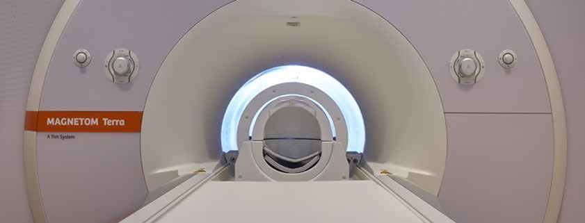
Through a Siemens Healthineers and Houston Methodist multiyear consortium agreement, the Research Institute is home to one of the world’s most powerful magnetic resonance imaging (MRI) machines – the Siemens 7 Tesla (7T) MAGNETOM Terra, which is the first 7T MRI of its kind in Texas and the first 7T MRI scanner approved for clinical use in the U.S. This agreement also emphasizes an interventional imaging partnership that allows clinicians and researchers to collaborate on developing new technologies for robotic- and image-guided navigation. The higher spatial resolution and improved contrast capabilities of the 7T MRI is being harnessed in clinical studies to more accurately localize the lesions that may be responsible for seizures in epileptic patients with focal cortical dysplasia, the most common cause of medically refractory epilepsy in children and the third most common cause of medically intractable seizures in adults. For epilepsy patients who are good candidates for surgery, equipping the surgeon with more detailed information on the lesion sites has the potential to significantly improve surgical outcomes. Houston Methodist’s collaboration with Siemens is also enabling safer, more autonomous imaging with intracardiac echocardiography, or ICE, which allows physicians to focus their efforts and time on delivering therapy rather than dividing their attention with acquiring images. The use of robotic- and AI-assisted ICE imaging may provide simple and intuitive procedural navigation. C. Huie Lin, MD, PhD, assistant professor of cardiology, is leading an effort to introduce image-based control for semi-autonomous robotic ICE imaging and to optimize the physician-robot interface. The 7T is also allowing physicians to better monitor patients with small cerebral aneurysms over time and tailor their course of treatment as needed. Traditionally, follow-up for such patients is conducted with computed tomography (CT) angiographic or digital subtraction angiographic
examinations. These procedures use radiation and contrast load that pose a risk of renal failure and contrast reactions apart from the risks of cumulative radiation. Although the widely available 3T clinical MRI can circumvent these undesirable effects, it has its own limitations. The 7T clinical MRI scanner Diego Martin, MD, PhD may provide a good alternative as its superior spatial resolution and shortened T1 relaxation time of stationary tissue results in superior contrast to blood than current 3T clinical MRI and thereby may prominently display the lumen of the aneurysm and the parent vessel. Experienced endovascular neurosurgeons and neuro-interventional radiologists plan to compare magnetic resonance angiographic images with clinical standard CT angiographic or digital subtraction angiographic acquisitions to determine when the 7T provides a significant advantage.
A ribbon-cutting ceremony to celebrate the new imaging center and its capabilities was held on June 26, 2019. Leaders from Houston Methodist, Siemens and across the Texas Medical Center enjoyed tours and demonstrations of the most advanced, high-performance equipment for imaging research and innovation.
7T MRI Brain Scan 3T MRI Brain Scan
The array of advanced imaging technologies at the Research Institute Translational Imaging Center, the partnership with Siemens Healthineers and the strong interdisciplinary collaborations within our organization are all major reasons for my excitement in the opportunity to join and help lead our efforts to make imaging sciences a centerpiece of innovation. The 7T Terra whole-body MRI, in addition to our other MRI research systems, our advanced radiopharmacy GMP lab, cyclotron, PET facilities and our imaging scientists are vital building blocks that support development of a world-class translational research center. ”
– Diego R. Martin, MD, PhD
Professor and Chair, Department of Radiology
Houston Methodist


