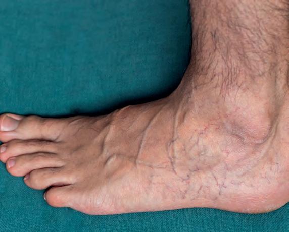
15 minute read
5 page CPD article
The Venous Foot Pump:
Modelling its function in gait
Advertisement
By Andy Horwood
Independent Clinical Gait Analyst, Researcher, & Lecturer Product Designer Healthystep Ltd Visiting Lecturer & Fellow Staffordshire University
INTRODUCTION
Venous return involves internal forces of the cardiovascular system and external forces derived from breathing, muscle contraction and gait. The venous foot pump is important in lower limb health, and systemic wellbeing. Its interaction with cardiovascular disease must be considered during patient management.
The Physiology of Blood Vessels and Haemodynamics
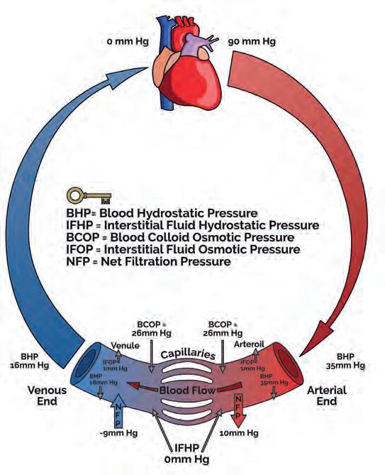
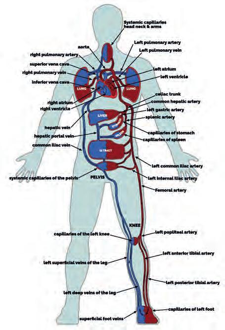
Systemic blood leaves the left side of the heart to enter large elastic-walled arteries. These divide into medium sized arteries, which in turn divide into arterioles. As arterioles enter tissues, they divide into countless microscopic vessels called capillaries, where exchanges of nutrients and waste occur between blood vessels and tissues. Before leaving the tissues, the capillaries unite to form venules, which in turn unite to form progressively larger veins, returning blood to the right side of the heart (Fig. 1). Blood pressure averages 90 mmHg in the proximal aorta, but returns to the heart at pressure close to 0 mmHg (Lie et al, 1989; Lee et al, 2013).
Figure 1
Schematics of the cardiovascular system (left) and capillary exchange (right). Images www.healthystep.co.uk
Arteries are lined with simple squamous epithelial cells overlaid with elastic tissue, a layer of elastic fibres within smooth muscle, and an outer layer composed of elastic and collagen fibres. Sometimes a further elastic layer separates the middle layers from the external coating. Collagen fibres wind around the artery in two helices with opposite pitch (Murphy, 2014), giving arteries heart pump-linked elasticity (storing potential energy) and recoil (applying force through kinetic energy). When the ventricles eject blood, the arteries expand to accommodate the extra blood, and as the ventricles relax, the vessels recoil, pumping blood forward. Smooth muscle only controls stiffness and compliance of the artery wall in response to demands (Liu et al, 1989).
Large arteries (aorta, common carotid, vertebral, and common iliac arteries) are termed elastic arteries. They are thin walled, have little smooth muscle, but containing many elastic fibres. Medium sized arteries feed blood to tissues from the large vessels to the arterioles. Arterioles have little elastic tissue, but extensive smooth muscle that plays a key role in controlling blood flow and pressure, through vasoconstriction and dilatation. Capillaries are extensive in metabolic tissues such as skeletal muscle, nerves, kidneys, lungs, and liver. Few are found in tendons and ligaments, and usually none are present in cartilage and epidermis. Capillaries are composed of a single layer of endothelial cells on a basement membrane to allow blood-tissue gas and nutrient exchange to take place.
Venous Anatomy
At rest, around 60% of blood volume is within veins and venules, but this reduces under sympathetic nerve impulses that constrict veins, releasing more blood for skeletal muscle during exercise. Venules unite and drain capillaries after nutrient/waste exchange is complete. Blood pressure entering venules, is around 16mmHg, decreasing towards the heart. Veins’ walls are extremely thin compared to the arteries, although the connective tissue outer layer is thicker. Unlike arteries, they are not subjected to high pressures, and as a consequence are stiffer and less elastic. Veins contain bicuspid valves that prevent reflux of blood to help overcome the effects of gravity (Meissner, 2005).
There are three groups of veins: superficial veins running close to the skin, deep veins running deeper within the muscles and body tissues, and the perforating veins that link the superficial and deep veins together. The superficial veins drain through perforating veins towards the deep veins. Valves in the perforating veins orientate to prevent blood flowing back towards the superficial veins and are most common in the lower limbs (Meissner, 2005). In the foot, deep veins flow towards the superficial veins with reversed orientated valves (Bojsen-Møller, 1999; Meissner, 2005; Ricci et al, 2014).
Blood moves via hydrostatic pressure (Lee et al, 2013), where fluid flows from areas of high-pressure (arteries) towards areas of low-pressure. Tissue fluid enters blood via osmotic pressure, which draws fluid out of the interstitial spaces (Fig. 1). Fluid balance can be disturbed through increasing blood hydrostatic pressures due to cardiac failure, vein valve dysfunction, or blood clots. Protein loss from burns, malnutrition, or liver and kidney disease will change the osmotic blood pressure, keeping fluid in tissues. Excess interstitial fluid is called oedema. Blockage or damage to the lymphatics can results in extremely disfiguring severe local oedema.
To avoid leg oedema, gravity must be overcome. The valves of the veins prevent reflux towards the feet but efficient venous blood flow requires three other mechanisms. Changes in thorax and abdomen pressures during breathing create a respiratory pump action. During inspiration, the diaphragm moves downward decreasing pressure in the thoracic body cavity, while it increasing pressure in the abdomen. Blood is pulled towards the lower chest cavity pressure superiorly. On expiration, the valves prevent the blood flowing back, as the pressures reverse (Takata and Robotham, 1992; Miller et al, 2005). Muscle activity creates skeletal muscle pumps, moving blood as muscles contact. Ground reaction forces (GRF) on the foot during stance and gait create a foot pump. These techniques and hydrodynamic flow rely on healthy functional vein valves (Horwood, 2019).
Venous Return from the Lower Limbs
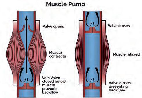
Lower limb deep veins exist in pairs (venae comitantes), and are more complicated and variable than their corresponding arteries, with which they share their name (Meissner, 2005; Ricci et al, 2014; Ricci, 2015). Superficial veins (e.g. great and small saphenous) run above the deep fascia, linked to the deep veins by an average of 64 perforated veins between the ankle and the groin (Meissner, 2005). This allows blood to flow by aspiration into the deep veins (Ricci, 2015). Within the deep veins of the calf lie intramuscular venous sinuses that act as collecting sites for the muscular pump of the calf. Muscular pumps work by compressing stiff vein walls running through the muscles and their fascial compartments, producing integrated flow out of the lower limbs (Meissner 2005; Ricci et al, 2014; Ricci, 2015). On contraction, muscles tighten the fascia and compress the veins raising pressures within the muscle compartment. Proximal valves open and blood is milked into the next section of vein towards the heart, with the valves preventing reflux on muscle relaxation (Fig. 2). This creates volumetric pumps which can reach pressures of over 200 mmHg in the calf, making it the most powerful lower limb venous pump (Meissner, 2005; Ricci, 2015).
Gait biomechanics and cardiovascular physiology meet directly in the action of the foot pump (Horwood, 2019). Blood pools in the plantar vault (arch) and heel when the leg is vertical and non-weightbearing (Gardner and Fox, 1983), thus tending to gather blood when sitting. Having perforating veins valves that shut towards the deep veins and open towards the superficial ones, means blood runs from deep to superficial in the foot (Bojsen-Møller, 1999; Meissner, 2005; Ricci et al, 2014). In the plantar heel, the veins mainly run transversely. Hence on heel loading, the blood is squeezed towards the sides of the foot and not towards the forefoot (Bojsen-Møller, 1999).
The plantar surface of the foot deforms under GRF during gait, allowing the heel and forefoot to be used as ‘compression pumps’, expelling blood into calf veins in concert with the action of plantar intrinsic muscles pumping (Broderick et al, 2010; Corley et al, 2010). GRFs acting on deep plantar veins at each step works like a hydraulic pump, with the valves in the perforating veins preventing reflux to the deep foot veins on offloading (Horwood, 2019). Each stroke of the foot and calf pump in ‘static’ weightbearing on the foot is estimated to move approximately 33ml blood into the popliteal vein at the knee, around 20% of this flow arising from the veins passing the ankle (Broderick et al, 2010). Higher gait forces likely increase this (Ricci et al, 2014), causing blood pressure in the superficial and deep veins of the leg to rise abruptly with each step (Bojsen-Møller, 1999). The foot, calf, and thigh muscle pumps overcome gravity induced pressures of around 90 mmHg in standing, and around 20 mmHg during walking (Reeder et al, 2013). Any failure in any one of these pumps through valve dysfunction or vein obstruction will cause a compromise in venous return.
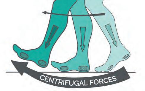
Figure 3 Centrifugal and gravitational forces pooling blood in feet during swing phase. Image www.healthystep.co.uk.
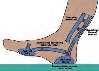
Ankle motion help propel blood during the stance phase (Ricci, 2015). Ankle dorsiflexion (under eccentric calf muscle contraction) draws blood out of the superficial veins via those perforating the fascial envelope, taking blood to the deep calf veins, to propel it up the leg (Gardner and Fox, 1983; Ricci, 2015). The foot and calf venous pumps can therefore be modelled together, providing distinct periods of passive and active component activity during gait (Horwood, 2019). During swing phase, the foot is nonweightbearing, permitting gravity and swing centrifugal forces (Fig. 3) to temporarily pool venous blood in the feet (Horwood, 2019). The presence of blood pooling in the plantar foot at heel strike may even have a small effect on the mechanical behaviour of the plantar fat pad (Aerts et al, 1995; Gefen et al, 2001; Weijers et al, 2005). Heel strike initiates the passive foot pump as its venous plexus is compressed (Fig. 4), driving the blood to the heel margins through the transversely orientated heel veins, up towards the foot and calf’s superficial veins (Horwood, 2019).
Figure 4 The foot pump at heel strike. Image www.heathystep.co.uk.
At forefoot contact, the forefoot venous plexus undergoes compression, driving blood flow towards the superficial and proximal veins of the midfoot (Fig. 5). This process is aided by natural motions of pronation, increasing soft tissue compression by enlargement of the surface contact area (Horwood and Chockalingam, 2017; Horwood, 2019). During single-limb support phase, blood pumping continues under the GRF-body weight induced foot compression, until activation of the triceps surae and other ankle plantarflexors initiate engagement of the skeletal calf muscle pump (Horwood, 2019). This activity continues through late midstance in concert with active plantar foot intrinsic muscles (Horwood, 2019). Despite decreasing compression on the heel, increasing compression of the forefoot maintains compression pumping (Horwood, 2019). With foot vault lowering, the foot stiffens (Bjelopetrovich and Barrios, 2016) facilitating and improving propulsion energetics producing a large forefoot GRF (Cunningham et al, 2010; Usherwood et al, 2012).
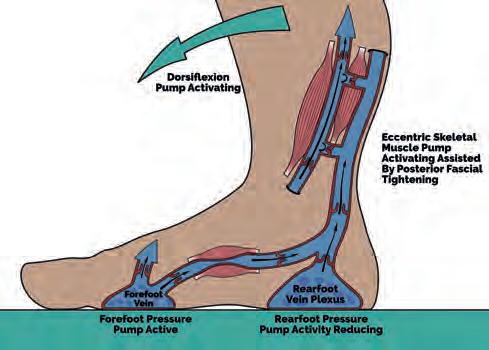
Figure 5 The foot pump at midstance. Image www.healthystep.co.uk.
At heel lift (Fig. 5), plantarflexion power expresses shortening calf muscle fascicle length, maintaining muscle pumping and generating high GRF compression forces on the forefoot. The offloaded heel’s venous plexus may start to refill again in readiness for the next heel strike.
Active muscular pumping from the plantar intrinsic muscles will continue until the forefoot is unloaded (Horwood, 2019).
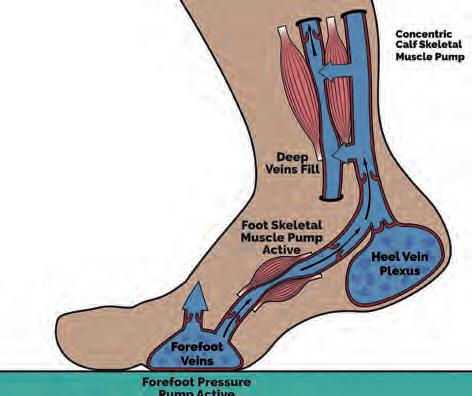
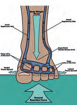
Figure 6 The foot pump during terminal stance. Image www.healthystep.co.uk.
Gait perturbations may influence venous return, for patterns of foot compliance and stiffening are likely to be significant in driving the active and passive elements of the foot pump (Horwood, 2019). There could be conflicts between the mechanisms of the foot and calf pumps function that prevent harmonious unifunctional lower limb pumping, while the foot pump efficiency itself may have implications in therapy for leg ulcers that use foot-free bandaging (Reeder et al, 2013; Ricci, 2015). Deficient venous return (venous reflux) can lead to chronic venous insufficiency. This can result from blockage of veins (such as a blood clot/thrombus), valve malfunction, or failures of the respiratory musculature, skeletal muscles, and/or foot pumps. Most venous reflux affects superficial and perforating veins, but sometimes just deep veins or all three vein-types are affected (Labropoulos et al, 1996). Leg veins dysfunction causes aching, foot-ankle swelling, skin changes, and ulcers (Labropoulos et al, 1996), which can be mild or incapacitating (Brandjes et al, 1997; Reeder et al, 2013). Skin changes, including discolouration from venous eczema, varicose veins, and itchy thrombophlebitis. Left untreated, venous reflux can increase the risk of serious infections in the feet and legs. Blood clots can form in sluggish flow of veins, causing thrombosis, causing local damage at the thrombus site. Blood clots risks breaking up in loose clumps that can then flow to small arteries in the lungs or the brain to block them. This can result in pulmonary embolism and strokes.
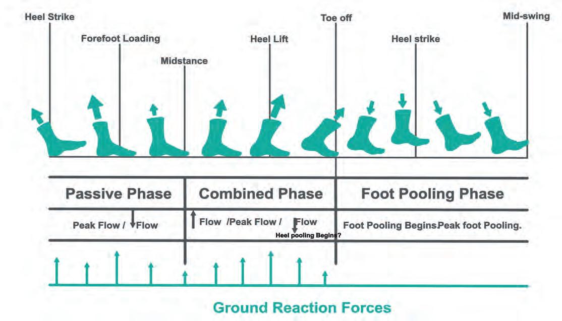
Fig 7 The activity of the foot and calf pumps in venous return during gait. Image www.healthystep.co.uk.
The foot may also suffer from valve incompetence. Gardner and Fox (1983) reported a case of intravascular thrombi in the foot presenting clinically as plantar heel pain. It is unknown how common a phenomenon this is because the foot is not examined routinely for thrombus at post-mortem. Deep vein thrombosis (DVT) or venous thromboembolism (VTE) is common in the calf. A hot red swollen (painful) calf without a significant known injury event should be considered a DVT until proven otherwise, requiring urgent intervention. Even in seemly healthy individuals, long periods of sitting and inactivity are associated with emboli formation especially after surgery (Assareh et al, 2014) but also during long-distance flying (Hughes et al, 2003). Around 60% of people who develop a DVT go on to develop post-thrombotic syndrome within 24 months, developing leg swelling, pain, and ulceration (Brandjes et al, 1997).
Clinical Solutions
Graduated compression stockings influence venous return pressures. They apply gradually decreasing pressure the higher they go up the leg (hence the name ‘graduated’). Compression stockings reduce the risk of developing more serious post-thrombotic syndrome after a DVT by around 50% (Brandjes et al, 1997; Prandoni et al, 2004). They also improve venous ulcer healing rates (Reeder et al, 2013; Nelson and Bell-Syer, 2014). Compression hosiery is a well-proven biomechanical treatment in venous disease (Rabe et al, 2018). Combining compression stocking and lower limb exercises, including regular walking, can help manage venous insufficiency through biomechanical principles (Horwood, 2019).
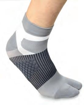
Figure 8 Modern ‘foot pump friendly’ graduated compression hosiery avoids toe compression restricting intrinsic foot muscle activity. Images www.healthystep.co.uk.
REFERENCES:
Aerts P, Ker RF, De Clercq D, Ilsley DW, Alexander RM. (1995). The mechanical properties of the human heel pad: a paradox resolved. Journal of Biomechanics. 28(11): 1299-1308. Assareh H, Chen J, Ou L, Hollis SJ, Hillman K, Flabouris A. (2014). Rate of venous thromboembolism among surgical patients in Australian hospitals: a multicentre retrospective cohort study. BMJ Open. 4(10): e005502. Bjelopetrovich A, Barrios JA. (2016). Effects of incremental ambulatoryrange loading on arch height index parameters. Journal of Biomechanics. 49(14): 3555-3558. Bojsen-Møller F. (1999). Biomechanics of the heel pad and plantar aponeurosis. (Chapter 9.) In: Disorders of the Heel, Rearfoot, and Ankle. [Eds.: Ranawat CS, Positano RG.] Philadelphia: Churchill Livingstone. pp. 137-143. Brandjes DPM, Büller HR, Heijboer H, Huisman MV, de Rijk M, Jagt H, et al. (1997). Randomised trial of effect of compression stockings in patients with symptomatic proximal-vein thrombosis. The Lancet. 349(9054): 759-762. Broderick BJ, Corley GJ, Quondamatteo F, Breen PP, Serrador J, ÓLaighin G. (2010). Venous emptying of the foot: influences of weight bearing, toe curls, electrical stimulation, passive compression, and posture. Journal of Applied Physiology. 109(4): 1045-1052. Corley GJ, Broderick BJ, Nestor SM, Breen PP, Grace PA, Quondamatteo F, et al. (2010). The anatomy and physiology of the venous foot pump. The Anatomical Record. 293(3): 370-378. Cunningham CB, Schilling N, Anders C, Carrier DR. (2010). The influence of foot posture on the cost of transport in humans. Journal of Experimental Biology. 213(5): 790-797. Gardner AMN, Fox RH. (1983). Venous pump of the human foot Preliminary report. Bristol Medico-Chirurgical Journal. 98(3): 109-112. Gefen A, Megido-Ravid M, Itzchak Y. (2001). In vivo biomechanical behavior of the human heel pad during the stance phase of gait. Journal of Biomechanics. 34(12): 1661-1665. Horwood AM, Chockalingam N. (2017). Defining excessive, over, or hyperpronation: A quandary. The Foot. 31: 49-55. Horwood A. (2019). The biomechanical function of the foot pump in venous return from the lower extremity during the human gait cycle: An expansion of the gait model of the foot pump. Medical Hypotheses. 129: https://doi. org/10.1016/j.mehy.2019.05.006 Hughes RJ, Hopkins RJ, Hill S, Weatherall M, Van de Water N, Nowitz M, et al. (2003). Frequency of venous thromboembolism in low to moderate risk long distance air travellers: the New Zealand Air Traveller’s Thrombosis (NZATT) study. The Lancet. 362(9401): 2039-2044. Labropoulos N, Touloupakis E, Giannoukas AD, Leon M, Katsamouris A, Nicolaides AN. (1996). Recurrent varicose veins: Investigation of the pattern and extent of reflux with color flow duplex scanning. Surgery. 119(4): 406-409. Lee BK, Lee HY, Jeung KW, Jung YH, Lee GS. (2013). Estimation of central venous pressure using inferior vena caval pressure from femoral endovascular cooling catheter. American Journal of Emergency Medicine. 31(1): 240-243. Liu ZR, Ting CT, Zhu SX, Yin FC. (1989). Aortic compliance in human hypertension. Hypertension. 14(2): 129-136. Meissner MH. (2005). Lower extremity venous anatomy. Seminars in Interventional Radiology. 22(3): 147-156. Miller JD, Pegelow DF, Jacques AJ, Dempsey JA. (2005). Skeletal muscle pump versus respiratory muscle pump: modulation of venous return from the locomotor limb in humans. Journal of Physiology. 563(3): 925-943. Nelson EA, Bell-Syer SEM. (2014). Compression for preventing recurrence of venous ulcers. Cochrane Database of Systematic Reviews. 2014(9):CD002303. https://doi.org/10.1002/14651858.CD002303.pub3 Prandoni P, Lensing AWA, Prins MH, Frulla M, Marchiori A, Bernardi E, et al. (2004). Below-knee elastic compression stockings to prevent the postthrombotic syndrome: a randomized, controlled trial. Annals of Internal Medicine. 141(4): 249-256. Rabe E, Partsch H, Hafner J, Lattimer C, Mosti G, Neumann M, et al. (2018). Indications for medical compression stockings in venous and lymphatic disorders: An evidence-based consensus statement. Phlebology: The Journal of Venous Disease. 33(3): 163-184. Reeder SWI, Maessen-Visch MB, Langendoen SI, de Roos K-P, Neumann HAM. (2013). The recalcitrant venous leg ulcer - a never ending story? Phlebologie. 42(6): 332-339. Ricci S, Moro L, Incalzi RA. (2014). The foot venous system: anatomy, physiology and relevance to clinical practice. Dermatologic Surgery. 40(3): 225-233. Ricci S. (2015). The venous system of the foot: anatomy, physiology, and clinical aspects. Phlebolymphology. 22(2): 64-75. Takata M, Robotham JL. (1992). Effects of inspiratory diaphragmatic descent on inferior vena canal venous return. Journal of Applied Physiology. 72(2): 597-607. Usherwood JR, Channon AJ, Myatt JP, Rankin JW, Hubel TY. (2012). The human foot and heel-sole-toe walking strategy: a mechanism enabling an inverted pendular gait with low isometric muscle force? Journal of the Royal Society: Interface. 9(75): 2396-2402. Weijers RE, Kessels AGH, Kemerink GJ. (2005). The damping properties of the venous plexus of the heel region of the foot during simulated heelstrike. Journal of Biomechanics. 38(12): 2423-2430.


