Basic Science


ORTHOPAEDIC OUTCOMES &RESEARCH
VOLUME 6
Jefferson Health’s Department of Orthopaedic Surgery includes a robust basic science program devoted to uncovering the mechanisms that promote bone health and optimal recovery from orthopedic disease or injury.
Through their work in the laboratory, Jefferson Health scientists are identifying novel methods to enhance bone healing, minimize surgical complications such as scarring and infection, and stave off the intervertebral disc degeneration that is common with aging.

While discovery is at the heart of what the scientists do, they are also focused on translating their findings into clinical solutions for patients in need of orthopedic care. Those efforts are bolstered by the involvement of practicing surgeons on research teams.
What follows is a sample of recent research published by Jefferson Health orthopedic scientists.
Dear Colleagues,
Jefferson Health’s Department of Orthopaedic Surgery is pleased to share the latest in our series of Orthopaedic Research and Outcomes reports. This forward-looking volume focuses on the important work of scientists in our Division of Orthopaedic Research
The orthopedic care offered at Jefferson is informed by all phases of research—from the laboratory to clinical trials. We are especially grateful for our team of orthopedic scientists who are exploring the root causes of orthopedic disease and injury and proposing novel treatments and prevention approaches.
Patients coming to Jefferson clearly recognize the outstanding care they receive in our exam rooms and operating suites, but they also benefit from what goes on behind the scenes in our orthopedic research laboratories.
A tour through that space would show dedicated scientists who are targeting aging cells to prevent disc degeneration and low back pain. Others are focused on mechanisms to enhance healing from injury and prevent scarring. As you know, a dreaded complication of hip or knee replacement surgery is periprosthetic joint infection. Jefferson Health scientists are developing antimicrobial therapies that could improve the quality of post-operative recovery and reduce the need for longer hospital stays or readmissions.
Jefferson Health’s orthopedic scientists don’t work in isolation, instead collaborating with clinicians to find paths to bring laboratory and pre-clinical discoveries into clinical testing. I invite you to read about their work in the pages ahead.
I also invite you to learn more about all facets of our orthopedic program by visiting our website, JeffersonHealth.org/Ortho. To refer a patient, please call, 215-503-8888
Thank you for your interest.
Sincerely,
Alexander R. Vaccaro, MD, PhD, MBA
Richard H. Rothman Professor and Chair Department of Orthopaedic Surgery, Jefferson Health Sidney Kimmel Medical College, Thomas Jefferson University

1-800-JEFF-NOW | JeffersonHealth.org/Ortho | Transfers: 1-800-JEFF-121 | Referrals: 215-503-8888 3 BASIC SCIENCE
From the Laboratory of Ryan Tomlinson, PhD
The TrkA Agonist Gambogic Amide Augments Skeletal Adaptation to Mechanical Loading
The periosteal and endosteal surfaces of mature bone are densely innervated by sensory nerves expressing neurotrophic tyrosine kinase receptor (TrkA), the high-affinity receptor for nerve growth factor (NGF).
In previous work in mice, Jefferson Health scientists demonstrated that inhibition of TrkA significantly diminished load-induced bone formation, while, on the other hand, administration of exogenous NGF significantly increased bone formation following loading. Taken together, the research suggests there is a therapeutic potential for leveraging NGF-TrkA signaling to improve the anabolic response of the skeleton to mechanical load. However, previous research found that administration of NGF induces long-lasting pain that limits its usefulness to improve skeletal health in patients.
Building on that prior knowledge, Jefferson Health scientists led by Ryan Tomlinson, PhD, used a mouse model to test the effect of gambogic amide (GA), a recently identified robust small molecule agonist for TrkA, on hyperalgesia and load-induced bone formation. Behavioral analysis of the mice was used to assess pain up to one week
after axial forelimb compression. The researchers found that, contrary to their expectations, GA treatment was not associated with diminished use of the loaded forelimb or sensitivity to thermal stimulus.
The researchers also did a series of in vitro experiments using mouse cells to study the direct effect of GA administration on osteoblasts. They observed that GA induced the significant upregulation of the osteoblastic differentiation markers that indicate support for bone formation, whereas treatment with NGF did not affect these markers.
“The results from our study indicate that GA may be a potential novel therapeutic for increasing bone formation rate following loading,” the researchers reported in Bone “Importantly, we observed a significant increase in relative periosteal bone formation rate following axial forelimb compression that was not associated with increased thermal or mechanical sensitivity.”
While they said use of GA needs further study, the researchers said it may prove to be “sufficient to prevent fatigue injuries in at-risk individuals.”
4 Jefferson Health | Orthopaedic Outcomes & Research BASIC SCIENCE
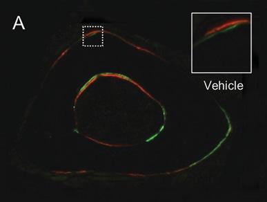
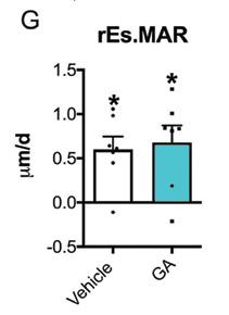
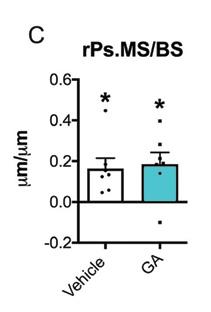
GA increased periosteal bone formation following axial forelimb compression. A,B) Calcein (green) and alizarin red (red) fluorescent bone formation markers were injected following 3 days of axial forelimb compression. Relative (loaded–non-loaded) periosteal bone formation parameters C) rPs.MS/BS, D) rPs.MAR, E) rPS.BFS/BS, F) rES.MS/BS, G) rES.MAR, H) rES.BFR/BS were quantified. *p < 0.05 vs. non-loaded, +p < 0.05 vs. vehicle by two-way ANOVA. n = 7.


Source: Ryan Tomlinson, PhD



1-800-JEFF-NOW | JeffersonHealth.org/Ortho | Transfers: 1-800-JEFF-121 | Referrals: 215-503-8888 5
Periprosthetic joint infection (PJI) can be a devastating complication of joint replacement, and figuring out how best to treat or prevent it is key to improving patient outcomes. That goal could be helped along by the development of better laboratory models that mimic bacterial behavior in the setting of PJI.
Jefferson Health researchers and others have shown in previous work that synovial fluid, the lubricious fluid within the joint space, causes aggregation of many bacterial species, including methicillin-resistant or methicillin-susceptible Staphylococcus aureus (MRSA or MSSA). These and other pathogens assemble a proteinaceous matrix in synovial fluid, which is coated by a polysaccharide slime that is characteristic of biofilms.
In new research reported in Frontiers in Microbiology, Jefferson Health scientists led by Noreen Hickok, PhD, set out to identify proteins in synovial fluid that are important for that aggregation and determine the concentration ranges of the proteins. The team identified albumin, fibrinogen and hyaluronic acid as being critical proteins, which can, when combined, reproduce the
effects of synovial fluid on Staphylococcus aureus , including MRSA. The protein combination was tested for its ability to cause marked aggregation, antibacterial tolerance, preservation of morphology, and expression of the phenol-soluble modulin (PSM) virulence factors. Bacteria such as MRSA are able to use the joint environment to change the way they behave so as to resist antibiotic treatment and suppress the production of virulence factors that normally would allow the immune system to respond to infection.
Using that process, the researchers created a viscous fluid that they say models the bacterial behavior that occurs in vivo in synovial fluid. The new medium will be useful for laboratory testing of potential therapies for PJI as well as joint injections in the absence of implants.
“Use of this fluid can help better model bacterial behavior of new antimicrobial therapies…and serve as a starting point to study host protein-bacterial interactions characteristic of physiological fluids,” the paper concluded. A follow-up study is being done.
6 Jefferson Health | Orthopaedic Outcomes & Research
From the Laboratory of Noreen Hickok, PhD Staphylococcus Aureus Floating Biofilm Formation and Phenotype in Synovial Fluid Depends on Albumin, Fibrinogen and Hyaluronic Acid
BASIC SCIENCE
fluid aggregate
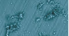
(Top) Using

morphology in samples retrieved from an infected knee replacement [periprosthetic
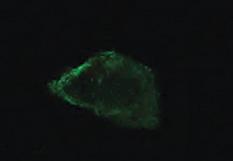
infection (PJI)] or formed on titanium using methicillin-susceptible Staphylococcus aureus (MSSA) in human synovial fluid (SynF)

Broth (TSB). (Bottom) Floating aggregates retrieved from an infected knee replacement
Trypticase
formed with MSSA in human synovial fluid. (B) Sodium dodecyl sulfate–polyacrylamide gel electrophoresis (SDS-PAGE) gel stained with Coomassie Blue. The gel shows the composition of human synovial fluid and of synovial fluid
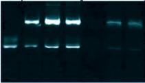

from three separate samples of synovial fluid, as compared to fibronectin (Fn), fibrinogen (Fg),
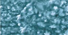


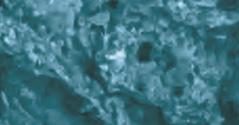
Western blot analysis of MSSA aggregates from synovial fluid for Alb (left)
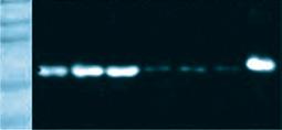
controls.
chain are indicated, as compared with human synovial fluid, Fg, and Alb standards.
and
1-800-JEFF-NOW | JeffersonHealth.org/Ortho | Transfers: 1-800-JEFF-121 | Referrals: 215-503-8888 7 Synovial
morphology and composition. (A)
SEM, biofilm
joint
or
Soy
or floating aggregates
MSSA aggregates
albumin (Alb), and serum
(C)
and for Fg (right). The γ γ dimer and α
Lanes represent human synovial fluid samples
resulting aggregates from different samples. (D) MSSA aggregates stained for Alb, Fg, or secondary antibody alone (Control). All images are representative images of at least three samples. Source: Noreen Hickok, PhD Retrieved from PJI Human SynF TSB Floating Aggregate Abherent Biofilm 30 mm x3.0k 30 mm x3.0k 30 mm x3.0k 30 mm x3.0k 30 mm x3.0k A MW 250 150 100 75 50 37 MW 250 150 100 75 50 37 SynF1 SynF2 SynF3 Fn Fg Alb Serum SynF1 SynF2 SynF3 Human SynF AggregatesB 500 mm 500 mm 500 mm Alb Fg Control D Fg γ γ dimer α chain b chain γ chain MW 150 100 75 50 37 Fg SynF3SynF4SynF5 SynF3SynF4SynF5 AggregatesC MW 150 100 75 50 37 SynF6SynF7SynF8 SynF6SynF7SynF8 Alb Aggregates Alb
From the Laboratory of Andrzej Fertala, PhD
Mechanisms of Reducing Joint Stiffness by Blocking Collagen Fibrillogenesis in a Rabbit Model of Posttraumatic Arthrofibrosis
Posttraumatic fibrotic scarring is a significant problem that alters the proper function of injured tissues. Current methods to reduce posttraumatic fibrosis rely on anti-inflammatory and anti-proliferative agents with broad intracellular targets. As a result, their use is not fully effective and may cause unwanted side effects.
A Jefferson Health research team led by Andrzej Fertala, PhD, previously demonstrated that extracellular collagen fibrillogenesis is a valid and specific target to reduce collagenrich scar buildup. Prior studies showed that a rationally designed antibody that binds the C-terminal telopeptide of the α2(I) chain involved in the aggregation of collagen molecules limits fibril assembly in vitro and reduces scar formation in vivo.
Building on that research, the team utilized a clinically relevant arthrofibrosis rabbit model to study the broad mechanism of the anti-
scarring antibody. They analyzed the effects of targeting collagen fibril formation on the quality of healed joint tissues, including the posterior capsule, patellar tendon and subchondral bone.
In an article in Plos One, the researchers reported that “our results show that blocking collagen fibrillogenesis not only reduces collagen content in the scar but also accelerates the remodeling of healing tissues and changes the collagen fibrils’ cross-linking.”
While more work is needed to move the anti-fibrotic technology toward clinical application, the researchers concluded that “this study demonstrated that targeting collagen fibrillogenesis to limit arthrofibrosis affects neither the quality of healing of the joint tissues nor disturbs vital tissues and organs.”
8 Jefferson Health | Orthopaedic Outcomes & Research
BASIC SCIENCE
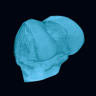

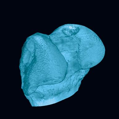
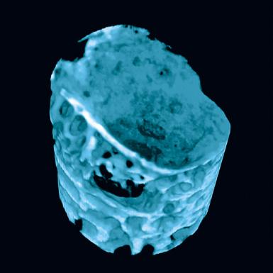
1-800-JEFF-NOW | JeffersonHealth.org/Ortho | Transfers: 1-800-JEFF-121 | Referrals: 215-503-8888 9 Representative computer tomography images of the uninjured (A) and injured (C, arrow) bones. Panels B (uninjured bone) and D (injured bone) indicate subchondral bone regions analyzed here to determine the extent of mineralization in the injured area (C&D). Source: Andrzej Fertala, PhD DC A B 5 mm 5 mm 1 mm 1 mm
From the Laboratory of Makarand Risbud, PhD
Long-Term Treatment with Senolytic Drugs Dasatinib and Quercetin Ameliorates Age-Dependent Intervertebral Disc Degeneration in Mice
Intervertebral disc degeneration is highly prevalent within the elderly population and is a leading cause of chronic back pain and disability. Despite the staggering medical and societal costs of treating the multitude of pathologies associated with intervertebral disc degeneration, there are few available clinical interventions. No pharmacologic approaches to prevent age-related disc degeneration exist, and surgeries for symptomatic relief of low back pain have suboptimal outcome. The nation’s deadly opioid epidemic has highlighted the dangers of addiction that can stem from prolonged use of painkillers post injury or surgery.
Recent studies have suggested that senescence is an important contributor to the progression of disc degeneration.
Studies of human tissue and mouse models have shown an increased incidence of senescent cells during intervertebral disc aging and degeneration.
Using a mouse model, Jefferson Health scientists led by Makarand Risbud, PhD, evaluated the therapeutic potential of small molecule drugs called senolytics, which target senescent cells. They used a combination of Dasatinib and Quercetin (D + Q) to determine if the drugs could prevent or slow the age-dependent progression of disc degeneration. The researchers also evaluated whether
the timing of the start of treatment made a difference.
Mice were treated with weekly injections of D + Q at 6, 14 and 18 months of age and their disc tissue was then analyzed at 23 months. The study found that, compared to mice given a placebo, the treated mice showed a lower incidence of degeneration, and treatment resulted in a significant decrease in senescence markers p16INK4a, p19ARF and SASP molecules IL-6 and MMP13.
“Treatment also preserves cell viability, phenotype and matrix content,” the researchers reported in Nature Communications. “Although transcriptomic analysis shows disc compartment-specific effects of the treatment, cell death and cytokine response pathways are commonly modulated across tissue types.”
The researchers said the therapy appears to be most effective when given to younger mice, before there is a lot of disc degeneration. They said the approach warrants further pre-clinical studies in large animal models of disc degeneration.
“Systematic administration of D + Q posits an exciting therapeutic approach to treating disc degeneration, without the inherent risks associated with invasive surgical intervention,” they concluded.
10 Jefferson Health | Orthopaedic Outcomes & Research
BASIC SCIENCE
NP and AF Cell
Cell Viability
Disc Matrix
Collagen
Circulatory Levels
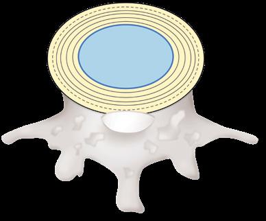
Schematic summarizing the overall beneficial effects of
to alleviation of intervertebral disc degeneration during aging.
Source: Makarand Risbud, PhD
1-800-JEFF-NOW | JeffersonHealth.org/Ortho | Transfers: 1-800-JEFF-121 | Referrals: 215-503-8888 11
D + Q contributing
Phenotypical markers Senescence markers SASP
1, COMP CS NP Fibrosis Collagen degradation Aggrecan degradation Collagen X
IL-1b IL-6 TNF-α IL-16 IL-17C IL-17E/IL-25 IL-21 IL-31 NP AF Vertebra
Physician
Jefferson Health Department of Orthopaedic Surgery Philadelphia, PA 19107
Referrals: 1-800-JEFF-NOW Patient Transfers: 1-800-JEFF-121 JeffersonHealth.org/Ortho CS 22-2073


