McKelvey Engineering


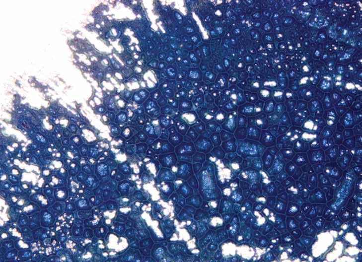

The vivid blue image, dubbed the “blue mosaic,” shows lithium metal coated on a stainless-steel current collector. A thin layer of lithium is compressed under high pressure between two plates and then viewed under a microscope. The subsequent features display an intricate web-like pattern of lithium (dark blue/grey) covering the current collector (silver).
A major challenge facing solid-state batteries is the growth of dendrites through the solid electrolyte during charging, rendering them unusable. While the exact reason for such growth have remained elusive, web-like patterns like the one illustrated have been shown to evolve between grains of the electrolyte during dendritic penetration. Understanding how these patterns form and their true origin, brings us one step closer toward unlocking the full potential of all-solid-statestate battery designs.
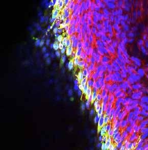


These are immunohistochemistry images of collagen gels seeded with fibroblasts, an in vitro model system that is used to study conditions involving fibrosis and contracture in connective tissues.
These gels have been treated with anti-fibrotic drugs in order to inhibit cell contractility. Each color corresponds to a different marker of cell morphology. Images of gels treated with different drugs are compared to understand how cells alter their structure in response to different treatments.
The Musculoskeletal Soft Tissue Lab is developing improved treatments for post-traumatic joint contracture, a condition in which the range of motion of the affected joint is severely reduced following an injury such as a fracture or dislocation. Novel physical and biological treatment strategies are currently being explored using both in vitro and in vivo models.
Rebecca Reals is a member of Associate Professor Spencer Lake's lab.
g lakelab.wustl.edu


Here, transient “flashes” of Nile red reveal the intricate nanoscale organization of amphipathic KFE8 (Ac-FKFEFKFE-NH2) peptide assemblies. In collaboration with the Rudra Lab, these experiments are the first demonstration of utilizing fluorophore orientation “spectra,” i.e., the 3D orientations of dye molecules, to resolve the complex nanoscale organization of self-assembled peptides in solution. Scale bars: 1 µm, 100 nm (inset).
The Lew Lab develops advanced fluorescence microscopes for imaging the positions and orientations of individual molecules.
Weiyan Zhou is a member of Associate Professor Matthew Lew's lab. g lewlab.wustl.edu





Xingyi Du PhD candidate in computer science
The image shows two examples of implicit surface networks computed using our method: the intersection of implicit surfaces for modeling a mechanical part (top) and the generalized Voronoi diagram of rotating lines (bottom). The three pictures in each example show the 1, 2 and 3D elements of the surface networks.
This collaboration with Adobe Research explores efficient and robust algorithms for computing implicit surface networks, which are widely used for modeling shapes with sharp features and interior structures, with applications in engineering and biomedicine.
Xingyi Du is a member of Professor Tao Ju's lab.
g cse.wustl.edu/~taoju/

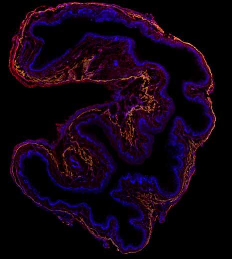
The image shows a cross-sectional view of the murine vaginal wall with fluorescently labeled cell nuclei (blue), elastin (red), and smooth muscle cells (yellow). Based on the microstructural organization, we can see three primary layers in the vaginal wall – an inner layer of dense cells (epithelium), an intermediate layer of elastin staining (subepithelium), and an outer layer of smooth muscle (muscularis).
The Bersi lab’s ongoing research in the field of women's health is focused on better understanding how different natural events — including birth, postpartum healing and aging — contribute to changes in the microstructural organization and mechanical properties of the vaginal wall and how this knowledge could be used to address conditions such as pelvic organ prolapse (POP).
Niyousha Karbasion is a member of Assistant Professor Matthew Bersi's lab.
g bersilab.wustl.edu

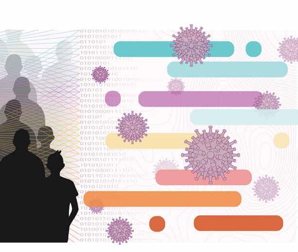

This image illustrates the idea of a meta-analysis that detected common trends in worldwide studies monitoring SARS-CoV-2 spreads by analyzing wastewater.
A new study from the Ling Lab will help facilitate the exchange of data and results between engineers and medical researchers and lead to a more robust understanding of the relationships between viruses moving through the engineered world and diseases spreading through populations.
David Mantilla-Calderon is a member of Assistant Professor Fangqiong Ling's lab.
g linglab.wustl.edu
Postdoctoral fellow in the Department of Biomedical Engineering
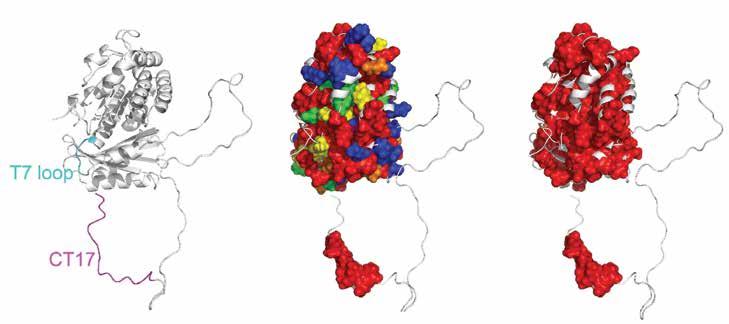
Here, protein sectors identified within FtsZ show covariation of the CT17 and most of the core domain surface, including the T7 loop.
Researchers in McKelvey Engineering and in the Department of Biology collaborated to discover how a small part of a key bacterial division protein functions at the molecular level. Antibiotics prevent bacteria from growing and multiplying, making cell division a particularly appealing target for drug development. To illuminate the mechanisms responsible for cell division in bacteria and facilitate the design of precision drugs, scientists discovered the regulatory role of the disordered region of the FtsZ protein from Bacillus subtilis.
Min Kyung Shinn is a member of Professor Rohit Pappu's lab.
g pappulab.wustl.edu

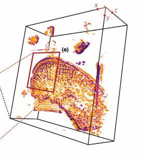

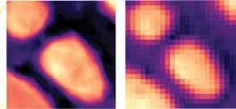

Neural fields are machine learning models that can be used to obtain continuous representations of images. This 3D image of diatom microalgae was rendered using a neural fields model trained on a set of 2D digital images captured using different illuminations. Since the neural field representation is continuous, we can render the sample on grid of any resolution.
The key to this work was the use of a neural field network, a particular kind of machine learning system that learns a mapping from spatial coordinates to the corresponding physical quantities. When the training is complete, researchers can point to any coordinate and the model can provide the image value at that location.
g cigroup.wustl.edu

This two-photon image shows a damaged tibialis anterior mouse tendon. The red signal is a geometry-dependent second harmonic generated measurement of collagen fibers, while the green and blue fluorescence signals are from excited dyes, representing elastin and denatured collagen respectively. Further processing is required to isolate true elastin fibers from the general green signal.
Elastin deficiency can arise due to genetics and aging, and researchers hypothesize that it reduces tendon resistance and response to cyclic loading, which leads to greater chance of injury. These images will help test the hypothesis by enabling researchers to view elastin and quantify tendon damage as shown by collagen fiber kinking and CHP damage staining.
lab. g lakelab.wustl.edu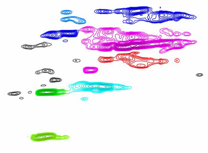
PhD candidate in energy, environmental & chemical engineering
The image shows a heteronuclear single quantum correlation (HSQC) nuclear magnetic resonance (NMR) spectrum of hybrid poplar lignin, which is a constituent of poplar biomass. Each of the peaks generated in an HSQC NMR chromatograph provides information about the types and quantity of monomers and linkages present in our lignin sample.
Research in the Foston Lab seeks to understand compositional and structural differences between lignins sourced from different plant species and how these features affect lignin depolymerization. The end goal is to rationally engineer conversion processes that take advantage of the unique features of lignins as feedstocks to access new products.
Aditya Ponukumati conducts research in Associate Professor Marcus Foston's lab. g fostonlab.wustl.edu


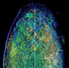

These images depict 3D optical coherence tomography (OCT) scans of fallopian tube tissues and the inversed intensity en face maps derived from normal and malignant fallopian tube fimbriated end scans. Different color marks different imaging depth. Morphological changes observed from these images suggest the feasibility of using inverse intensity map to assist fallopian tube malignancy detection.
Research in the Optical and Ultrasound Imaging Laboratory focuses on the understanding of optical properties of human ovarian and colorectal cancers. Luo has developed an optical coherence tomography catheter to help with ovarian and colorectal malignancy evaluation (featured on the inside cover of the Journal of Biophotonics in June 2022).
This work is in collaboration with the Departments of Pathology & Immunology, Obstetrics & Gynecology and Radiology at the School of Medicine. The project is funded by National Cancer Institute (CA237664).
Hongbo Luo conducts research in Professor Quing Zhu's lab. g opticalultrasoundimaging.wustl.edu


Class of 2024
For a summer fellowship in the Lake Lab, Maggie Yang was awarded supplement funding from the National Science Foundation (NSF). During the fellowship, Yang built the Mueller Matrix polarimetry imaging system with Leanne Iannucci, mentor and PhD candidate. Using Autodesk Fusion, Arduino and circuitry, Yang was able to create this final prototype as a contribution towards her mentor's research. In Fall of 2022, Yang began an independent study under the mentorship of PhD candidate Shawn Pavey, researching ways to optimize cell viability in tendon explants from mice using confocal microscopy, which was used to obtain the calendar image.

Undergraduate students work side by side in the lab with some of the best faculty in engineering, medicine, and the sciences to solve problems.
g engineering.wustl.edu/academics/undergraduateresearch