
5 minute read
AI and AR are always hot topics in ophthalmology... how soon are robots going to replace retinal surgeons?


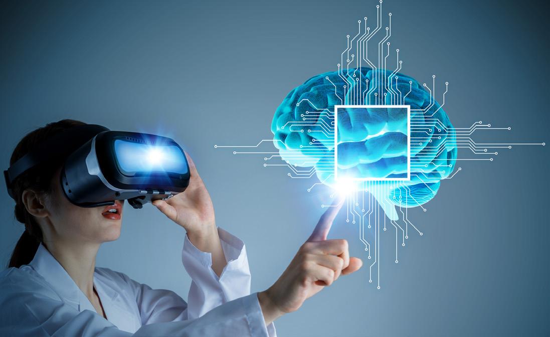
Optimizing Outcomes through AI and AR
by Tan Sher Lynn
Experts discussed how artificial intelligence (AI) and augmented reality (AR) are being used in ophthalmology to optimize surgical procedures and outcomes in a session called Technology Symposium: Updates in Artificial Intelligence (AI), Augmented Reality (AR) and Robotics held on the first day of the (virtual) American
Society of Retina Specialists 38 th Annual
Scientific Meeting (ASRS 2020).
AI for diabetic retinopathy screening
It is estimated that 600 million people worldwide will get diabetes by 2040, with a third having diabetic retinopathy (DR). 1 With the advent of deep learning, various companies have developed automated artificial intelligence diabetic retinopathy screening systems (AIDRSS) which can help reduce the incidence of preventable blindness worldwide.
Dr. Aaron Lee from the University of Washington (USA), and colleagues compared the performance of seven AIDRSS in real-world setting. The five companies which participated in the study are ADCIS (Saint-Contest, France), AirDoc (Beijing, China), Eyenuk (California USA), Retina-AI Health (California, USA) and Retmarker (Coimbra, Portugal).
A total of 311,604 retinal images for Seattle and Atlanta, as well as the original VA teleretinal grades, were extracted from the national VA Teleretinal Imaging Screening Program. The dataset were run through seven AI algorithms (labeled A-F) from the five companies and their performances were compared to the original VA grades.
“The VA grader has 100% sensitivity for moderate non-proliferative diabetic retinopathy (NPDR), severe NPDR and PDR and results from the arbitrated dataset showed that algorithms E, F, G was statistically similar to VA grader for moderate NPDR or higher,” Dr. Lee said.
“We concluded that algorithms varied in performance despite having regulatory approval and/or being clinically deployed,” he added. It is therefore important, highlighted Dr. Lee, to understand AI models in the context of the underlying prevalence of disease.
“We believe that external, independent validation with real-world imaging is critical prior to deployment even after algorithms reach regulatory approval,” he shared.
AR for macular degeneration
Age related macular degeneration (AMD) is the most common cause of vision loss in older adults in the United States. There were 13M people with AMD in the US and 30M worldwide in 2019 and this number is predicted to increase up to 22M in the US and 288M worldwide. 2-3
The novel augmented reality (AR) headset Oculenz (Ocutrx Vision Technologies, CA, USA) is set to change the lives of these patients. high or low light surroundings and applications in low vision, medicine and education,” said Dr Dr. Linda Lam, associate professor of ophthalmology of the USC Keck School of Medicine, USA.
Especially designed for patients with advanced macular degeneration with central scotama(s), the Oculenz headset provides a perceived de-emphasis of the scotoma, enhanced reading of letters in a word and improved recognition of familiar faces. “Its simultaneous localization and mapping software offers instant location mapping and object identification with 7 million objects identified,” added Dr. Lam. The device is anticipated to be available in early 2021.
Deep learning applications in retinopathy of prematurity
Retinopathy of prematurity (ROP) is a leading cause of childhood blindness worldwide, but screening is labor intensive and clinical diagnosis is subjective and qualitative. Dr. J. Peter Campbell from the Oregon Health & Science University (USA) talked about translational applications of deep learning in ROP.
“In terms of image analysis, AI can now automate the classification of all ROP components, including zone, stage and disease for multiple groups. AI can provide real-time segmentation of images to aid in acquisition and interpretation of images,” he said.
Deidentified images (RetCam; Natus Medical Incorporated) captured after clinical examinations between July 2011 and December 2016 were assessed as part of the Imaging and Informatics in ROP (i-ROP) cohort study. A deep learning system was used to classify the probability of an image having a reference standard diagnosis of plus disease, and converted to an automated 1 (most benign) to 9 (most severe) scale using published methods. Quantitative scale values were analyzed for 5255 clinical examinations in 871 infants and reported using descriptive statistics. Inter- and intra-examiner variability in diagnosis, and differences between hospitals were assessed using the quantitative scale.
Dr. Campbell concluded that deep learning may facilitate quantitative diagnosis of retinal diseases like ROP. The ROP vascular severity scale corresponds with current clinical classifications of disease severity and may enable quantitative disease monitoring in the future. “Blindness from ROP is largely preventable with appropriate primary, secondary and tertiary prevention. To maximize the potential of AI’s value, we need to develop systems of care,” he said.
Robotics in ophthalmology
Today, robots are revolutionizing the various fields of medicine. Speaking about how robotics helps to optimize surgical steps in ophthalmology, Dr. Richard Rosen from the New York Eye Infirmary of Mount Sinai (USA) said: “Robotics provides high positional stability as well as micrometer precision and accuracy in XYZ. This removes any time constraint on drug delivery within the retinal space, whether intracannular or subretinal. It also allows for a detailed analysis of each phase of a surgical procedure and its optimization.”
Three main robotic approaches currently developing in ophthalmology are the assistive handheld device by the Johns Hopkins Group, co-manipulation by the UCLA Group and telemanipulation by the Einthoven Group, reported Dr. Rosen.
Further, Dr. Rosen demonstrated the advantages of high precision robotics in retinal delivery over manual technique through a study of the PRECEYES Surgical Robot (PRECEYES BV, Eindhoven, The Netherlands) in conjunction with the PRECEYES Microscope with Integrated OCT.
“The surgical robot features tremor filtering and motion scaling, which enhances human performance by an order of magnitude to the precision of less than 10 microns. The arm allows for intuitive motion with positional stability and memory. Comparing between the accuracy and precision between human and robotic performance on a dynamic test, we found that the robot is able to perform for at least an order of magnitude better than the human,” he said.
In short, noted Dr. Rosen, robotic assistance removes time constraints of delivery, allowing high precision positioning for controlled subretinal drug delivery. Robotic assistance also controls tremor, reducing fatigue and helps avoid inadvertent injury in tight spaces. “Robotic-assisted ocular surgery shows promise for advancing the surgeon’s ability to perform more complex maneuvers necessary for the next generation of retinal interventions,” added Dr. Rosen.
References:
1 Yau JW, Rogers SL, Kawasaki R, et al. Global prevalence and major risk factors of diabetic retinopathy. Diabetes Care 2012;35:556–564.
2
3 National Federation of the Blind. 2018 Annual Report. Bright Focus Foundation (2016)

