
35 minute read
WHAT’S UP, DOC? Investigating the effects of ectasia after LASIK and SMILE

from CAKE Magazine Issue 04: The ebook version ('The Stand-Up Issue', AIOC 2020 Edition)
by Media MICE

This isn’t exactly what we meant when we said “Rabbit Model”...
by Joanna Lee
Laser-assisted in-situ keratomileusis (LASIK) has come a long way since Jose I. Barraquer Moner first tried to reshape the cornea with a stromal sculpting method in 1948. In 1990, the use of a microkeratome opened the doors to make the contemporary LASIK surgery a reality. 1 Today, the latest method in refractive surgery is SMILE or small incision lenticule extraction. During the procedure, an intrastromal lenticule is created with a femtosecond laser; and compared to LASIK, SMILE has shown to better preserve the biomechanical structures of the eye postoperatively. That’s because LASIK’s vertical cut (to create the flap) increases the risk of structurally weakening the cornea 2 – and it’s this weakening that exposes the cornea to ectasia.
18 Recently, a team in Singapore investigated the effects of ectasia in the cornea after LASIK and SMILE. The goal was to find out if SMILE would result in less ectasia compared to LASIK, as detailed in their paper “Corneal stability of LASIK and SMILE when combined with collagen cross-linking”, published in Translational Vision Science & Technology 3 . Another objective of the study was to look at one of the latest techniques – collagen cross-linking (CXL), which has been noted to stop the progression of keratocconus 4 when the procedure is done together with SMILE or LASIK, also known as SMILE Xtra and LASIK Xtra.
Although there has not been much evidence of concomitant CXL’s benefit on the structure of the cornea, there has been, however, emerging support for SMILE Xtra and LASIK Xtra 5 . Thus, the team investigated if CXL could help protect the cornea after undergoing these procedures.
Careful Hops to a (Studied) Conclusion
Corresponding researcher of this investigation, Dr. Jod Mehta from Singapore National Eye Centre, shared how they used a rabbit model to induce ectasia.
“Corneal ectasia is a weakening or protrusion of the cornea due to biomechanical weaknesses,” said Dr. Mehta. According to him, ectasia is caused primarily by keratoconus, while the secondary type of ectasia sometimes arises after LASIK or refractive surgeries.
“Previous studies have shown that it is the vertical incision in the cornea, such as after-flap creation, that weakens the cornea the most – hence causing ectasia. With SMILE, the vertical incision is much smaller than after LASIK, therefore SMILE does not prevent ectasia, but it may reduce it. However, this requires more evidence,” he continued.
What the Rabbits Taught Us
“We used a rabbit model to induce ectasia by removing a lot of stroma tissue,” shared Dr. Mehta. Fourteen rabbits were involved in the experiment in accordance to ethical guidelines. The team compared the occurrence of ectasia between LASIK and SMILE treatments and also looked at the effects of concomitant CXL.
The rabbits were divided into 4 groups: SMILE, SMILE Xtra, LASIK and LASIK Xtra. Save for one rabbit which had corneal infection, 6 eyes underwent SMILE, while SMILE Xtra was performed
on 5 eyes. Another 6 underwent LASIK, while 5 eyes had LASIK Xtra.
The rabbits underwent bilateral surgery as the procedure does not interfere with their daily activities. Under anesthesia, the animals were examined preoperatively, and postoperatively at weeks 2, 4 and 6.
“We used a model of intentional ectasia induction to mimic a clinical scenario of performing LASIK/SMILE on someone who then went on to develop ectasia,” continued Dr. Mehta.
For CXL treatment, the researchers used Vibex Xtra protocol after LASIK Xtra and SMILE Xtra. “Collagen CXL is a treatment consisting of riboflavin and ultraviolet A (UVA) light. It strengthens the cornea by increasing the cross linking of the collagen fibrils,” explained Dr. Mehta.
CXL – Xtra Strength for the Cornea?
Following this, investigations and analysis were carried out on the rabbits’ eyes using slit lamp biomicroscopy photography, anterior segment optical coherence tomography (AS-OCT), corneal topography and in vivo confocal microscopy.
“The study showed that there was a significant difference in posterior elevation between LASIK and LASIK Xtra (with CXL),” said Dr. Mehta, adding that overall, LASIK alone induced the highest posterior elevation, that is, the induction of ectasia. Meanwhile, the addition of crosslinking provided a certain strengthening of the cornea that was more apparent in LASIK Xtra than in SMILE Xtra.
Trials on humans would be difficult to conduct due to the small percentage available of human eyes with ectasia. Dr. Mehta said: “We used a model of intentional ectasia induction to mimic a clinical scenario of performing LASIK/SMILE in someone who had then went on to develop ectasia. Since this study would be impossible to do clinically due to the small numbers of ectasia cases, and ethically, we don’t treat people to induce ectasia, this model gleans some good information.”
”– Dr. Jod S. Mehta
Significant Outcomes Despite the Challenges
During the investigation, the researchers were mindful of the particular challenges that presented in rabbit eyes. “There is no ideal model of ectasia in any animal, so this had to be created in a controlled manner in this rabbit model,” said Dr. Mehta. “The other challenge was that the rabbit eye has amazing wound-healing properties, so even if you induce ectasia as we did, and if you leave it long enough, the cornea will self-heal, and the ectasia will flatten.”
He added that they had expected the latter to occur at some point during the follow-up stage but were unsure of the exact time. However, on the whole, they were pleasantly surprised at the outcomes of this research.
“The results really showed a clear effect in the LASIK eyes that underwent LASIK and CXL compared to LASIK alone, but not such a profound effect in the SMILE eyes, that underwent SMILE and CXL,” he said.
The implications of this research for the risks of ectasia associated with LASIK, LASIK Xtra, SMILE and SMILE Xtra could help shed light in the postoperative management of these procedures.
“I think it really adds some evidence to the biomechanical stability in patients who are undergoing LASIK and CXL, which would have been very difficult to show through any clinical study,” concluded Dr. Mehta.
References 1 Reinstein DZ, Archer TJ, Gobbe M. The History of LASIK. J Refract Surg. 2012;28(4):291-298. 2 Knox Carwright NE, Tyrer JR, Jaycock PD, Marshall K. Effects of variation in depth and side cut angulations in LASIK and thin-flap LASIK using a femtosecond laser: A biomechanical study. J Refract Surg. 2012;28(6):419-425 3 Konstantopoulos A, Liu Y, Teo EP, Nyein CL, Yam GH, Mehta JS. Corneal stability of LASIK and SMILE when combined with collagen cross-linking. Transl Vis Sci Technol. 2019;8(3):21. 4 Wittig-Silva C, Chan E, Islam Fm, Wu T, Whiting M, Snibson GR. A randomized, controlled trial of corneal collagen cross-linking in progressive keratocconus: three-year results. Ophthalmology. 2014;121(4):812-821. 5 Seiler TG, Fischinger I, Koller T, Derhartunian V, Seiler T. Superficial corneal crosslinking during laser in situ keratomileusis. J Cataract Refract Surg. 2015;41(10):2165-2170.
About the Contributing Doctor
Professor Jod S. Mehta graduated from St. Thomas’s Medical School, University of London, UK, in 1995. He is currently head of corneal and external eye disease service and senior consultant of refractive service at the Singapore National Eye Centre. He is also deputy executive director and head of the Tissues Engineering and Stem Cells Group, at the Singapore Eye Research Institute. He is a member of the Royal College of Ophthalmologists in the UK, the American Academy of Ophthalmology, the Singapore-Malaysian Ophthalmology Society, and the Asian Corneal Society. He is a professor at the Duke-NUS Graduate Medical School, Singapore, an adjunct professor at the School of Material Science & Engineering and School of Mechanical and Aerospace Engineering, Nanyang Technological University, as well as Adj. Prof. at the Yong Loo Lin School of Medicine, Department of Ophthalmology, National University of Singapore. He has won 24 awards in the UK and around the world for his clinical and research work. [Email: jodmehta@gmail.com]

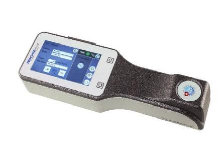
The latest 2017 PXL Systems are out now! Made in Switzerland by PESCHKE
PXL Platinum 330 PXL Sapphire 318
Highlights: Variable treatment options at different energy levels Continuous, interval and LASIK modes Self-calibrating and self-adjusting 5” Color touch screen The PXL Platinum additionally offers: Highlights: Variable energy level settings Continuous, individually customable pulsed and LASIK radiation modes Self-calibrating and self-adjusting Colour touch screen PXL Platinum 330 PXL Sapphire 318
Eye tracking with adjustable real time camera view Bluetooth communication interface + treatment PDF report Integrated ultrasound contact Pachymeter optional CXL treatments are easy to perform, safe for the patient, and can be combined with other medical therapies. SAFE –EFFECTIVE - FLEXIBLE These fully portable and ergonomically designed systems come with an adjustable table mount and a sturdy transport case. These are open systems, i.e. no activation cards, no barcodes, no treatment fees, and the treatment protocols are included. They are considered being the best high-end portable and user-friendly devices on the market according to Swiss, European and global users. No other portable system comes with an eye trackerand custom treatment mode. Our PXL systems contain a built-in communication technology, allowing to communicate across systems (i.e. export of generated treatment reports). CXL –The Experience In recent years corneal cross-linking has become the standard procedure for treating patients with progressive keratoconus and other ectatic corneal diseases because of its effectiveness and lack of serious side effects. A large number of major clinical studies has proven the effectiveness of CXL and the lack of serious side effects. More than 85%ofthe eyes treated with CXL showed a significant increase in BCVA. CXL is the only effective non-invasive treatment to stop progressive Keratoconus and other ectatic disorders (such as PMD and iatrogenic ectasia) and has a regularisation effect on corneal topography. In addition to its role in treating ectatic corneal diseases, CXL has an established place in the management of infectious keratitis. UV light has long been known for its ability to kill different micro-organisms (such as bacterial and fungal). Since keratitis in humans is an important cause of blindness, and antibiotic resistance is an increasing problem worldwide, CXL proves to be an extremely valuable possibility to manage the condition with a satisfactory Outcome. PXL Sapphire 318 The PXL Platinum offers: Eye tracking with adjustable real time camera view Bluetooth communication interface + treatment report creation Ultrasound contact Pachymeter optional CXL treatments are easy to perform, safe for the patient, and can be combined with other medical therapies. SAFE –EFFECTIVE - FLEXIBLE CXL –The Experience In recent years corneal cross-linking has become the standard procedure for treating patients with progressive keratoconus and other ectatic corneal diseases because of its effectiveness and lack of serious side effects. A large number of major clinical studies has proven the effectiveness of CXL and the lack of serious side effects. More than 85%of eyes treated with CXL showed a significant increase in BCVA. Six months after the procedure cylinder was reduced in the majority of patients. CXL is the only effective noninvasive treatment to stop progressive Keratoconus and other ectatic disorders (such as PMD and iatrogenic ectasia) and has a regularisation effect on corneal topography. The PXL systems come in a sturdy transport case
The PXL systems come in a sturdy transport case Ergonimic, flexible table mount In addition to its role in treating ectatic corneal diseases, CXL has an established place in the management of infectious keratitis. UV light has long been known for its ability to kill different microorganisms (such as bacterial and fungal ones). Since keratitis in humans is an important cause of blindness, and antibiotic resistance is an increasing problem worldwide, CXL proves to be an extremely valuable possibility to manage the condition with a satisfactory outcome…….Contact Peschke to learn more! Ergonimic, flexible table mount

Anti-infectives Update NTERIOR SEGMENT Fighting Smarter Optimizing the Current Landscape of Ocular Anti-infectives
by April Ingram
The landscape of ocular infections is changing as pathogens evolve. In addition, how these patients are managed – which differs among regions, and even between practices – is changing as well. To treat patients, doctors need to take the limited clues (and lack of cultures) observed at presentation and act with the speed and precision of bomb diffusion specialist. So, how can optimizing today’s antiinfectives contribute to the best outcomes for our patients . . . and what does the future hold?
Prof. Shigeru Kinoshita of Kyoto Prefectural University of Medicine is a cornea surgeon who, for 40 years, has had a special interest in ocular surface biology and treating corneal infection. Prof. Kinoshita has noticed a shift in the microorganisms they are treating in Japan, similar to other regions. “In Japan, we have many microorganisms and used to target gram-negative bacteria, but nowadays, our patients are mainly gram-positive. We aren’t certain why, but I feel it has much to do with the aging population in developed countries,” he shared.
An observation confirmed by Asia Cornea Society Infectious Keratitis Study (ACSIKS), surveyed the demographics, risk factors, microbiology and outcomes for infectious keratitis in Asia. The study reported that the most common microorganism isolated in both Japan and South Korea was Propionibacterium acnes, a gram-positive bacterium. Prof. Kinoshita added: “In countries, such as India, China or the Philippines, there are more fungal infections. Singapore and Taiwan have high contact lens usage, and therefore more contact lensrelated infections.”
ACSIKS data agrees that wearing contact lenses is the highest risk factor for infectious keratitis in Singapore, Taiwan and Japan, and even higher for females in these regions. Pseudomonas aeruginosa is on the rise and extremely
‘No to mutants!’: Strategy against microbial resistance. Not an X-men movie slogan.

dangerous as more people are wearing contact lenses, and young people are wearing them for longer periods (Stop sleeping in your contacts!). Prof. Kinoshita’s first choice is always levofloxacin, over moxifloxacin or gatifloxacin, for these patients. “If they are younger patients, use it [levofloxacin]. Contact lens wearers – always be thinking pseudomonas, and definitely use it,” he emphasized.
Prof. Kinoshita described how he manages these patients. “Initially, all we have is the clinical manifestation, before getting the results of the culture, to know which pathogenic bacteria is which. You don’t always know, but you have to treat – so we cover with both, that’s why we choose levofloxacin for the first seven days. It works for grampositive and -negative and we feel safe enough to use it for Pseudomonas aeruginosa.”
Prof. Kinoshita, and his colleagues at Kyoto Prefectural University of Medicine, check the bacterial flora when they first see a patient, and they also try to culture some of the bacteria from the conjunctival specimen. This way they can determine if they are up against a gram-positive or -negative bacteria, and they also try to have all the data of the MIC. Refresher: MIC is the minimum inhibitory concentration of an antimicrobial agent – it measures the activity an antimicrobial agent has against an organism and the lowest concentration that it will inhibit growth of the microorganism. MIC has been used extensively to classify bacteria as resistant to an antibiotic. A second metric of resistance is the mutant prevention concentration (MPC), the minimum concentration restricting the growth of the least susceptible, single-step mutant of a bacterial isolate. The inhibitory concentration between MIC and MPC is the window (mutant selection window) where the evolution of resistance can occur.
Resisting the antimicrobial resistance is futile. Resistance is a wellrecognized and ongoing problem across all infectious diseases. Once upon a time, only one type of MRSA was known and feared . . . now, there are four. Resistance patterns and mutations of organisms have now seen MRSA leave the hospital (a.k.a. health care-associated MRSA) and venture out into the community (community-associated MRSA). Due to the misuse and overuse of antibiotics, there is an increase of failure in previously efficacious treatments and a rise in antimicrobial resistance worldwide.
Prof. Kinoshita has also witnessed the evolution of drug resistance: “We used to have several kinds of antimicrobials to treat infection, now we only have one,” he said. “Resistance is a real problem, therefore we try not to use antimicrobial agents on a continuous basis. We extensively treat with levofloxacin, and often will use it for a specific duration of time and then stop, in order to prevent resistance.” It might also be considered if altering how drugs are administered, even on an individual basis, could slow development of resistance.
Elderly patients can bring a full range of challenges and need to be considered differently when it comes to anti-infectives. As immune systems gradually diminish and are compromised more easily, older people are especially susceptible to infection – and the bacteria in this population may develop resistance to antibiotics quicker. Prof. Kinoshita shared: “Always think about the age of the patient and the health of the patient. I profile the patient, it is important to determine if they are compromised, or if they are diabetic. Usually up to 50 or 60 years I feel it is safe enough to use a quinolone. For those over 60, we use quinolone less frequently, because we find we often have to modify treatment to another agent because this population easily develops the mutation for resistance.”
How can we apply what we know about MPC? Although mutant prevention sounds like a good tactic for the characters in a sci-fi movie, it may provide a strategy to minimize antimicrobial resistance. Knowing that when drug concentrations are below the MIC, neither the original nor first step resistant bacterial cells are inhibited, and they also do not become selectively resistant. At concentrations between the MIC and the MPC, the mutant selection window (MSW) growth of first step resistant mutants is blocked, but amplification of resistant subpopulations takes place. Finally, at concentrations higher than the MPC, no selective amplifications take place, because the original and first step resistant cells are annihilated. Then why aren’t we always dosing above the MPC? Optimizing this concept, there are data from in vitro studies and animal models to support using the MSW to improve antimicrobial dosing regimens. Unfortunately, the MSW/MPC ‘sweet spot’ varies for every fluoroquinolone/pathogen combination. There is ongoing study, and much debate about the potential impact of MPC on fluoroquinolone resistance. Prof. Kinoshita is following this discussion closely. “I think it is very important, but still conceptual. In vitro data shows some truth, but there is a lack of data that can be applied in the clinical situation, especially when we have to consider the age and overall health of the patient,” he explained.


AIOS GOVERNING COUNCIL and LOCAL ORGANIZING COMMITTEE Welcome You All To the largest Scientific feast in the world of Ophthalmology

The key to optimizing treatment is selecting an anti-infective depending on relative penetration and concentration. The principal treatment strategy for bacterial infections is to administer antiinfectives at high concentrations. The rationale is that high concentration will kill all microbes quickly, so that there is no chance for resistance to develop, while lower concentration treatments can help with resistance but putting selection pressure on resistant strains. Prof. Kinoshita explains how concentration and penetration factor into his clinical decisions: “We have either 0.5% or 1.5% levofloxacin and 1.5% is our choice for most infections. We’ve found the concentration to be almost the same or better with levofloxacin than moxifloxacin, but the effectiveness has to do with the penetration. When we see corneal infection, high penetration is needed,” he said. “We do also use the 0.5% for

prophylactic treatment, 0.5% is less toxic to the corneal epithelium.” When asked about the next generations of anti-infectives, Prof. Kinoshita said: “I haven’t found much difference in 3rd and 4th generation. It doesn’t matter if someone says, ‘this is a 3rd or 4th generation agent’. Its effectiveness, that is the most important. Is it a quinolone that treats gram-positive and also pseudomonas aeroginas? That is what matters.”
References 1. Drlica K, Zhao X. Mutant selection window hypothesis updated. Clin Infect Dis. 2007;44(5):681- 688. 2. Hansen GT, Blondeau JM. Mutant prevention concentration as a strategy to minimize antimicrobial resistance: a timely concept but will its acceptance be too late? Therapy. 2005;2(1):61-66. 3. Khor WB, Prajna VN, Garg P, et al; ACSIKS Group. The Asia Cornea Society Infectious Keratitis Study: A Prospective Multicenter Study of Infectious Keratitis in Asia. Am J Ophthalmol. 2018;195:161-170.
About the Contributing Doctor
Prof. Shigeru Kinoshita is a professor and chairman of ophthalmology at Kyoto Prefectural University of Medicine, Kyoto, Japan. Prof. Kinoshita is a cornea and refractive surgeon with a special interest in the close relationship between the cornea and glaucoma. Prof. Kinoshita established, along with Richard Thoft, the concept of centripetal movement of corneal epithelium. This shed new light on the importance of the limbal epithelium and contributed to the development of corneal stem cell theory. Over the last 40 years, his primary interests have been focused on the research and development of new therapeutic modalities for the cornea. To this end, Kinoshita’s group has established systems to transplant cultivated mucosal epithelial stem cells and cultivated corneal endothelium.

by Brooke Herron
My earliest childhood memory is going to the eye doctor when I was 8 . . . life before that was a blur. – ophthalmic joke
At first glance, it wouldn’t seem like comedy and ophthalmology could have much in common. In fact, other than the quirky one-liner above, an Internet search reveals a startling lack of eyeball-related humor online. Of course, this makes sense as surgery is serious business. Eliciting laughs and removing cataracts are decidedly different things. However, there is one commonality that surgeons and comedians both share – the ability to improvise.
Improvisation (or improv) refers to creating an action without pre-planning. Generally, improv is used in comedy, and requires the ability to think, process and retain information quickly. That same ability is also necessary in the OT, especially when complications occur. As such, in this CAKE cover story, we look at case studies in improvisation, with tips on how to proceed when the unwanted surprise of a complication arises.
Keep Calm and Operate On
In comedic improvisation, calming skills like meditation and deep breathing are key. That’s because performers must remain calm, but act fast, with complete awareness of their environment.
The same can be said for surgical improvisations. When a complication arises, Dr. David Lubeck, a cornea, cataract and refractive surgery specialist from Chicago, Illinois, USA, says that

before any improvisation can begin, he takes a moment to focus with some deep, meditative breathing.
“It’s what I do, and it’s what I was taught a long time ago. When a complication happens, I immediately begin a short breathing mediation,” he explains. “I stop the procedure and mitigate anything that is pressing or urgent. This then allows me a moment

to stop thinking about the case, to do a very quick (but very effective) mediation, and then come back to it.”
Dr. Lubeck says this brief moment of quiet, takes him from initial feelings like anxiety and panic, to being clearheaded and focused – and this opens the door for improvisation to mitigate the complication. “Until you’re clearheaded, you can’t improvise,” he explains. “Once
and differently than their intended design, or that the instrument itself may have multiple applications.
“Understanding the flexibility and the potential of your resources is part of the broad base of knowledge that surgeons need to have,” he says. “For all the pre-planning you can do on a case-by-case basis, you really just have to have these improvisation strategies in your head, deep in your mechanistic thinking.”
He recommends having a general, internalized strategy for handling different situations and understanding all the resources available, as well as their different uses.
you have the ability to think laterally. You can assess everything you have at hand to help you, such as are your staff, who’s alongside you and how capable they are; your instrumentation; and the capabilities of the hospital where you’re operating (which varies a lot).”
Dr. Lubeck says it’s important to understand that certain pieces of instrumentation can be used creatively
Dress Rehearsal: Unstable Lenses in Cataract Surgery
A complication that Dr. Lubeck says happens frequently, but can still catch surgeons off-guard, is an unstable lens. “In these cases, there’s often no indication. But the moment you begin working on the cataract and the capsulotomy, you find the lens is unstable and you know things are not going to go as planned.” When CAKE magazine interviewed Dr. Lubeck, he had experienced this exact complication earlier in the same day. “Invariably, what happens is, the cases you think are going to be difficult often end up being straightforward and simple, and the cases that look like they’re going to be straightforward can completely surprise you – like today. I had no indication that it was going to be a loose lens . . . there was no sign of it whatsoever.”
When this complication occurs, his first step (following a brief meditation) is to ask if his assistant is familiar with an unstable lens. Their answer is usually ‘no’, so Dr. Lubeck talks them through it: “A loose lens means that I may have trouble extracting the lens, we may drop the lens, we may have to do a vitrectomy . . .”
Then he begins his mental – or improv – checklist, asking questions
”– Dr. David Lubeck

like: Are there capsule hooks? A vitrector? More visco-elastics? Once he knows what resources are available, a strategy is devised and the surgery proceeds. All of which occurs in two to three minutes.
He says that when complicated cases are approached this way, they largely turn out well, and the sense of accomplishment is far greater. “I have much greater sense of satisfaction and sense of accomplishment as a surgeon when these complicated cases turn out well, as compared to the normal cases.”
Curtain Call: The Case of the Skewered Lens

As a pediatric ophthalmologist based in Pittsburgh, Pennsylvania, USA, Dr. Kanwal Nischal has many patients referred to him with Marfan syndrome, a genetic disorder that affects the connective tissue. When this affects the eyes, it can cause the lens to dislocate spontaneously and move out of the central position.
“This happens because the zonules are weak, so their lenses move
upwards and laterally,” explains Dr. Nischal. “Of course, that’s a problem: The lens in no longer central and it’s not focusing light properly.”
During these surgeries, he says that he normally removes the entire lens without the assistance of a vitreoretinal surgeon, as it’s something he can do safely. However, in one case, due to the position of the lens, Dr. Nischal was worried that the lens was going to drop into the capsular bag – and without a vitreoretinal surgeon on hand, there could potentially be a problem. Thus, his case in improvisation began. The lens in the left eye was dislocated superiorly and laterally, so Dr. Nischal went in at 9 and 2 o’clock, and used two 30-gauge needles to skewer and fixate the lens. “Then you put the infusion in and with the vitrector, you remove the lens material and little bit of the vitreous, so you’re left with nothing in the eye,” he explains, adding that these cases are rehabilitated with a contact lens, with the option for an implant at age 21.
“Here, my improvisation was taking two needles and skewering the lens so it couldn’t fall into the back of the eye while I was removing it. And the patient’s result was good,” he says. Dr. Nischal says this isn’t the only instance when he’s used this improvisation: “I’ve done it before in adult cataract surgery, in patients with a posterior polar cataract,” he says, “It’s not something that someone showed me, it’s an improvisation to stabilize the lens while I remove it.”
Avoiding Improv in Cases of Expulsive Hemorrhage
Some complications are so severe that surgeons plan in advance for them. For example, when approaching infant corneal transplants, one complication that Dr. Nischal always takes into consideration is an expulsive hemorrhage.
Although they can occur during any intraocular operation, in this case, once the cut is made to remove the diseased tissue and the eye is opened, for example, a change in intraocular
”– Dr. Kanwal Nischal

pressure can cause the choroid to suddenly fill with blood. And if this continues, it can result in the extrusion of the all the eye’s contents. In these cases, the surgeons must act rapidly to help save the eye.
“It’s the worst thing that can happen to an ophthalmic surgeon. If you were to go into any of the conferences and ask somebody, ‘have you ever had an expulsive hemorrhage?’, they would either say ‘yes’ and it was horrible, or they would touch wood 1,000 times,” says Dr. Nischal, who is most well-known for performing corneal transplants in infants.
Dr. Nischal says he had one case of expulsive hemorrhage in an infant’s eye in 2001, and since then he takes many precautions. The first thing was to change the anesthesia. “The carbon dioxide (CO 2 ) level has to be below 30mmHg. The lower the CO 2 , the less positive pressure in the eye, and when you open the eye, there’s less of a change from high pressure to low pressure,” he explains, noting that this complication occurs from the sudden intraocular pressure (IOP) change.
At the moment of his initial cut, he checks the blood pressure and pulse with the anesthesiologist. And if either of them rises, the eye could be at risk, so he
proceeds using the ‘sandwich technique’. Dr. Nischal says he’s employed this technique in worrisome cases just five times throughout his career.
“Normally, in a cornea transplant, the diseased cornea is removed and the donor cornea is placed and sown in,” he says. “In this sandwich technique, every time there’s a 90-degree cut, a suture is put in. You cut the host, but you’ve put in four sutures as you’ve done it.”
Next, the donor tissue is placed on top the host, but with a filling of viscoelastic to protect the donor’s endothelium. In the quadrants where there are no sutures in the host, you place them, but before the fourth is placed, the previous sutures in the host are cut. This allows the host tissue to be removed from underneath the donor tissue, and then the fourth suture can be applied.
“It’s complicated, but what you’ve essentially done is you’ve removed the host tissue without ever exposing the eye completely to an absolutely open globe,” explains Dr. Nischal, adding that this is the most technically detailed plan he undertakes to avoid a complication.
“I think what happens is that people don’t talk about expulsive hemorrhage. So in your training, no one tells you what you should do when it happens,” he
says. “If you’re lucky, you come across someone whose had it. Most people, when it happens, they are so stunned that it’s too late to do anything.”
Innovations Reduce Improv in the OT
According to Dr. Lubeck, over the last decade, continued innovation in devices, instrumentation and surgical techniques has helped reduce the amount of ‘improv’ in the OT, while making the likelihood of complications lower, and the prevention of complications significantly higher. Specifically, he notes that fluidics in phaco machines, femtosecond lasers and improvements to ultrasonic energy delivery are the top innovations that have contributed to this trend. Another game-changer is intraoperative OCT (iOCT). “Integrated intraoperative OCR has made a huge difference for me,” says Dr. Nischal. “It has reduced my complications in the sense that I can do – for example – an endothelial keratoplasty in a cornea that’s completely opaque, because I can see through it with the OCT.”
“I can operate using OCT on what I can’t actually see in the surgical field,” he continues. “It not only allows you to take on more complicated surgeries more safely, but it flattens the learning curve. When I do pediatric cataract surgery with my fellows, I can now show them all the steps and what happens to the tissues. I can see vitreous if it’s in the anterior chamber, I don’t have to stain it. It’s a game changer.”
However, there can be downsides, even with technology that helps improve safety and reduce the rate of complications. Dr. Lubeck says that, of course, lowering complications is a good thing. However, if surgeons don’t experience them, especially in training, they don’t develop improvisational strategies – and therefore, can’t access those skills when needed.
“I think it’s great that we have fewer complications and that the risk of complications is drastically less than when I was trained,” he explains. “But I think the likelihood that the young surgeons will have the breadth of improvisational skills is less. Surgeons learning in this current environment, with vastly improved technology, won’t need to develop broad improvisational skills.”
Final Thoughts on Improv
So, when it comes to improvisation and complications, what tips do experienced surgeons have for the younger generation?
Dr. Nischal notes that one thing he teaches his fellows is to look at the whole eye. “Often, we are so taken by the moment, we forget to look at the whole eye. So, I always say to my fellows: ‘Don’t just concentrate at the end of your instrument, obviously you need to know where it is . . . but you have look at the whole picture,” he explains, adding that he himself had to be trained to do this.
“In many ways, it’s very Zen, very Buddhist. If you just concentrate on one point, you miss the bigger picture. And that’s when you miss the complication that’s about to happen,” he says.
For example, when performing anterior vitrectomy and posterior capsulorhexis in children, Dr. Nischal says to look at the iris. “Rather than waiting until the end to see if there’s any vitreous in the wound, while you’re doing your vitrectomy, you should be looking at the iris. If you see the iris quiver or flutter, there’s vitreous hitting it,” he says. “But if you’re so taken by looking at the end of your vitrector – and not looking at the whole eye while you’re operating – you’ll miss that.” A final tip? Take a moment to pause and assess. “When you realize a complication is arising, do a hard stop and do whatever it takes to stop its progression,” says Dr. Lubeck. For example, when a complication occurs during cataract surgery, he says filling the anterior chamber with visco (specifically) usually buys the surgeon some time. “Then, you can take a step back and break the neurologic response of panic and fear, so you can then access the rational and calm parts of your brain.” These tips, along with each surgeon’s intrinsic framework of knowledge, experience, training and support from technology, contribute to the overall success of overcoming complications. And with improvisation, a clear head and quick decision making, unwanted surgical occurrences can be abated and visual outcomes can be saved or maintained. And that’s one big reason to cue the applause!
About the Contributing Doctors
Dr. David Lubeck is a cataract, corneal and refractive surgeon who has practiced in Chicago, Illinois, USA, for 30 years. He completed his medical training at Northwestern University and then his ophthalmology residency at the University of Illinois Eye and Ear Infirmary in Chicago. A fellowship in corneal surgery was served at Flinders Medical Center, Adelaide, Australia. He is an assistant clinical professor of ophthalmology at the University of Illinois Eye and Ear Infirmary, Chicago. Dr. Lubeck is compelled by the methodology and psychology of surgical learning and practice development. His primary focus is to make current surgical principles, techniques, and instrumentation accessible to physicians at all levels of experience. Curricula that he has developed have been integrated into surgical teaching programs in several different countries. [Email: dr.lubeck@arboreyecare.com]
Prof. Kanwal “Ken” Nischal is chief of the Division of Pediatric Ophthalmology and Strabismus at UPMC Children’s Hospital of Pittsburgh, director of Pediatric Program Development at the UPMC Eye Center, medical director for Telemedicine Services at Children’s Hospital of Pittsburgh, associate medical director for UPMC International and professor of ophthalmology at the University of Pittsburgh School of Medicine. His focus is on evidence-based protocol-led clinical care with clinical outcome measures as a source of clinical research. His main areas of clinical research are Anterior Segment Developmental Anomalies Affecting the Cornea, Lens and Trabecular Meshwork. He has published widely in Pediatric Cataract, Glaucoma and Cornea and Craniofacial Anomalies. He has developed an ocular genetic service at Children’s Hospital of Pittsburgh and is co-founder of WSPOS. [Email: nischalkk@upmc.edu]
COVER STORY My dad was given a selfie stick as a Christmas present . . . it turns out that he can now hold it far enough away to read text messages.


Q A


Which kind of humor do ophthalmologists appreciate the most?


Eye-rony.


QWhy did the gangster have to see an eye doctor?
AHe had glock-oma.
These jokes are as ‘cornea’ as it gets . . . all sourced from the finest online eyeball comedians. When is a lens not a lens? When it’s aphakic. When is a lens REALLY not a lens? When it’s ‘a-fake-ic’. A man goes to the eye doctor. He sits down and the receptionist asks him why he is there. The man complains: “I keep seeing spots in front of my eyes."


Q A


What do you call a blind stag?


No Eye-Deer The receptionist asks: “Have you ever seen a doctor?" to which the man replies:
“No, just spots."
HOYA Launches Joint Venture with GeMax in China
Chinese physicians and patients will have broader access to HOYA Surgical Optics (HSO) products, thanks to a joint venture with GeMax, a specialty intraocular lens (IOL) service provider. Since 2006, GeMax has been a distributorpartner for HSO and has a strong market position in China, covering 630 hospitals in 32 provinces. The joint venture will be called HOYA GeMax Medical and the deal is expected to close by the end of March 2020.
Due to its high population, cataract is a major public health concern in

China. According to HSO CEO John Goltermann Lassen, “this venture will allow us [HSO] to be a stronger partner and meet the growing needs in China, a market of strategic importance to our organizations”.
Combining sales and distribution into one unified business will provide Chinese surgeons and patients more access to the entire HSO portfolio of IOLs across the AF-1 and Vivinex platforms.
HSO Vice President Global Sales Mads Bjerre Andersen said they are

excited to embark on this journey with GeMax: “We have worked together for more than a decade and this next step will help take our business to new heights in China.”
“This is a natural progression given the strong partnership and mutual trust between our organizations and we’re looking forward to strengthening our position in China together,” shared Xia Xintao, general manager of Gemax.
The official signing ceremony took place in January 2020.


