by Tan Sher Lynn and Hazlin Hassan
Prof. Frank G. Holz, President of the 20th Congress of the European Society of Retina Specialists (EURETINA 2020) started his welcome remarks by expressing his feelings of being heartened by the dedication of medical teams who continue to deliver care to patients at great personal risk even as the pandemic continues to cause devastation across the globe.
According to Prof. Holz, hot topics will be discussed during the congress and expert faculties will address multiple new therapeutic


developments in both medical and surgical aspects of the retina. More importantly, Prof. Holz shared how he has initiated, together with the board of EURETINA, a strategic roadmap to outline plans and priorities for the society over the next five years.


“A broad range of impactful projects have been identified, including the overhaul and development of the society’s digital offerings.
on Page 3

CAKE AND PIE MAGAZINES’ DAILY CONGRESS NEWS ON THE ANTERIOR AND POSTERIOR SEGMENTS 03 | 10 | 20 cataract • anterior segment • kudos • enlightenment 2ISSUE posterior segment • innovation • enlightenment &
Cont.
>>
J o i n u s f o r t h e v i r t u a l e x p e r i e n ce a n d l e a r n m o r e a b o u t t h e l a t e s t t e c h n o l o g i e s i n e y ec a r e We h a v e a n e x c i t i n g p r o gr a m l i n e d u p, i n c l u di n g t h e u n v e il i n g o f o u r a l l - n e w v i r t u a l s h o w r o o m ! R e g i s t e r he r e: ww w. z e i s s .c o m /e sc r s - e u r et i n a - r e g is t r a t i o n Z EISS O p ht hal m i c V i r tu al E x p e r i e n ce E S C R S - E U R E T I N A S p o t l i g h t 2020 O c t o ber 1 – 4, 2020 Join us for the virtual experience and learn more about the latest technologies in eyecare. We have an exciting program lined up, including the unveiling of our all-new virtual showroom! Register here: ZEISS Ophthalmic Virtual Experience ESCRS-EURETINA Spotlight 2020 October 1 – 4, 2020 Join us for the virtual experience and learn more about the latest technologies in eyecare. We have an exciting program lined up, including the unveiling of our all-new virtual showroom! Register here: www.zeiss.com/escrs-euretina-registration ZEISS Ophthalmic Virtual Experience ESCRS-EURETINA Spotlight 2020 October 1 – 4, 2020 A Warm Welcome, Promising Therapies for Inherited Blindness and Lessons from Great Cataract Surgeons Published by piemagazine.org cakemagazine.org Matt Young CEO & Publisher Robert Anderson Media Director Hannah Nguyen Production & Circulation Manager Gloria D. Gamat Chief Editor Brooke Herron Editor International Business Development Ruchi Mahajan Ranga Brandon Winkeler Writers Andrew Sweeney Hazlin Hassan Joanna Lee Olawale Salami Sam McCommon Tan Sher Lynn Maricel Salvador Graphic Designer Media MICE Pte. Ltd. 6001 Beach Road, #19-06 Golden Mile Tower, Singapore 199589 Tel: +65 8186 7677 | +1 302 261 5379 Email: enquiry@mediamice.com www.mediaMICE.com
EURETINA 2020 Virtual Congress
Missed our EURETINA 2020 symposia? Watch the video recordings to hear our global experts talk about optimizing outcomes in patients with nAMD and DME.
Optimizing patient outcomes in nAMD: The key principles


Chair: Prof. Paolo Lanzetta
Hear from Dr. Michael Stewart, Dr. Nancy Holekamp, and Prof. Nicole Eter as they present the latest evidence and discuss hot topics in nAMD management.
Video recording
Vision in focus: Delivering optimized patient outcomes in DME in the real world


Chair: Prof. Ian Pearce


Listen to Prof. Jean-François Korobelnik, Dr. Varun Chaudhary, and Prof. Francine Behar-Cohen as they share expert guidance for optimizing management of patients with retinal vascular diseases.

Video recording
“Join us as we examine the latest evidence from clinical trials and realworld practice to address key challenges in retinal disease management and ensure we are optimizing outcomes for our patients.”
Prof. Paolo Lanzetta
Additionally, we warmly welcome you to the Bayer Networking Lounge, which will provide you with an opportunity to connect with experts in the treatment of retinal diseases.
Learn more about Bayer Ophthalmology by visiting our online channels:

03 Oct 2020 | Issue #2 2 &
DME, diabetic macular edema; nAMD, neovascular age-related macular degeneration. The summary of product characteristics (SPC) of aflibercept solution for injection as approved by the European Commission can be found here BAYER and the Bayer Cross are registered trademarks of Bayer. © Bayer AG 2020. All rights reserved. Bayer (South East Asia) Pte Ltd, 2 Tanjong Katong Road Paya Lebar Quarter 3 #07-01, Singapore 437161. September 2020 | PP-PF-OPHT-ALL-0046-1 | PP-EYL-SG-0184-1 (09/20)
Research funding opportunities will be increased and we will also expand the portfolio of benefits available to young retinal specialists, starting with the introduction of a new retina mentorship program,” he said. Then he introduced Professor Stanislao Reedsville as the speaker of the keynote address, dedicated to the founder and first president of EURETINA, Dr. Gisbert Richard.
Gisbert Richard Keynote Lecture
Prof. Stanislao Reedsville of the University of Florence has developed various innovative devices for vitrectomy and won multiple awards, including the Innovative Surgeon Award 2013 and the Alfred Mann Foundation Award for Scientific Achievement.
In his speech titled The Restless Fight against Inherited Blindness, he noted that various types of genetic mutations cause retinal dystrophies, and 30% of these mutations are still unknown. Different stages of retinitis require different treatment. The initial stage where there are no symptoms except night blindness involves pharmacologic treatment, the intermediate stage with visual field constriction involves gene therapy, the advanced stage where visual field is severely constricted involves stem
cell transplantation, while the end stage with no light perception requires retinal prosthesis.
“Currently, it is estimated that 36 million individuals are blind. Visual prostheses can provide benefit to those with severe vision loss, especially if no other medical treatment options exist. There are different retinal prostheses approaches, such as the epiretinal (Argus II, IRIS I/ III), intraretinal (Nano Retina), subretinal (Alpha, PRIMA), suprachoroidal (Bionic Eye, NIDEK) and subretinal with 3D electrodes (Pilar),” he said.
As work on developing better prostheses continues, he noted that future prostheses need to address issues regarding the number of electrodes and spacing, sharpen visual acuity and expand visual field area, reduce size and surgical invasiveness, and offer simple, non-invasive (wireless) power delivery.
Moving into the future, Professor Reedsville said that the NR600 System by Nano Retina is a sophisticated and elegant solution which can restore the sight of people who lost their vision due to retinal degenerative diseases.

“The miniature Nano Retina device, the NR600 Implant, replaces the functionality of the damaged photoreceptor cells and creates the
electrical stimulation required to activate the remaining healthy retinal cells. It consists of two components; a miniature implantable chip and a set of eyeglasses worn by the patient. It features an IOL-like fixation at the ciliary sulcus, while the posterior fixation involves hermetically sealed capsule with microelectrodes, avoiding NFL stimulation and providing direct stimulation to the ganglion cells, resulting in high resolution images.
“It is indeed an exciting time in regards to care for patients with inherited retinal diseases. Today, clinicians can provide treatments that can potentially prevent blindness for some patients with inherited retinal disease and soon we will have more and more effective ‘weapons’ to fight this battle. As scientists, researchers, surgeons and doctors, we are fortunate to live in this era,” he said.
38th ESCRS Congress and Ridley Medal Lecture
Meanwhile, over at the 38th Congress of the European Society of Cataract & Refractive Surgeons (ESCRS 2020) virtual platform, congress President Prof. Dr. Rudy MMA Nuijts welcomed the delegates and presented this year’s Ridley Medal Lecture awardee…
Sometimes the greatest lessons can be learned from the developing world.
This was what David Chang, MD, clinical professor at the University of California, San Francisco, shared during the Ridley Medal Lecture at the European Society of Cataract and Refractive Surgeons (ESCRS) virtual congress on Friday (Oct 2).
It remains a challenge to tackle the growing backlog of cataract blindness which is the leading cause of global blindness.
“The problem is that there are so few qualified cataract surgeons...that we need to maximize their productivity by allowing them to do rapid surgery at very high volume but it has to work well for the terribly advanced cataracts that they face and at a very low complication rate. And it has to be cost effective and affordable,” said Dr. Chang.
3 CAKE and PIE magazines’ Daily Congress News on the Anterior and Posterior Segments
Cont. on Page 15 >>
>> Cont. from Page 1


2 18 November 2022 | Issue #1 SHOW DAILY by DIGITAL MARKETING + ADVERTISING + VIDEO PRODUCTION + MEDICAL WRITING + EVENTS Request our 2023 Media Kit Now! Write enquiry@mediamice.com for a copy HQ Office: 6001 Beach Road, #09-09 Golden Mile Tower, Singapore 199589 Phone: +65 8186 7677 Satellite Office: 2 Nuoc Man 2 Street, Da Nang City, Vietnam 50506 Phone: +84 868 063 773 E-mail: enquiry@mediamice.com Web: www.mediamice.com
Symposium Highlights
The Bigger Picture in Clinical Practice
by Olawale Salami
In a Satellite Symposium at the virtual EURETINA 2020 congress, Allergan (an AbbVie company) presented ‘The Bigger Picture in Clinical Practice’, and here are some of the highlights…
Getting the best results from dexamethasone implants in DME: No DRILs required
biomarker. Therefore, patients with these three biomarkers are the best candidates for dexamethasone implants.
“Besides, we found that dexamethasone implant was well suited for both naïve and refractory cases of DME, and vision gain more significant in suitable naïve as compared to refractory eyes and was generally well tolerated,” noted Prof. Iglicki.
Ozurdex® shines in patients with DME, RVO and noninfectious uveitis
“We
Diabetic macular edema (DME) remains an ever-increasing challenge globally, and the International Retina Group has been at the forefront of conducting clinical studies designed to answer pragmatic questions that impact patient care. Prof. Matias Iglicki presented a summary of some of the most essential published results from the group’s work in recent years.
According to Prof. Iglicki, the pathogenesis of DME is driven by two pathways: an inflammatory and an ischemic pathway. “While dexamethasone implants have been shown to target both pathways, antiVEGFs target only the ischemic pathway. Therefore we explored possible biomarkers in the OCT that could serve as functional outcome predictors in DME patients treated with dexamethasone,” he explained.
Furthermore, Prof. Iglicki noted that they found patients with presence of subretinal fluid, absence of hyperreflective foci and the absence of DRIL (disorganization of retinal inner layers) gained better vision. If you find DRIL on a patient’s OCT, that is a bad
— Prof. Laurent
The RELDEX-2 study aimed to answer the question: Does Ozurdex® provide long-term visual improvements in patients with DME? The investigators studied 227 eyes of 152 patients treated with Ozurdex®, shared Prof. Laurent Kodjikian. They found that the average gain in BCVA during the follow-up period was about 12 letters, and over 80% of these improvements occurred after the first injection, noticeable from as early as 2 months. “Furthermore, we observed DME resolution, shown as the absence of cystoid macular spaces on OCT in 37% of eyes. We found very favorable long term outcomes when using Ozurdex®, which gave us the confidence of long term benefits in the treatment of DME,” reported Prof. Kodjikian.
Prof. Kodjikian also summarized the SAFODEX-2 study. “We were interested in understanding the long-term incidence and risk factors for ocular hypertension in patients who received Ozurdex® injections,” said Prof. Kodjikian. “We followed-up almost 500 eyes, for over
3 years and found ocular hypertension in about a third of patients, which was controlled by ocular treatment alone in 97% of cases,” he reported.
In patients with uveitis, Prof. Kodjikian explained that in the RUVDEX study, 152 eyes were followed-up for 19 months and is the to date, the largest cohort of patients with uveitis treated with Ozurdex®, for mostly macular edema. “Our results showed that the maximum mean VA gain was +12 letters and a functional response of >5 letters was seen in 64 % of eyes,” he emphasized.
Furthermore, recent studies have also shown that in treatment naïve patients with retinal vein occlusion who present with macular edema, treatment with Ozurdex® showed excellent responses.
Time and money: Dexamethasone treatment In Swedish DME patients
“We had less visits and less per patient costs when we treated DME patients with dexamethasone.”
— Dr. Inger Westborg
Dr. Inger Westborg shared insights on a recent DME cost evaluation project in Sweden. She explained that in the national DME program in Sweden, both anti-VEGFs and dexamethasone implants are available for patients. “We asked ourselves an important question: is it possible to increase clinic capacity without increasing cost,” she said.
To answer this, her team conducted interviews and cost calculations in five clinics across the country.
“We found that the total number of visits per patient within 1 year was higher for patients treated using anti -VEGFs, while the total costs for treatment and visits were less with dexamethasone compared to all the anti-VEGFs,” shared Dr. Westborg.
“With COVID-19, the DME guidelines in Sweden have now recommended switching DME patients to dexamethasone implants for extended injection interval,” she added.
5 CAKE and PIE magazines’ Daily Congress News on the Anterior and Posterior Segments
“We found dexamethasone implants well suited for both naïve and refractory cases of DME, vision gain greater in suitable naïve as compared to refractory eyes, and was generally well tolerated.”
— Prof. Matias Iglicki
found very favorable long term outcomes when using Ozurdex®, which gave us confidence of long term benefits in the treatment of DME.”
Kodjikian
Making a real difference in the fight against vision loss
The Vision Academy Fighting to save sight
The Vision Academy is a unique, independent group of over 90 world leaders in ophthalmology across 36 countries, working to develop knowledge and provide guidance where none currently exists.
The group has unrivaled knowledge of patients’ needs and tackles key issues that no other program is addressing, by:
Identifying and researching impactful clinical challenges
Expertly reviewing clinical evidence to develop clear clinical guidance
Developing multichannel educational programs to support local clinical practice
Mentoring the next generation of retinal experts
To learn more, visit www.visionacademy.org
03 Oct 2020 | Issue #2 6 & VISION ACADEMY people | research | education
Follow us on LinkedIn
As a leader in ophthalmology, Bayer is committed to transforming management of retinal disease. The Vision Academy, sponsored by Bayer, is fighting to save sight around the world.
September 2020 | MA-PFM-OPHT-ALL-0208-1
I am proud to be part of this ambitious initiative. The Vision Academy is tackling key issues that no other program is addressing, for the benefit of patients.
Professor Anat Loewenstein Co-chair, Vision Academy Steering Committee
Understanding nAMD Treatment Better
While there is no cure for wet or neovascular age-related macular degeneration (nAMD), there are treatment options to prevent further disease progression. During the Bayer Satellite Symposium entitled: Optimizing Patient Outcomes in nAMD: The Key Principles during the virtual EURETINA 2020 congress, experts discussed current treatment paradigms, extending treatment intervals and the importance of fluid.
Key points in drug dynamics, fluid and treatment intervals
Following the three main presentations, session chair Paolo Lanzetta (Italy) provided a summary of key learnings, before discussing practical clinical insights with the expert panel.
Dr. Michael Stewart (USA) kicked off the symposium with a talk on Considering drug dynamics: what is the relevance for clinical practice?
In summary, Dr. Lanzetta said: “According to the studies presented, aflibercept has a higher VEGF binding affinity and relative potency, and a longer intraocular half-life than other
by Brooke Herron
anti-VEGF agents. In a vitreous PK model, aflibercept has a longer VEGF suppression time than brolucizumab.”
Next, Dr. Nancy Holekamp (USA) discussed Fluid in focus: advancing our understanding. “Intraretinal fluid (IRF) has a negative association on visual outcomes, whereas some amount of subretinal fluid during a proactive treatment may be tolerated, as it does not have a negative impact on function,” continued Dr. Lanzetta.
The final presentation was from Dr. Nicole Eter (Germany) on Proactive and practical: extending intervals with aflibercept. Dr. Lanzetta explained: “Regarding the regimen, a proactive treat-and-extend (T&E) approach with aflibercept, results both in visual gain and extended intervals. The intervals may reach 12 or more weeks and even 16 weeks, which translates into a reduced burden.”
Expert exchange: practical insights from the clinic

During the symposium, much study data was presented. In the final moments, Dr.
Lanzetta asked each expert to translate the data into practical clinical advice.
On safety, Dr. Stewart says that aflibercept, ranibizumab and bevacizumab have very low incidences of ocular inflammation, relatively equivalent incidences of endophthalmitis, and almost non-existent vasculitis. He then mentioned brolucizumab, which has reported incidences of adverse events like retinal vasculitis. “Cummatively, it’s about one per 1,000 injections. That may sound small, but when you compare that to aflibercept, it’s less than one in one million,” shared Dr. Stewart.
“This may be a problem that is very difficult to fix due to the very nature of the drug and the concentration going into the eye,” he continued. Abicipar was also recently denied FDA approval due to a high rate of intraocular inflammation.
“We have three safe drugs, but now as we’re pushing the limit, we’re starting to look at complications that we haven’t previously seen — to me, this is a significant concern that every physician needs to look at very carefully and discuss with patients in great detail before embarking on the use of these new drugs,” concluded Dr. Stewart.
“Do you tolerate some subretinal fluid in clinical practice?” was the next question asked by Dr. Lanzetta to Dr. HoleKamp.
“Yes,” she replied. “I think that all the arrows are essentially pointing to the same direction — that in these post hoc analyses, if we tolerate subretinal fluid, it’s not damaging to vision. And really, to everyone’s surprise, [in certain post hoc analyses] subretinal fluid actually produced eyes with better vision than eyes with resolved or full resolution of fluid,” shared Dr. Holekamp, adding that now is the time for a more sophisticated view on fluid management.
Moving on to T&E in clinical practice, Dr. Lanzetta asked of Dr. Eter if she extends up to 16 weeks with aflibercept.
“In my daily routine, I have a lot of patients with an extension of 12 weeks, and I also have some patients on 16 week intervals,” said Dr. Eter. She explains that every patient should be regarded as an individual case, but that extensions up to 16 weeks are possible.
7 CAKE and PIE magazines’ Daily Congress News on the Anterior and Posterior Segments
Anti-VEGFs can stop nAMD progression in its tracks.
A Spotlight on:
by Olawale Salami
changes, according to Dr. Pilotto, involve an increased volume of inner and outer retinal layers and thickening of neuronal cells.
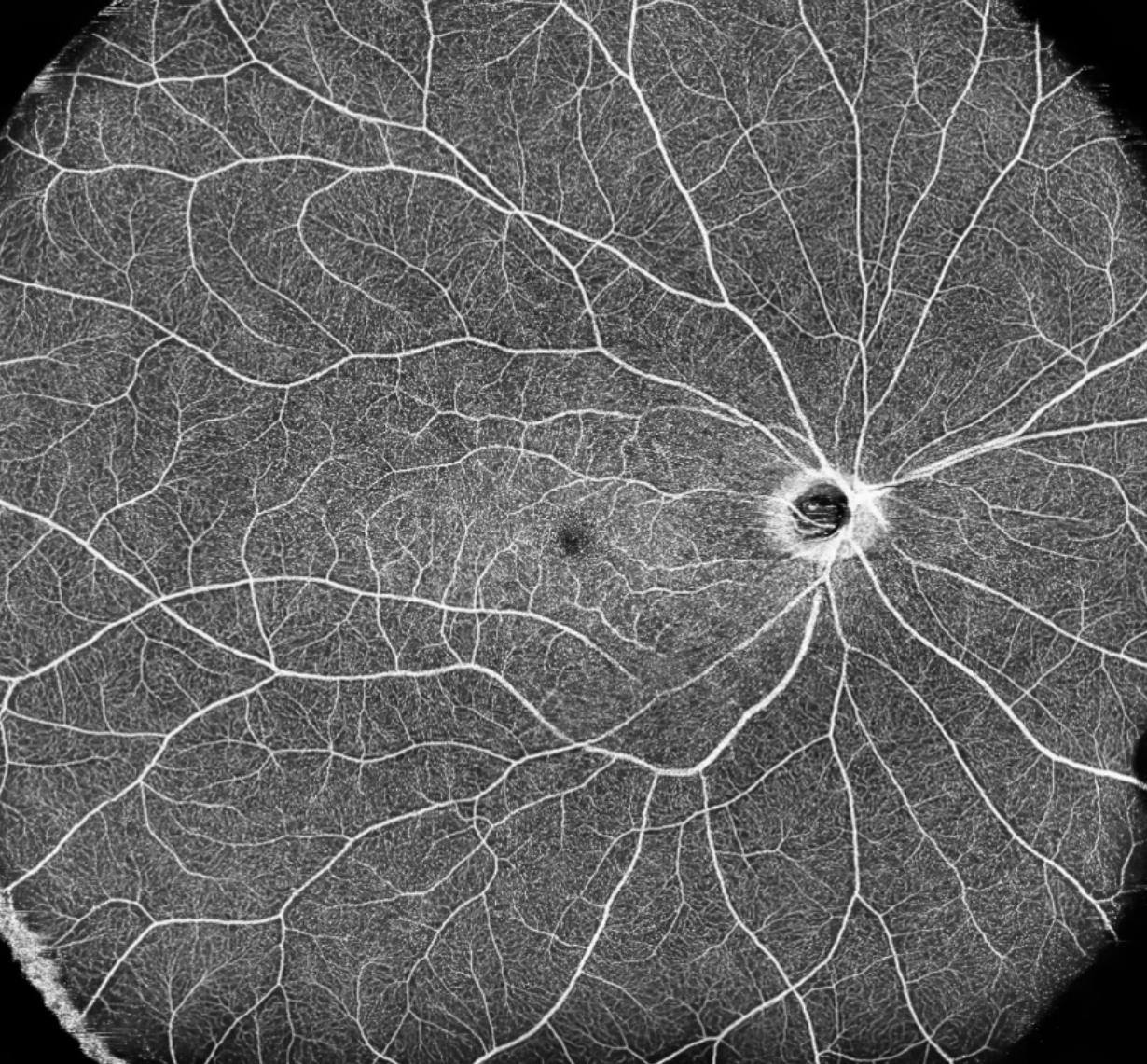
“Optical coherence tomography (OCT) has also allowed us to study retinal vascular changes in very young children with diabetic retinopathy, and we have seen early changes in the foveal avascular zone, before the development of retinopathy,” she shared.
“Single vascular plexuses are also involved early while the intermediate capillary plexus changes are strongly linked with the levels of HBA1c and mean glycemia,’ said Dr. Pilotto.
develop. “It is a common procedure for people with diabetes to need a vitrectomy, and studies have shown that about one in six patients will need a vitrectomy over a 2-year period,” shared Dr. Laidlaw.

At the virtual EURETINA 2020 congress, Dr. Elisabetta Pilotto from the University of Padova in Italy, presented her research into early detection of retinal changes in type 1 diabetic children. According to Dr. Pilotto, the incidence of type1 diabetes is on the increase globally, and given that up to 60% of patients develop diabetic retinopathy within the first two decades after the onset of diabetes, screening by fundus examination has been recommended by the American Academy of Ophthalmology (AAO).
Diabetic retinopathy in children
Regarding the pathogenesis, Dr. Pilotto explained that retinopathy in diabetic children is characterized by very early diffuse involvement of retinal structures and flow changes, with differential involvement of retinal plexuses. These
In her concluding remarks, Dr. Pilotto stated: “With advances in imaging systems, we are now able to detect imaging biomarkers which serve as early signs of retinal involvement, even in very young diabetic children.”
When do we call the surgeons?
In his presentation, Dr. Alistair Laidlaw, consultant vitreoretinal surgeon at St. Thomas Hospital in London, United Kingdom, summarized the main indications for surgery in patients with diabetic retinopathy.
According to Dr. Laidlaw, proliferative diabetic vitreoretinopathy lies at the heart of complications seen in patients. Laser treatment effectively controls proliferation; however, surgery is needed when vitreous traction and hemorrhage
He emphasized that the pathology of changes are divided into different phases. The first phase is the ischemic drive, in which there is a proliferation of pre-retinal vessels into the posterior vitreous. At this stage, laser or antiVEGF drugs may not wholly arrest the proliferation, and these leave behind the vascular scaffold in the vitreous. The remaining damaged vitreous then contracts, pulling on the retina, leading to the second phase, called the angiogenic phase. Here, the contraction of the vitreous induces fibrous metaplasia and retinal traction.
“There are two main indications for vitrectomy in diabetic retinopathy,” said Dr. Laidlaw. “Firstly, there are reactive indications, and the common one is recurrent or non-clearing hemorrhage,” he explained. And many of these patients will require a vitrectomy if there is hemorrhage that continues for more than 6 weeks, highlights Dr. Laidlaw. “Secondly, macular traction requires vitrectomy to avoid total loss of vision. And thirdly, tractional macular edema is an essential indication for vitrectomy,” he explained. Furthermore, he also noted that there is evidence to suggest that tractional macular edema may be less responsive to anti-VEGF treatment than patients with standard macular edema.
Proactive indications for vitrectomy are more contentious, and these include florid proliferative diabetic retinopathy with extensive new vessel formation and macular edema. “Overall, diabetic vitrectomy remains a pretty safe procedure,” concluded Dr. Laidlaw.
03 Oct 2020 | Issue #2 8 &
“With more than 120 successful surgeries performed in Israel and USA, including vitrectomy, endolaser treatment and membrane peeling, it has great potential to enhance our surgical ability and treat our patients better,”
— Dr. Elisabetta Pilotto
“There is evidence to suggest that tractional macular edema may be less responsive to antiVEGF treatment than patients with standard macular edema.”
— Dr. Alistair Laidlaw
The EURETINA Guidelines for Retinal Disease Management
by Tan Sher Lynn
The EURETINA 2020 virtual congress kick-started its first session with experts offering various insights in managing retinal diseases…
Professor Ursula SchmidtErfurth from the Medical University of Vienna, Austria, and colleagues have developed a fully automated algorithm (called the Vienna fluid monitor) to detect and quantify intra- and subretinal macular fluid via deep learning. This method was validated in 1200 eyes, three diseases [choroidal neovascularization (CNV), diabetic macular edema (DME) and retinal venous occlusion (RVO)] and two optical coherence tomography (OCT) devices, and has an accuracy of 90-96%.

Through various studies done using this algorithm, the investigators concluded that fluid volumes can be precisely identified, localized and measured by deep learning. “The Vienna fluid monitor offers quantification of fluid in terms of type, dose and regimen; and demonstrated that change in central subfield thickness (CSFT)/central retinal thickness (CRT) does not correlate with fluid in neovascular age-related macular degeneration (nAMD), fluid matters in all compartments, intra-retinal fluid (IRF) has a negative impact on best corrected visual acuity (BCVA), and sub-retinal fluid (SRF) has a better prognosis but benefits from retreatment. We think that objective and accurate fluid quantification is the future for optimized management of nAMD, which is a leading cause of blindness,” she said.
Speaking on diabetic retinopathy (DR) severity, Dr. Shoba Sivaprasad from the Moorfields Eye Hospital, London, said that while panretinal photocoagulation (PRP) remains the standard of care for proliferative diabetic retinopathy (PDR),
DR severity scale based on multimodal imaging is likely to change in the future. “Adequate PRP should be done and patients followed-up for at least 5 years. Anti-VEGF is a non-destructive diseasemodifying treatment for PDR, but we must be aware that it does not improve capillary non-perfusion (CNP). As such, we need new therapies for CNP in terms of treatment and prevention,” she said.
Prof. Anat Loewenstein from the Tel Aviv Medical Centre, Israel, talked about the novelties in intraoperative vitro-retinal imaging using Heads-Up 3D Display and Head Wearable Device. The 3D system has great value for teaching and educational purposes as all the staff in the OR can have exactly the same 3D view and depth perception as the surgeon. The head wearable device features an ultra-resolution camera, real time data accessibility and multiple application potential. It uses head gestures to flip screens, control focus, zoom and other functions. It can accommodate multiple virtual displays and can be customized to the surgeon’s needs. “With more than 120 successful surgeries performed in Israel and USA, including vitrectomy, endolaser treatment and membrane peeling, it has great potential to enhance our surgical ability and treat our patients better,” she said.
According to Dr. Caroline Klaver from the Erasmus Medical Centre, the Netherlands, 50% of young people in Europe have myopia and it was predicted that 10% of the world’s population will have high myopia. “1 in 3 high myopes will develop severe visual impairment, mostly due to macular degeneration, glaucoma and retinal detachment,” she said.
Understanding that early childhood onset of myopia predisposes one to high myopia, Dr. Klaver and her colleagues invented the 20-20-2 rule as a practical recommendation for parents: “After 20 minutes of near work, look at the distance for 20 seconds and play outside for at least 2 hours per day.”
They also developed a strategy for myopia control after finding out that the axial length of myopes grow at about 0.34 mm per year. “We prescribed children with axial growth rate above the 75th percentile with high dosage of antropine (0.5%) as well as multifocal photochromatic glasses. Out of these children, 73.4% were able to adhere to the treatment for the entire study period of 3 years. A 75% reduction of growth compared to pre-treatment rate was observed. As conclusion, we think that high dose antropine is the European response to the myopia pandemic,” said Dr. Claver.
9 CAKE and PIE magazines’ Daily Congress News on the Anterior and Posterior Segments
“With more than 120 successful surgeries performed in Israel and USA, including vitrectomy, endolaser treatment and membrane peeling, it has great potential to enhance our surgical ability and treat our patients better,”
— Prof. Anat Loewenstein
Highlights from the European Virtual Forum
 by Olawale Salami
Ahead of the virtual ESCRS and EURETINA congresses, the Ophthalmology Futures Forum, featuring the Retinal Virtual Forum and European Virtual Forum, was held on September 30 and October 1, respectively. These forums are innovation meetings that are clinician-driven, with particular focus on new technology, entrepreneurial ventures, market access and other aspects of commercialization in the ophthalmic healthcare market. Presented by scientists, physicians, regulators, reimbursement specialists, corporate leaders, venture capitalists and other investors supporting the advancement of eye care globally, the forums cover all aspects of global innovation in ophthalmic devices, diagnostic and pharmaceuticals…
Pain in dry eyes: Finding the right biomarkers
“Pain in patients with dry eye is subjective, and signs and symptoms of dry eye don’t always correlate,” emphasized Dr. Ashley Brisette, while presenting at the recently held European Virtual Forum. She shared her experience with other panelists,
stating that, “There is that subset of patients, where you’ve treated the dry eye but they’re still symptomatic from the pain perspective, despite objective improvements in measurable metrics. It’s important to explore other modalities for treating their pain.”
The panel chair, Dr. Christophe Baudouin raised an interesting point on the role of biomarkers in assessing primary end points of dry eye therapeutic development.
“Surrogate endpoints for dry eye trials are needed. We need to re-evaluate the relevance of biomarkers, how they correlate with improvement in patient comfort. This makes their assessment as surrogate end points very complicated,” explained Dr. Baudouin. Further, Dr. Brisette gave more emphasis in the design of new treatments. “It will be important to ensure that those patients who experience pain are included as a subset, and that new treatments are evaluated based on their pain relief properties as end points,” she added.
Pivoting from keratoconus to COVID-19 testing: The story of Avellino
“Since 2019, a lot has changed since the launch of Avagen, which is the first and only commercial genetic test that identifies high risk genes for keratoconus and the TFGßi mutation in corneal dystrophies,” said Mr. Eric Bernabei, chief sales and marketing office of Avellino Precision Medicine (CA, USA).
According to Mr. Bernabei, after the successful launch at the AAO in 2019,
03 Oct 2020 | Issue #2 10 &
“There is that subset of patients, where you’ve treated the dry eye but they’re still symptomatic from the pain perspective, despite objective improvements in measurable metrics. It’s important to explore other modalities for treating their pain.”
— Dr. Ashley Brissette
and all pre-study initiation activities were concluded, then came the outbreak of COVID-19, which spurred Avellino to develop PCR tests to support the operational teams in China and South Korea. “We used our existing technology to take advantage of the global challenge, and pivoted from a rare, genetic eye disease company, to a coronavirus testing company,” he said.
“We had to suspend our eye genetic testing services to meet the increasing shortage of COVID-19 tests in the United States. All our physicians understood that we had to make these changes which would be beneficial to them and their patients, a lot of whom are elderly, in the long term,” shared Mr. Bernabei. Discussing the quality standards of Avellino’s COVID-19 PCR tests, Mr. Bernabei stated that the test developed had to meet or exceed standards. “The validation showed that our test sensitivity and specificity met the gold standards. Since March 2020, we’ve performed over 350,000 COVID-19 tests,” he shared.
adjustment, helping to preserve eyesight and reduce treatment cost.
“In a pandemic, you need to continue to provide superior performance either in the technology or service you are providing, concluded Mr. Bernabei.
Glaucoma monitoring, 24/7? Introducing the Implandata Eyemate® system
The challenge with glaucoma management today is that patient monitoring can only happen at the eye care centers. Currently, in between office visits, it is impossible for the physicians to know the status of disease or patients’ compliance to treatment. Therefore, remote monitoring of care will be key for real time therapy control and
So how can this be achieved? According to Dr. Max Ostermeier, the solution Implandata Ophthalmic Products GmbH (Hannover, Germany) introduced is the Eyemate® system and it is empowering clinicians to control glaucoma by remote monitoring of patients. So how does this system really work? “It consists of proprietary biosensors and mobile data collection solutions for continuous monitoring of glaucoma at home. All the data collected is analyzed, generating treatment recommendations for patients, allowing real time personalized service,” explained Dr. Ostermeier, CEO and founder of Implandata.
“Today, the Eyemate® is the only validated and CE-marked data driven system allowing round-the-clock monitoring of glaucoma by patients and clinicians,” said Dr. Ostermeier. Furthermore, he noted that patients are empowered with a smartphone application where they can visualize how they’re responding to treatment. “And this has been shown to improve compliance and patient satisfaction,” Dr. Ostermeier said.
Endothelial cell loss in tube surgeries for glaucoma: Going posterior
Endothelial cells (ECs) are vital towards corneal clarity and integrity and their preservation is particularly important in glaucoma treatment.
Dr. Chelvin Sng, senior consultant at the Department of Ophthalmology of the National University Hospital (NUH) in Singapore, and CAKE Magazine Advisory Board member, commented on this during the panel discussion. “In tube surgeries, there are some factors responsible for EC loss, one of
which is the mechanical factor. Recent evidence shows that the sulcus tubes was associated with lower endothelial cell loss compared to tubes placed in the anterior chamber,” shared Dr. Sng. She also noted that the endothelial cell loss depends on the tube entry site, and in spite of this, not all anterior chamber tubes are bad for ECs; and it’s acceptable that the tube is placed behind the Schwalbe’s line. “I routinely perform intraoperative gonioscopy to ascertain the tube entry position and ensure repositioning of the tube if it is too anterior,” she explained. Commenting on current publications in this subject matter, Dr. Sng noted: “Results from some of the recent studies suggest that there may be non-mechanical factors implicated in EC loss, as even the sulcus tubes result in cell losses >150 cells.”
“The level of IOP, the aqueous flow dynamics, inflammation and other factors which we do not completely understand may also play a significant role. We may be able to reduce endothelial cell loss with a more posterior tune position, but it seems unlikely that we can completely eliminate it after tube surgery,” concluded Dr. Sng.
Editor’s Note:
The European Virtual Forum 2020 took place on October 1, 2020. Reporting for this story also took place during the Forum.
11 CAKE and PIE magazines’ Daily Congress News on the Anterior and Posterior Segments
“We used our existing technology to take advantage of the global challenge, and pivoted from a rare genetic eye disease company, to a coronavirus testing company.”
— Mr. Eric Bernabei
“Today, the Eyemate® is the only validated and CEmarked data driven system allowing round-the-clock monitoring of glaucoma by patients and clinicians.”
— Dr. Max Ostermeier
“We may be able to reduce endothelial loss with a more posterior tune position, but it seems unlikely that we can completely eliminate it after tube surgery.”
— Dr. Chelvin Sng
03 Oct 2020 | Issue #2 12 & AI-powered performance OCT
AI Imaging Cutting Edge, But Still Limited
by Sam McCommon
EURETINA 2020 DAY 1 hosted a fascinating discussion on the role and future of artificial intelligence (AI) and deep/machine learning algorithms in ophthalmology. If you want to talk about the cutting edge, this is a good focal point.
Ophthalmology is now the busiest of all medical specialties, accounting for 10% of specialist clinic visits according to Dr. Pearse Keane. While that certainly means business is booming for many ophthalmologists, it can sometimes be overwhelming. And one thing is for sure: All these visits lead to lots and lots of data.
So it’s no surprise that AI or machine/ deep learning algorithms are quickly coming to the forefront of ophthalmic discussions. If AI and algorithms can help sort through all this data, it’ll not only free up ophthalmologists’ time — it’ll also be another valuable tool for all sorts of things, especially on the diagnostic end.
Diagnostic potential
One of the most promising aspects of AI or algorithmic learning is its diagnostic potential. In many cases — notably in detecting diabetic retinopathy (DR) — algorithms have proved equal to or even more accurate than human screeners in detecting the disease.

The goal of AI here is not to replace doctors or human screeners, but to simply help. This can be especially valuable when sifting through mountains of data and images that may pass through a busy ophthalmic clinic. As these algorithms learn, the more data is fed in, the better they are at their job.
For example, Dr. Raphael Snitzman, from Bern, Switzerland, noted that an AI system can match or outperform human graders in catching some biomarkers in detecting age-related macular degeneration (AMD). What’s interesting, however, is that the AI often outperformed human graders when
it came to difficult cases — giving a diagnosing doctor an edge.
There’s simply an enormous amount of data out there. One of the best examples was provided by Dr. Roy Schwartz. He drew on the 85,000 OCT scans available in the UK Biobank’s database to help develop machine learning techniques that help identify genetic associations that increase the risk for reticular pseudodrusen. In short, that’s a whole lot of data.
Should we feel threatened? No way. The AI is just a tool here, and advancing tool use defines human civilization. After all, you can’t use your hand to chop a tree, can you?
Limiting factors
After all, there are still significant limitations to this technology. For example, Dr. Tunde Peto, an expert in using AI for DR screening, noted that the parameters for an algorithm have to be changed between patients and settings — i.e. between different populations and demographics — in order to be most effective. So, human intervention and monitoring is crucial for proper outcomes to be possible.
Another major concern is image quality. In fact, Dr. Keane pointed out that image quality will be one of the most important topics of discussion over the next several years. This is because, for AI to be its most effective, the images need to be near-perfect. Imperfect images from which the human eye and brain can deduce conclusions don’t work for machines. He alluded to a Thai study in which 20% of images had to be discarded because they weren’t high quality enough for the machine learning tools to use, even though human graders could measure them.
One reassuring bit of information came from an audience question. When asked, “When will Google take over?” the panelists laughed — slightly uncomfortably — and noted that the data is fairly well distributed. Those that control the data will control the future, and for now the data is largely decentralized. So, for now, we can rest easy and not worry about a “Skynet” of DR or AMD diagnoses.
13 CAKE and PIE magazines’ Daily Congress News on the Anterior and Posterior Segments
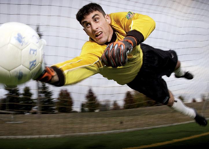
Total Football, and Why Science Will Always Win
 by Andrew Sweeney
To learn the lessons of coronavirus, ophthalmology must also learn from football. Yes, you read that right, we aren’t letting the spirit of the Netherlands affect us too much… but as the Dutch embrace the concept of ‘total football’ so too must the ophthalmology industry.
That at least is the gist of one of the first symposia of the ESCRS 2020 congress, ‘Covid-19: And then Everything Was Different’. Chaired by president of the ESCRS Prof. Dr. Rudy MMA Nuijts, the session was dedicated to understanding the full effect of coronavirus on
ophthalmology and what lessons could be learned. Clocking in at slightly under 30 minutes, it was short, succinct, and at times emotive.
This was because the symposium heard from a number of ophthalmologists from across Europe who had been affected by coronavirus. From Dr. Lucio Buratto in Italy, who saw firsthand the chaos that erupted in his country as the frontline of Europe’s struggle with the virus, to Dr. Bahram Bodaghi in France who warned of his country’s second wave, the stories were personal and revelatory. The mood was perhaps best summed up by Dr. Sheraz Daya, calling in from the United Kingdom.
“It worries me that we could head into another shutdown again. What coronavirus illustrates is that we’re all living on one planet and we’re all affected by this. We have to work together as a global family,” Dr. Daya said.
And it was the subject of working to overcome coronavirus that the symposium then moved on to cover. Dr. Marcel Levi, chief executive of University College London Hospitals, presented
his research and views on tackling coronavirus. Taking a left field yet highly innovative approach, Dr. Levi used the analogy of football to make his point.
Dr. Levi described how football teams used to use the ‘catenaccio’ approach, where each player was highly specialized yet inflexible. The Dutch development of this was ‘total football’ where each player was less specialized yet more adaptable, and able (to a variable extent) of playing in their colleagues’ roles. Dr. Levi believes this is the response required of coronavirus.
Learning from the Dutch masters to combat coronavirus
Dr. Levi argues that medicine currently has too much of a focus on specialization which ended up limiting its overall effective response to coronavirus. While stating that specialization is not necessarily a negative, Dr. Levi argued that there needs to be a move away from the subspecialists to what he described as a super-specialist. This would represent professionals who retain specialization but can adapt quickly to scenarios like the coronavirus pandemic.
He also argued that doctors increasingly need to step into leading management roles, as there is a disconnect between management and medical staff. This is especially acute in certain countries, particularly the United Kingdom which has a health system with a top heavy management structure.
Finally, Dr. Levi summed up and considered the future of ophthalmology deriving from coronavirus. Happily for all of us, he is optimistic about the future. The lockdown catastrophes of previous lockdowns won’t be repeated as we’re better prepared, and scientific development is reaching new levels already.
“I believe that science will always win and technology will help us out of this crisis, the best bet now is a vaccine. In Europe there are at least three or four promising vaccine trials. Technology works for coronavirus, so too will vaccines,” Dr. Levi said.
Positivity, optimism, and a love of football, kudos!
03 Oct 2020 | Issue #2 14 &
“It worries me that we could head into another shutdown again. What coronavirus illustrates is that we’re all living on one planet and we’re all affected by this. We have to work together as a global family,”
— Dr. Sheraz Daya
But one person has succeeded in achieving all these.
Dr. Chang cited the example of Dr. Govindappa Venkataswamy who devoted his life to curing blindness due to cataracts by creating the Aravind Eye Hospital in India, which treats poor people for free or at subsidized costs, and has performed more eye surgeries than any other eye hospital.
A single surgeon here can do as many as 16 cases an hour, with about 300-400 cases in one day, at a cost of USD10 per case. The eye hospital saves money by having its own manufacturing company for supplies.
“Resource-rich countries like mine in the United States can still learn a lot from resource-poor settings such as in southern India,” said Dr. Chang.
Five compelling lessons from what Dr. Chang described as the “Greatest Team
of Cataract Surgeons” at the Aravind Eye Hospital are:
• Lesson 1: Manual Small Incision Cataract Surgery (M-SICS) is more affordable and scalable.
• Lesson 2: M-SICS is often safer for mature cataracts. Comparing the safety of phacoemulsification versus M-SICS, he noted that there was no difference in rates of surgical complications with experienced surgeons, but complication rates were higher with phacoemulsification for least experienced surgeons. M-SICS is safe for experienced surgeons but is the safer technique for less experienced cataract surgeons.
• Lesson 3: Square optic edge reduces posterior capsule opacification (PCO) regardless of intraocular lens (IOL) material.
• Lesson 4: Endophthalmitis prophylaxis (intracameral moxifloxacin) is safe and effective.
• Lesson 5: Many operating room (OR) regulations unnecessarily increase costs and waste. Aravind reuses phaco tubing, gowns, and gloves.
In 2017, the carbon footprint of phacoemulsification was 20 times higher in the United States and the United Kingdom compared to Aravind Eye Hospital. The endophthalmitis rate in 2019 at Aravind was 0.01% and in the American Academy of Ophthalmology (AAO) IRIS data registry, 0.04%.

Editor’s Note:
The combined ESCRSEURETINA 2020 congress was supposed to be held in Amsterdam, The Netherlands. Because COVID-19 happened, reporting for this story took place in virtual Amsterdam instead.
INNOVATIVE OPHTHALMIC SURGICAL INSTRUMENTS, SYSTEMS, LIQUIDS AND GASES.


15 CAKE and PIE magazines’ Daily Congress News on the Anterior and Posterior Segments PRECISION MADE IN GERMANY. WWW.GEUDER.COM
>> Cont. from Page 3
IOLs in Children Benefits, Complications and More
by Sam McCommon
DAY 1 at the virtual 38th Congress of the European Society of Cataract & Refractive Surgeons (ESCRS 2020) hosted a panel presentation followed by a spirited discussion on pediatric intraocular lenses (IOLs) — and the nuanced practices that must accompany such treatments.
There are plenty of questions that accompany IOL treatments for children, especially for infants. What is the right age for IOL implants? Are IOL implants right at all for certain ages? What complications are there for children who undergo IOL implants? Are there procedures that mitigate potential complications?
Special considerations for children
It may not come as a surprise to readers that children, well, grow. What may be surprising is the rate at which their eyes grow. For example, the majority of a child’s eye’s axial length occurs within the first two years of their life, as presented by Dr. Ramesha Kekunnaya. Similarly, there are massive changes in a child’s eye’s keratometry past six years old. These changes in eyes leave surgeons with significant questions to ponder when considering inserting IOLs.

Dr. Kekunnaya presented long-term results from the study of IOL treatment on aphakic children. And the results weren’t great: only 25% achieved
excellent visual acuity in the treated eye, while 44% had poor vision in the treated eye. So, IOL treatments clearly aren’t always effective.
So, what can be done?
But clearly something must be done for aphakic children — unless blindness is an option, which no parent would ever wish upon their child. So, clearly there are some options. How do we do what’s best for the child?
A very important consideration seems to be age. On top of that, Dr. Kekunnaya noted several other factors to think about:
• Corneal diameter
• Laterality
• Intraoperative findings
• The family’s socioeconomic status (can they afford it?)
• Surgical experience (can the surgeon perform the task well?)
• Postoperative care (is the family near an ophthalmologist, and can they afford one?)
The most important takeaway is this: Using IOLs for children should be an individualized treatment, and no blanket statement can be made. It’s up to the
judgment of the physician, and risks need to be taken into consideration.
Glaucoma and IOLs in children
One such risk for a child who’s undergone an IOL implant is the potential to develop glaucoma later in life. According to Dr. Asimina Mataftsi, the earlier a child gets IOL implants, the higher the risk of glaucoma later. She initially introduced studies that evenly split the data on whether or not IOLs protect children from glaucoma, so clearly this is a controversial topic. But, according to her data, it’s readily apparent that implanting IOLs later in life helps prevent glaucoma as a complication.
Use of multifocal IOLs in children
Another topic discussed was the use of multifocal IOLs in children. This discussion was headed by Dr. Jagat Ram, a recipient of the fourth highest civilian award possible in India — so, the man has some significant credibility to bring to the table.
His conclusion was quite simple: Using multifocal IOLs in children is a viable and safe option. Neat! In the data he presented, photic phenomena like glare and halos were not even a concern for patients.
His conclusions came with one simple but important caveat, however: Multifocal IOLs should not be used in children under five years old.
Why even use IOLs for children?
Many may ask why surgically implanting IOLs for children who have cataracts or are aphakic. Can’t they simply use glasses or contact lenses?
Well, yes, hypothetically, they could. But as any parent knows, getting children to comply with such things on a regular basis is no easy feat — especially the contact lens part. And as eye development is closely tied to brain development, the better a child’s eyesight, the healthier their brain development will be.
03 Oct 2020 | Issue #2 16 &
Solutions for the Traumatized Cornea
 by Brooke Herron
The cornea can be damaged in various ways, and depending on the source, the treatment also varies. Therefore, during the ESCRS/ EuCornea Symposium: Dealing with the Traumatized Cornea, experts presented their insights on managing issues like corneal perforation,globe ruptures and laser refractive surgery in the posttraumatic cornea. Highlights from these presentations are included below.
Emergency care of corneal perforations
Dr. Bruce Allan (United Kingdom) says that corneal perforations are something that used to frighten him when he began his career in ophthalmology. “However, the principles of management are very simple,” he said.
For causes of corneal perforation, he shared that infections top the list, followed by autoimmune disease and persistent epithelial defects. Trauma is another cause, but that’s a bit more of a specialized area, said Dr. Allan.
One main point he emphasized was that
all corneal perforations are infective until proven otherwise. “You should always do a corneal scrape and microbiological investigation, and start with intensive topical antibiotic treatment,” said Dr. Allan.
The second principle he shared was that “we should always treat medically first.” This calms down inflammation and provides the best outcomes for the subsequent reconstruction. When there is a corneal perforation, a Cartella shield should be applied, followed by systemic antibiotics. “If you do these two things, then you’re protecting from the main cause of loss of sight.”
From there, he says to seal and reform the chamber (if you can) and suggests using the glue patch technique. “But in the future, I hope we’re going to have regenerative bio-glue soon to avoid keratoplasty,” he said further.
Laser refractive surgery in the post traumatic cornea
“When a large trauma happens to the cornea, it can have great implications
. . . but also smaller trauma can influence visual function, and can cause complaints in quality of life and quality of vision, like diplopia, and recurrent pain,” began Dr. Annette Geerards (The Netherlands).
She says that these cases can often be treated conservatively. However, refractory cases can be treated with laser surgery. Examples are scarring after foreign body removal and long-standing irritation after insignificant injuries.
“PTK might be the mode of treatment when other options fail,” she said. Phototherapeutic keratectomy (PTK) is an excimer-based procedure for treating anterior pathologies. In PTK, by ablating the corneal stroma, corneal clarity is improved. It also smoothens the surface and is less invasive than some other procedures.
“Indications for PTK might overlap, as problems with epithelial healing, opacities in the anterior stroma or irregular surface might be present, partly or completely,” continued Dr. Geerards. Side effects of the procedure are postoperative pain, refractive change, cost and disease recurrence.
Globe rupture: staged management of the posterior segment
Dr. Carlos Mateo (Spain) delved into the complicated surgical technique for managing globe ruptures. He says that failure or delay in diagnosis can result in a worse prognosis — this is due to the external pressure during the exam with more herniation of ocular content.
He also offered up some important points to consider in secondary care. Among them, time since trauma: “Don’t wait too long, apply the ‘100 hours law,’” said Dr. Mateo. He also noted that media transparency is very important: “You have to examine the cornea to know if it will be transparent enough to do ultrasonography. Remember, no light perception does not prevent surgery in severe trauma.”
In these cases, Dr. Mateo says the “worst enemies” are hypotony, epiretinal proliferation and retinal detachment with proliferative vitreoretinopathy, and corneal opacification.
17 CAKE and PIE magazines’ Daily Congress News on the Anterior and Posterior Segments
Highlights from the JCRS Symposium Controversies in Cataract and Refractive Surgery
by Olawale Salami
Debate Session 1
Gloves off: It’s SMILE vs. custom LASIK

The debate was a heated one with Dr. Timothy Archer from the London Vision Clinic (United Kingdom) arguing in favor of small incision lenticule extraction (SMILE) and against Dr. Vance Thompson, professor at the University of Dakota (USA), who was part of the development of both SMILE and LASI K.
In his opening statement, Dr. Archer stated: “From a patient perspective, SMILE is very attractive. Keyhole surgery is very appealing and is preferable to patients. The absence of a flap brings advantages to patients involved in contact sports and other outdoor activities.”
The additional benefits of SMILE, according to Dr. Archer, include reduced incidence of dry eye symptoms and superior biomechanics as compared to LASIK.
“SMILE is now a very mature procedure and learning curves for new surgeons today are very short,” Dr. Archer said.
“SMILE’s efficacy data is outstanding, with 95.5% of eyes we have treated achieved 20/20 visual acuity outcomes and 63% achieved 20/16 vision. For eyes with normal levels of aberrations, SMILE provides excellent visual outcomes while also bringing several other advantages,” he said.
However, in Dr. Thompson’s opinion, LASIK is the preferred procedure in many more scenarios. He noted that LASIK wins when patient fixation is unimpeded, low cylinder, and when topographically and wave-front guided corrections are needed, especially for the treatment of higher-order aberrations.
Furthermore, Dr. Thompson agrees that there are situations where SMILE is preferred; he noted that if correction is optimal and well centered, SMILE is his preferred lamellar approach.
Debate Session 2
After LASIK, which one takes the seat? Monofocal, EDof or multifocal IOLs?
There are several indications for IOLs in post-LASIK eyes, and the needs are
growing. Today, there are two schools of thought on the best IOL types to meet patient needs. The debate continued into the virtual auditoriums on DAY 1 of ESCRS 2020 congress.
Dr. Ruth Lapid-Gortzak from Amsterdam University, The Netherlands, knows her IOLs. “The message is quite clear, quality of vision decreases with multifocal IOLs. Monofocal IOLs are our gold standard. The challenge we face is that there are very few published papers on vision quality with monofocal IOLs,” she highlighted.
“We know that multifocal IOLs have numerous side effects, which are heightened in patients who require high visual acuity,” continued Dr. LapidGortzak.
“We looked at multifocal IOL implantation after previous corneal refractive laser surgery for myopia. In this series, we evaluated 77 eyes from 43 patients and found it risky using multifocal IOLs in lasered eyes,” she shared. “If a patient had laser for myopia of >6 D, our results were not satisfactory. I don’t think anyone wants to be the one to touch up a thin aberrated cornea,” added Dr. Lapid-Gortzak.
In her defense of monofocal IOLs, Dr. Lapid-Gortzak concluded: “There is a paucity of data about the quality of vision in monofocoal IOLs after laser surgery. We know from our experience that in myopes of > -6D, the results of multifocals become less predictable. This lack of predictability is worsened in hyperopes, who experience further loss of visual acuity.”
Dr. Jose Alfonso holds a contrary view and presented data from studies conducted by his team and other investigators. “After myopic photoablation, excimer laser induces positive spherical aberration, and options we recommend include trifocal, bifocal, non-diffractive EDof or intermediate enhanced Monofocal IOLs,” he explained.
“However, in case of previous hyperopic photoablation, we recommend implanting trifocal, non-diffractive EDof, or monofocoal neutral IOLs. In both cases, the optical lens selection is determined by the number of diopters previously corrected,” emphasized Dr. Alfonso.
03 Oct 2020 | Issue #2 18 &
Glaucoma Update
New Horizons in Therapy
 by Brooke Herron
trabeculectomy (SLT), minimally invasive glaucoma surgery (MIGS) or traditional surgery. “However, SLT is not effective in all patients . . . MIGS are becoming more widely used, and traditional surgery still has a very important place,” he shared.
“There are a number of considerations that can be made for the future of glaucoma treatment,” said Prof. Crowston. These include disease modification therapies, neuroprotective agents and drug-eluting products.
In summary, Dr. Crowston said: “The key take home message here is that many patients with glaucoma continue to progress while on treatment. Even in the early stages of disease, patients may be suboptimally controlled due to challenges with topical treatment.”
Changing dimensions in minimally invasive filtration surgery
During the Allergan Satellite Sponsored Symposium at the 38th Congress of the European Society of Cataract & Refractive Surgeons (ESCRS 2020), Prof. Jonathan Crowston (Singapore) and Dr. Herbert Reitsamer (Austria) discussed the limitations and changing dimensions in glaucoma therapy and surgery. This followed a welcome lecture from Associate VP and Head of Eyecare Global at Allergan (an Abbvie company), Charles Holmes, who talked about the company’s commitment to ophthalmology.
“2020 has been an extremely challenging year, both for practices and patients,” said Mr. Holmes. “The focus in this post-pandemic situation is to look at where we are with products in ophthalmology and lessen burden [on patients].”
Limitations of current glaucoma therapy
“Why are patients with glaucoma still progressing, despite therapy?” asked Prof. Crowston. He then revealed data that showed that 45% of patients experience glaucoma progression, even under topical treatment. Among the reasons for this, he says, is that administration, adherence, tolerability
and patient anxiety are key concerns.
In terms of administration challenges, one study found that less than one-third of patients could instill drops without touching the eye; in another study, in almost half of patients, the eyedrops fell onto the eyelid or cheek.
“One of the problems with improper use of drugs is that patients experience anxiety if things aren’t going as they’d like to,” said Prof. Crowston.
Adherence is another issue. “In glaucoma, we know that many patients do not take their medications appropriately,” he continued. Data shows that when patients miss two-thirds of their medications, there is a higher rate of glaucoma progression.
Tolerability of topical drops can also be problematic — 59% of glaucoma patients have symptoms of ocular surface disease (OSD) in one eye; 27% of those are severe.
“When we put patients on topical medications, we need to very quickly and repeatedly work out whether this is the best form of treatment,” he said.
Beyond topical drops, other treatment options include selective laser
The problem with glaucoma is we get the referrals too late and we need to take more serious measures to lower intraocular pressure (IOP), began Dr. Reitsamer.
“Sometimes drops just aren’t enough, so we need to do surgery — but we want to do the surgery that is the least invasive,” he said. During his presentation, Dr. Reitsamer focused on ab interno subconjunctival approaches, with the XEN45 and XEN63 gel stent implants.
He recounted study data that showed the XEN implant reduces drop burden: “Almost half of the patients were dropfree at 24 months,” he shared.
Further, he concluded that the XEN63 is a good option, with potentially lower IOP outcomes and less medications necessary than with the XEN45. “Early experience suggests that the new inserter provides good sealing around the implant, so there is less side flow around the implant, which also should provide less hypotony,” explained Dr. Reitsamer. “We definitely need further [prospective, randomized] clinical trials where we can compare the XEN63 with other procedures, preferably with trabeculectomy and other implants, to see if the safety profile is within limitations and within expectations.”
19 CAKE and PIE magazines’ Daily Congress News on the Anterior and Posterior Segments
CONTENT MARKETING + ADVERTISING + MEDICAL WRITING


03 Oct 2020 | Issue #2 20 & 6001
Road,
Golden Mile
199589 magazine posterior segment innovation enlightenment
Beach
#19-06
Tower, Singapore
