8 minute read
Ocular surface disorders are more than surface-deep


Scratching the Surface
Ocular Surface Disorders
by Sam McCommon
One of the first presentations that kicked off 37 th World Ophthalmology Congress (WOC2020 Virtual®) was the Diagnosis and Treatment of Ocular Surface Diseases live session. Some true nuggets of wisdom were shared by all presenters, three of which are discussed below.
The ocular surface microbiome
That the eyes are subject to infection is news to no one — but the type of bacteria involved in inflammation and infections may surprise some.
Prof. Shigeru Kinoshita’s discussion on meibomitis-related ocular surface disorders led to an interesting conclusion. Some 60% of patients who suffered from meibomitis-related keratoconjunctivitis (MRKC) showed bacterial colonization of the meibum by P. Acnes bacteria — the same bacteria that causes, well, acne. This is in comparison to just under 25% of healthy patients. Additionally, MRKC patients also showed colonization by other bacteria, including P. Aures, though Prof. Kinoshita noted it wasn’t as strong as P. Acnes.
“Eliminating bacteria like P. Acnes from the meibum is essential to reducing ocular surface inflammation,” Prof. Kinoshita concluded. He compared it to the now-known fact that the H. Pylori bacterium is responsible for gastritis, and when it is eliminated the gastritis goes with it. He recommended a combination of topical and systemic antibiotics to defeat the bacteria.
Dr. Vilavun Puangsricharern of Chulalongkorn University in Bangkok, Thailand, also discussed the ocular surface microbiome, but in a different context. Her presentation focused on Stevens-Johnson syndrome(SJS), a rare disorder of the skin and mucous membranes that causes chronic ocular disease in a large number of its survivors. The extreme version of Stevens-Johnson syndrome is toxic epidermal necrolysis (TEN).
The syndrome is associated with an increased risk of eye infection. Dr. Puangsricharem noted that under normal circumstances the ocular surface microbiome has relatively low species diversity compared to other microbiomes like the gut. Patients who suffered from SJS, however, had much greater species diversity in the ocular microbiome, especially more pathogenic bacteria. The implication is that a disruption of the microbiome can lead to colonization by more dangerous types of bacteria.
Ocular surface squamous neoplasia (OSSN)
Dr. Tsutomu “Ben” Inatomi of Kyoto Prefectural University of Medicine in Kyoto, Japan, shed light on a most unfortunate condition: cancerous tumor growth of the ocular surface. These growths occur in three phases: dysplasia, preinvasive carcinoma (referred to as in situ), or invasive carcinoma. One study showed that the vast majority (72%) of cases were in the limbus, though the eyelid and bulbar conjunctiva can also be affected.
Surgical excision is the gold standard for tackling this carcinoma according to Dr. Inatomi because it has a quick resolution and is both diagnostic and therapeutic. However, it does have its drawbacks. It can leave tumor cells remaining, leading to a potential recurrence. It can also lead to eye dysfunction due to the large amount of tissue removed. Furthermore, it can lead to stem cell deficiency and scarring.
Dr. Inatomi proposed several methods for reconstructing the ocular surface. An intriguing option is cultivated oral mucosal epithelial transplantation (COMET). COMET takes oral mucosal tissue and, through several steps, transplants it to the ocular surface. One of the major benefits? There is no risk of rejection since it comes from the patient’s own body.
He also recommends that if more than one third of the limbus is affected surgeons should perform a keratoepithioloplasty, which can provide stem cells to help with recovery.
On the note of recovery, Dr. Inatomi recommended both intra- and postoperative chemotherapy to resolve the carcinoma and prevent recurrence, especially if excision could not remove the entire tumor. When asked about preoperative chemotherapy, however, he posited it was not a good idea because it could make determining the margin of the tumor difficult.
Shigeru Kinoshita, M.D., Ph.D.
Kyoto Prefectural University of Medicine, Japan @WOC2020 Virtual®
Optimizing Outcomes with IOL Power Calculations
by Brooke Herron
Following cataract surgery, neither patients nor surgeons welcome a refractive surprise — thus, accurate intraocular lens (IOL) power calculations are crucial to outcomes. To dig deeper, Dr. Cathleen McCabe (USA), Dr. Chitra Ramamurthy (India), and Prof. Thomas Kohnen (Germany) offered their insights into these formulas during a session titled Progress in IOL Calculations on the first day of the 37 th World Ophthalmology Congress (WOC2020 Virtual®).
“In terms of accuracy, do we really calculate or estimate for intraocular lenses (IOLs)?” asked Prof. Kohnen — an interesting point to make during a session on power calculations. He said that it’s estimation for this reason: “The upper limit of accuracy is ±0.50D — even in the best scenario — and 90% of patients achieve this.”
He then discussed results from a study he co-authored that compared nine formulas to calculate power of a panfocal IOL in 38 eyes of 75 patients. “We saw that Barrett Universal II and Hill-RBF both had 80% at ±0.50D,” he explained, adding that only 60% of patients were within ±0.25D using Barrett’s; this dropped to 57.3% using Hill-RBF. Nevertheless, Prof. Kohnen and colleagues concluded that the Barrett II Universal formula and Hill-RBF achieved the best results. The older generations should not be used anymore, he continued. “I use the new formulas in all my patients because my outcomes are better. But we are not yet at 100%, and therefore, we still talk about estimation rather than calculation.”
Out with the old and in with the new, was also the message delivered by Dr. Ramamurthy. “With the advent of the newer biometers and newer formulas . . . today, if we use the right formula, every single patient can reach this target [within ±0.50D].”
Not only does the formula need to be right, its variables do too. It is important to customize and personalize the A-constant, anterior chamber depth and surgeon factor based on individual biometry techniques and equipment, using data from at least 30 cases, concluded Dr. McCabe.
WE STAND WITH THE WORLD OF OPHTHALMOLOGY
during this time of challenge, and also of hope. All around the globe we stand united with colleagues, organizations, ophthalmologists and industry, to make 2020 and beyond what it should be: Clearly, a better future.
In partnership with:
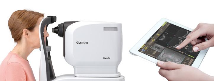
New Eye Care Realities Distancing safely while using your OCT device
Social distancing is the new reality in current eye care practices. However, most ophthalmic diagnostic devices require close proximity between patients and the scan operator, making physical distance an impossibility. This is reality is set to change with the new Canon Medical Xephilio OCT-A1 – where within just three clicks of a mouse, optical coherence tomography (OCT) scans can now be done a few meters away, from another room or even a remote location. Imagine the ease of mind your patients and staff members would experience with the safe distancing provided.
Three safer scenarios would now be possible with the following distancing solutions whenever your patients need to be examined with the Canon Medical Xephilio OCT-A1.
Solution 1: Operating the Canon Medical OCT at safe distance in the same room
The operator can control the OCT from a different desk that can easily be 2 meters or further from the patient. This is achievable using a longer monitor cable and a wireless keyboard and mouse (or by using a USB extension cable). The monitor’s cable could be up to 5 meters long while the USB extension cables could go up as long as 6 meters for the keyboard and mouse.
Solution 2: Operating the Canon Medical OCT remotely over a network in the same room
The operator can control Canon Medical Xephilio OCT-A1 from a tablet PC over a network. The tablet PC uses a remote desktop solution (e.g. TeamViewer) to communicate with the PC that controls the OCTA. This would offer the operator even more flexibility with the ability to still communicate with the patient while keeping a safe social distance.
Solution 3: Operating the Canon Medical OCT from another room or location
The Canon Medical Xephilio OCT-A1 can be operated over the hospital network or via the internet. This would require a remote desktop solution (e.g. TeamViewer) which would essentially enable the device to be operated from another room in the hospital or even a different location. Using this remote access solution, monitoring and communicating with the patient can even be done over popular video meeting software like Skype, for instance.
Safe physical distance is but only one of the key innovations the Canon Medical Xephilio OCT-A1 brings to the table apart from its exceptional imaging quality and efficiency.
The Canon Medical Xephilio OCT-A1 gives a cutting edge to imaging results with the first commercially available Intelligent Denoise capability, a deep learning feature that effectively removes noise while enhancing details within a single scan. A tomography scan with the Canon Medical Xephilio OCT-A1 can be done in just 2 seconds, resulting in less motion artefacts. This not only helps you save time and enhance your efficiency, but ultimately, the shorter examination time also increases your patient’s comfort.
Revolutionizing the future of OCT scan innovations, the Canon Medical Xephilio OCT-A1’s digital resolution of up to 1.6 µm enables excellent differentiation of
structures and individual layers of the
cornea and retina, thanks to Canon’s long history in recognized optical expertise.
In OCT imaging, optical axial resolution should not be confused with digital sampling resolution since axial resolution depends on the bandwidth and light source wavelength. The Canon Medical Xephilio OCT-A1 has a unique resolution in the market with a 3-micron native optical axial resolution combined with a spectral domain bandwidth of 100 nm, which anterior and posterior ophthalmologists appreciate very much due to the high imaging definition output.
Finally, the best part of Canon Medical Xephilio OCT-A1 is its ease of use with its three-click function to complete an exam. It offers a complete range of intelligent functions to enable fully automated examinations. This makes it easy for you to delegate the scanning operations to nurses or assistants.
All in all, safety in physical distance and deep learning imaging capabilities is now

a sharper, new reality.