7 minute read
Heart skips and bumps


Skips, thumps and bumps… should I worry?
By Dr Tim Gattorna, Cardiologist, Subiaco, Midland, Northam & Kalgoorlie
Palpitations are a common, subjective symptom resulting in frequent presentations in the primary health care setting. Although generally benign, they can occasionally be a manifestation of a potentially life-threatening arrhythmia. Appropriate evaluation is therefore required.
Cardiac or arrhythmic causes are the most common aetiology. Other causes include medical conditions (e.g. endocrine and metabolic abnormalities), psychiatric disorders, medication effects, and drug or other substances. It is important not to label a patient’s palpitations as secondary to panic/anxiety without
Key messages
Palpitations are frequent in the community History, examination and basic investigations can determine cause in many patients Case finding and management of AF is critical as is awareness of red flags.
a proper evaluation, as around 50% of this group will be diagnosed with an arrhythmic cause. A targeted and thorough history is required given the majority of patients present in sinus rhythm,
Table: Risk stratification
Skipped beats Thumping beats Short fluttering Slow pounding AND Normal ECG AND No Family History AND No Structural Heart Disease History suggests recurrent tachyarrhythmia Palpitations with associated AND/OR symptoms Abnormal ECG AND/OR Structural Heart Disease Palpitations during exercise Palpitations with syncope/ near syncope High risk of structural heart disease Family history of inheritable heart disease/SADS High degree AVblock
Low Risk Manage in Primary Care Refer to cardiology Refer to cardiology with urgency
Table published with permission of Primary Care Cardiovascular Journal
between episodes of palpitation. The description of skips, jumps and thumps may represent ectopic beats (atrial or ventricular); rapid onset/onset of racing heart may be consistent with a supraventricular tachycardia.
Ask about onset and offset (sudden or gradual), duration (momentary or sustained), frequency of episodes, triggers, any associated symptoms (e.g. syncope, breathlessness or chest pain), pre-existing cardiac issues and any family history of sudden cardiac death or cardiac conditions. Which investigations? This depends on the history, frequency and duration of episodes. A 12-lead ECG (if symptomatic at the time) is the gold standard. Ambulatory (Holter) monitoring is useful if frequent symptoms, whilst an event monitor is useful in less frequent symptoms. Loop recording (for recurrent syncope), echocardiography to evaluate heart structure and stress testing (if exercise induced or suspected IHD) can be helpful. Smart phone monitoring/apps may be the future.
continued on Page 39
By Dr Justin Ng, Cardiologist, Nedlands
Ventricular ectopic beats (VEBs) are common. Frequent (>60/hr or 1/min) in healthy subjects has an estimated prevalence of one to four percent in the general population. Over 10 percent on a Holter monitor is considered a high VEB burden. Monomorphic VEBs represent a focal arrhythmia arising from a single site. Generally, individuals with frequent monomorphic premature ventricular contractions (PVCs) have a benign clinical course. A portion have bothersome symptoms cardiomyopathy can develop in a minority of cases.
The clinical presentation of patients with frequent VEBs is variable. A large proportion are asymptomatic and are discovered on a routine ECG or during monitoring for an unrelated procedure. Other patients are mistaken to be bradycardic by palpation of the pulse and are referred for investigation based on this.
Individuals can be highly symptomatic complaining of palpitations, ‘missed beats’, ‘heavy beats’ and fluttering in the chest. Tiredness, dyspnoea, lightheadedness and mental clouding are other common complaints. Heart failure symptoms may be the initial presentation. Indications for treatment The majority of individuals with frequent VEBs have a benign
Key messages
Frequent monomorphic VEBs are common. The vast majority have a benign c course In high symptomatic burden or with evidence of VEB induced cardiomyopathy there are safe, effective therapies Ongoing clinical surveillance is important in conservatively managed asymptomatic individuals.
clinical course. A small proportion of individuals with a high burden of ventricular ectopy can develop left ventricular dysfunction and an even smaller proportion, VEB induced polymorphic ventricular tachycardia.
As such the indications for treatment include high symptom burden, VEB induced cardiomyopathy and rarely PVC induced polymorphic VT.
It is important to identify the those with structural abnormalities.
A 12 lead ECG is helpful to confirm the morphology and location of the VEB as well as to look for other evidence of structural heart disease such as Q waves, T wave inversion and epsilon waves anteriorly, suggestive of arrhythmogenic right ventricular dysplasia(ARVD). Echocardiography assesses structure and function.
A 24-hour Holter can quantify the burden and to see whether there is more than one morphology. Cardiac MRI may be warranted if there is any evidence of a wall motion abnormality, an abnormal ECG or multiple morphologies of PVCs making structural heart disease more likely. Given the small incidence of PVC induced cardiomyopathy clinical surveillance is appropriate even for asymptomatic patients.
In individuals with a high burden of symptomatic VEBs or individuals with evidence of VEB induced cardiomyopathy, treatment is recommended.
Beta blockers or calcium channel blockers remain first line options. Flecainide (once ischemia and abnormal LV function had been excluded) or sotalol are second line.
Catheter ablation with the aid of 3D mapping has a high success rate. It is generally reserved for patients who either cannot tolerate medical therapy or have failed medical therapy. The procedural risk is relatively low, and the curative rates are high.
Author competing interests – nil
continued from Page 38
Skips, thumps and bumps…should I worry?
Correlation of the rhythm at the time of symptoms is the key to the diagnosis (arrhythmic or not).
Thyroid function and full blood count as a baseline are recommended with further tests dependent on the history/findings.
Blood pressure and palpation of the pulse at each consultation is highly recommended as an irregularly, irregular pulse may be indicative of Atrial Fibrillation (AF). This needs confirmation with a 12-lead ECG. The diagnosis is important as AF is associated with a high morbidity and mortality, can be asymptomatic, and effective treatment including oral anticoagulation is available.
The management of palpitations depends on the cause and associated prognosis. Simple reassurance in many cases is appropriate, although medical therapy and referral to cardiac electrophysiologist may be required in some cases. Red flags include frequent, persistent and sustained episodes and significant associated symptoms (e.g. syncope, chest pain), abnormalities on resting ECG such as T-wave abnormalities, long or short QT interval and evidence of prior AMI (Q waves) or short PR interval (possible Wolff-Parkinson White syndrome).
IORT arrives
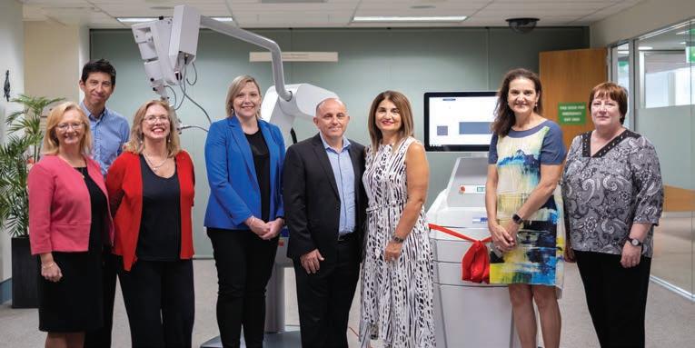
St John of God Subiaco Hospital has become the first private hospital in Australia to introduce Intraoperative Radiation Therapy (IORT) for breast cancer.
Breast surgeons Prof Christobel Saunders and Dr Lee Jackson, along with GenesisCare Radiation Oncologists Dr Yvonne Zissiadis and Dr Margaret Latham, are undertaking the IORT procedures and training of other breast surgeons is underway. The procedure is only suitable for certain women whose breast tumours are low-risk.
Christobel told Medical Forum that she had worked on IORT trials at University College London in the 1990s. There will be follow-up studies done on those patients, which will give a deeper picture of the efficacy of the therapy.
The surgical team operates to remove the tumour, add shielding
From left: SJG Subiaco CEO Prof Shirley Bowen, Dr Lee Jackson, Prof Christobel Saunders, SJG Foundation CEO Bianca Pietralla, donors Robert and Maria Carcione, Dr Yvonne Zissiadis and Dr Margaret Latham
to protect surrounding tissue, and then the radiation oncologists direct a calculated dose of radiation into the incision at the tumour site.
“Studies have shown that if radiotherapy is given at the same time as surgery for certain selected, low-risk breast tumours, there is equal local control. So, in other words, no higher local recurrence rates,” Christobel said. “IORT can help women avoid six or seven weeks of post-op therapy. That is great for every woman but especially those who live in the country, who we know have a 50% chance of choosing mastectomy because they don’t feel they can stay away from their homes and lives for that long. “But it’s important to reiterate it’s not for every patient, and it needs tumour pathology.”
Christobel said IORT was available at SCGH but because of scheduling and difficulty it was not often used. “The IORT means an extra 45 minutes in the operating theatre, but that avoids anywhere between three and seven weeks of daily radiotherapy afterwards,” she said.
The IORT equipment was funded by philanthropic support of a group of St John of God Foundation donors, including the Carcione Foundation.
Our Vascular surgeon is at the forefront of innovation and care in providing the following vascular services:
Aortic Aneurysm
Peripheral Arterial Disease
Thoracic Outlet Syndrome (TOS)
Arteriovenous Fistula or Vascular Access for Haemodialysis The Diabetic Foot and Ulcers
Varicose Veins and other Venous Disease
Pelvic Congestion Syndrome
Chronic Venous Disease and Deep Vein Thrombosis treatment