3 minute read
AI in Radiology


AI, machine learning and radiology
By Dr Brendan Adler, Radiologist, Wembley
Artificial intelligence (AI) will eventually extend universally into health care, but this has been perhaps faster in radiology due to the combination of rapid development of graphic analysis software and easy accessibility to large imaging datasets.
AI will become an integral part of requesting, performing and reporting diagnostic radiology tests. Analogous to the extensive data mining from social media platforms or improved voice recognition from home devices and facial recognition from widespread imagery, the development of picture archiving and communication systems (PACS) has provided a large body of labelled radiology data.
While AI encompasses a large group of software solutions, machine learning implies software solutions with the ability to independently learn and make predictions without being explicitly programmed to do so. As with humans, there is continual improvement with experience.
Key messages
AI will become integral to radiology workflow, organisation, data management and analysis AI may be incorporated as a diagnostic tool assisting tasks such as oncology follow-up Ethical and medicolegal issues will need addressing.
Given that substantial amounts of diagnostic radiology are largely pattern recognition, it is not surprising that it has been at the forefront of development of deeplearning techniques.
There have been substantial expectations versus actual practical solutions, but this is rapidly changing.
Multiple software can automate detection and interpretation of images, currently largely confined to less complex problems such as fracture assessment, pneumothorax, and mammography. Long-standing
Figure 1. AI Rad Companion Chest CT generated summary and images from ultralowdose unenhanced non-gated CT thorax (a) demonstrating automatic detection of small middle lobe micronodule in virtual bronchogram (b), automatically segmented and measured aortic diameters (c) and automatically segmented thoracic spine fracture assessment (d). Reproduced with permission from Siemens Healthineers.
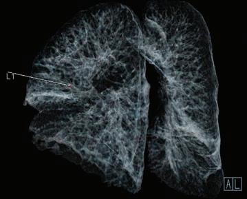
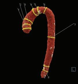
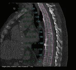
computer-assisted diagnostic tools (e.g. pulmonary nodules) are being improved to provide guidance as to malignancy risk. Post-processing of images is also under review. Much work is being done in automated segmentation and interpretation in evaluating brain parenchymal disease, cardiovascular abnormalities and liver metastatic disease. (Figure 1). These solutions will be combined with automated radiology analysis to provide standardised reporting.
Further solutions are available to improve current technologies such as diminishing noise in low-dose CT scans and optimising radiation dose. Intelligent scheduling, screening of referrals and automated triaging are being evaluated. There is huge potential for coordinating radiology imaging analysis with a patient’s clinical and pathological data in a digital health record. This may be significantly delayed until My Health Record becomes a truly digital archive and the population perceives benefit from inclusion.
While disrupting radiology practice, these techniques are unlikely to replace radiologists but improve what they currently do, as occurred with picture archiving and communication systems two decades ago. The term augmented radiology has been coined. There are also ethical and legal issues in regard to responsibility for the report. While driverless trains and trucks are currently being used, there are concerns analogous to aircraft, for which despite the ability currently to take off, cruise and land autonomously, pilotless commercial air travel is still some time away. Similarly, radiology reporting in the absence of any, albeit brief, review by the radiologist responsible for the report is unlikely to occur in the foreseeable future.