10 minute read
The era medicine
A look at the history of nuclear medicine at the University of Chicago
BY TIHA M. LONG, PHD
Advertisement
PHOTO BY JOHN ZICH
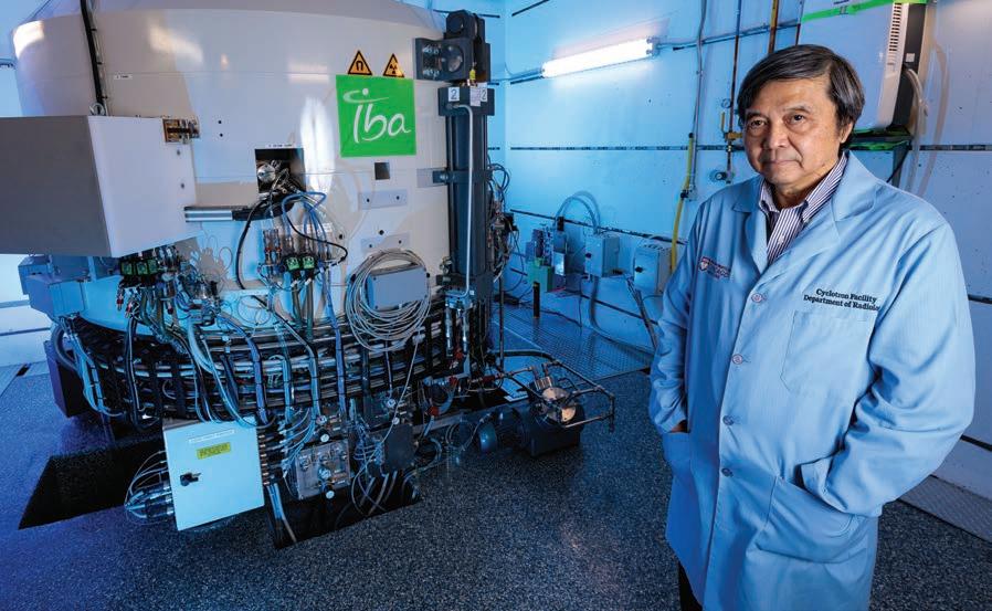
Chin-Tu Chen, PhD’86, led efforts for the installation of the IBA Cyclone 18 at the University of Chicago.
Recent publications
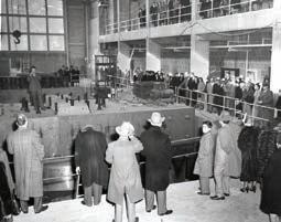
Chang et al. Angew Chem Int Ed. 2020. Persky et al. J Clin Oncol. 2020. Solanki et al. Pract Radiat Oncol. 2020. The nuclear era began in 1942 when the world’s first controlled, self-sustaining nuclear chain reaction took place at Stagg Field on the University of Chicago campus. In the decades that followed, University scientists made historic advances in harnessing this technology in the diagnosis and treatment of disease.
Early nuclear medicine’s A-Team
In 1954, the Argonne Cancer Research Hospital—the largest facility ever built for the purpose of cancer research and treatment using nuclear medicine— opened its doors. The facility attracted four scientists whose work would launch the field of modern nuclear medicine.
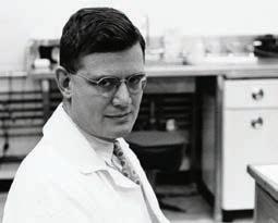
Katherine Austin Lathrop, Professor Emerita in the Department of Radiology, brought her biochemistry background to the University in 1945 as a member of the Manhattan Project, the secret research program to develop the atomic bomb. She became a pioneer in the development and testing of radiopharmaceuticals—radioisotopes used for the diagnosis and treatment of diseases.
Paul Harper, MD, completed his residency at the University and joined the Departments of Surgery and Radiology in 1953. Harper worked closely with Lathrop to investigate medical applications of radioisotopes.
Mathematician Robert Beck, a longtime faculty member in the Department of Radiology, joined the team in 1957 and began working on imaging instruments that could detect the signals from radioisotopes. Beck served as the assistant director of Argonne Cancer Research Hospital from 1963 to 1967 and became director of the Franklin McLean Memorial Research Institute, the center that evolved from the Argonne Cancer Research Hospital.
Alex Gottschalk, MD, completed his residency at the University of Chicago, then returned in 1964 to join the Radiology faculty and became director of the Argonne Cancer Research Hospital in 1967.
These scientists developed an imaging technique using a radiotracer labeled with technetium-99m
(99mTc), the most commonly used medical radioisotope today. Clinical 99mTc imaging is used millions of times worldwide every year to detect cancer and other diseases.
After numerous advances in nuclear medicine in just two decades, the installation of new equipment and the development of powerful technologies allowed for rapid and expansive growth of the field.
The state’s first and only academic medical cyclotron
The first cyclotron at the University of Chicago, installed in 1968, opened the door to creating numerous types of radioactive compounds for research, diagnosis and therapy.
A cyclotron is a stout, cylindrical-shaped particle accelerator. It speeds up charged particles from the center outwards along a spiral path. It operates by maintaining a static magnetic field that keeps particles within a perpendicular central circular plane. At the same time, electrical charges oscillate between the two semicircles that make up the central circular disk, causing the acceleration of the particles in a spiral motion. When particles reach the limits of the circumference, they are deflected to an exit point through a beam tube that can be aimed at a target. The beam of charged particles colliding with a material creates positron-emitting isotopes, types of atoms that are unstable and emit radiation. These radioisotopes have diagnostic and therapeutic utility, for example, as radiotracers for medical imaging.
The original cyclotron was decommissioned about 30 years after it was installed, in 1997. Interest in the program dimmed for almost two decades but would re-emerge.
PET: Precise and sensitive imaging
Beck, with Chin-Tu Chen, PhD’86, Associate Professor of Radiology, and his colleague, Malcolm Cooper, MBChB, had been key figures in the installation of a positron emission tomography (PET) facility in 1981—an early nonclinical brain PET scanner and the first system in the state of Illinois. In 2004, the University of Chicago became one of the first local institutions to install a clinical PET scanner for the routine care of patients.
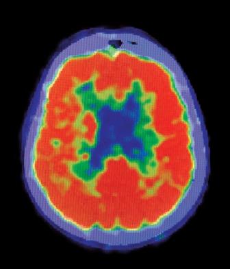
PET imaging uses radioactive materials that can detect events in the body at the molecular level, including the presence of biomarkers, and metabolic and enzymatic processes. A radioactive solution called a radiotracer is injected into the circulation system. The radiotracer interacts with molecules within specific tissues or cells of the body. The emission of positron particles produces gamma rays from precise locations in the body, and these rays can be detected and processed by a scanning machine recording images.
The far-reaching idea to combine PET with magnetic resonance imaging (MRI) and computerized tomography (CT) marked another jump forward. Chin-Tu Chen, with Charles Pelizzari, PhD, and George Chen, PhD, then faculty members in the Department of Radiation and Cellular Oncology, teamed up to figure out how to correlate PET with CT and MRI imaging to produce 3D information in a clinically relevant time frame. Progress through the late 1980s led to the PET/CT scanner becoming a 2000 Time magazine “Medical Science Invention of the Year.”
“MRI provides a picture of the anatomy, and PET tells you the functional information,” Chen said. “By lining up these two types of images, we can understand exactly where the activity is happening.”
A new era of research and medicine
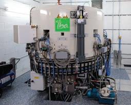
After two decades without a cyclotron at the University, Chin-Tu Chen led efforts for the installation of a modern facility in 2017. The IBA Cyclone 18, a massive 27-ton instrument, sits in a secure vault below the ground enclosed by thick slabs of concrete. It is surrounded by sterile rooms for production of radiotracers, drug dispensing and quality control.
The University of Chicago Cyclotron Facility, directed by Richard Freifelder, PhD, currently produces a new FDA-approved investigational drug, fluorothymidine, a PET radiotracer used to monitor response to cancer therapy, and numerous other experimental compounds for research and medicine.
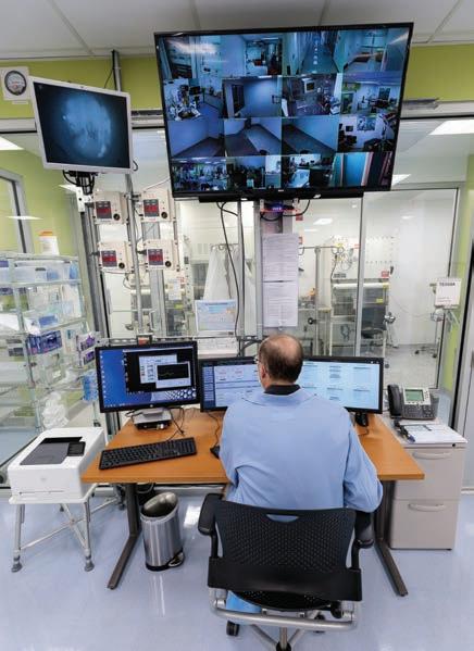
Richard Freifelder, PhD, director of the cyclotron facility, works in the control room.
PHOTO BY JOHN ZICH
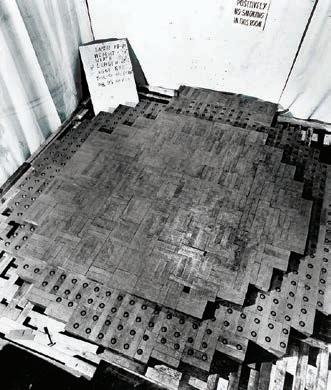
First nuclear reactor under construction beneath Stagg Field.
1942
Few institutions can offer full access to radiopharmaceuticals and PET scanning. Due to the short half-life of most types of radiotracers, a cyclotron must be close by or on site with the PET imaging facility. UChicago has these advanced capabilities, which are invaluable tools for cancer diagnosis and treatment.
PET is used to image primary and metastatic tumors and to monitor responses to therapy. It can supply information to physicians to assist with the characterization of tumors and treatment decisions. PET is an exquisitely sensitive tool that is able to detect biochemical events that may appear ahead of any detectable tumor.
Recent advances include the creation and testing of a first-in-class, activity-based PET radiotracer. This accomplishment required the combined expertise of a medical physicist, a biological chemist and a team of researchers with the Cyclotron Facility. Raymond Moellering, PhD, Associate Professor in the Department of Chemistry, and Chen created a novel radiotracer that can specifically label aggressive cancer cells in breast cancer tumors in whole body imaging.
The common approach to tumor detection involves the glucose analog 18F-FDG, a nonspecific glucose molecule that can enter any fast-growing tissue. It detects cancer cells simply because they metabolize increased glucose to produce energy enabling them to grow faster than noncancerous cells. Elevated concentrations of 18F-FDG in tumor cells allows them to be detected anywhere throughout the body.
This new, more precise approach combines the technology of PET imaging with the creation of a novel chemical probe with covalent activity. The recently developed radiotracer detects activity of the enzyme neutral cholesterol ester hydrolase (NCEH1), allowing for the direct visualization of active NCEH1, which is present in aggressive triple negative breast cancer.
In addition to the capability to image aggressive tumors, the researchers were able to make new discoveries using the NCEH1-activity radiotracer. They found that the levels of NCEH1 were higher in the leading edge of the tumors where growth and metastasis occur. Similar results were seen in a prostate cancer model.
“This imaging technology could help clinical teams determine whether a cancer is aggressive and inform treatment decisions,” Moellering said. “NCEH1 is elevated in many different types of
1959 1968
Nuclear medicine at the University of Chicago
1942
World’s first nuclear reactor creates a controlled, selfsustaining nuclear chain reaction
1945
Research on radiotracers begins
1954
Argonne Cancer Research Hospital opens
1959
Scientists demonstrate the use of localized radiation to treat cancer
1963
First technetium99m (99mTc) brain scan
1968
UChicago installs first cyclotron (decommissioned in 1997)
1981
Positron emission tomography (PET) research facility opens
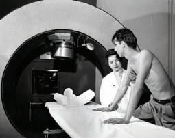
PHOTOS FROM THE UNIVERSITY OF CHICAGO PHOTOGRAPHIC ARCHIVE, SPECIAL COLLECTIONS RESEARCH CENTER, UNIVERSITY OF CHICAGO LIBRARY Paul Harper, MD, and colleagues investigated the medical application of radioisotopes.
1985–2005
aggressive cancer, so if it works to track aggressive breast cancer, it may have utility in tracking many other types of aggressive cancer.”
Managing treatment decisions
For types and stages of cancer that are difficult to detect, PET imaging may provide an option to see what other imaging techniques miss.
Sonali Smith, MD, Elwood V. Jensen Professor of Medicine, is an expert in the treatment of lymphoma. She has led recent clinical studies at UChicago, in collaboration with other academic groups, to elucidate the role of PET-directed therapy in the management of lymphoma. In a recently published study, patients with early stage diffuse large B-cell lymphoma (DLBCL) were scheduled for PET scans following their initial cycles of standard chemotherapy to determine how to proceed with treatment. Patients who had a negative PET scan proceeded with only one cycle of chemotherapy, whereas a positive result by PET scanning, indicating that cancer was still present, required radiation plus directed radioimmunotherapy.
Both patient groups from the study had positive outcomes. These results showed that PET-directed therapy was helpful for guiding treatment decisions.
“Overtreatment has been an issue in limited-stage diffuse large B-cell lymphoma,” Smith said. “The use of PET-directed therapy may allow for a reduction of the numbers of cycles of toxic drugs and reduce negative side effects, which will be welcomed by patients.”
PET imaging may benefit patients with advanced prostate cancer as well. Stanley Liauw, MD, Professor of Radiation and Cellular Oncology, and colleagues evaluated how PET contributed toward treatment decisions for patients with recurrent advanced prostate cancer referred for radiation therapy.
Prostate cancer recurrence after prostatectomy is difficult to localize by conventional methods. Liauw and colleagues investigated the use of PET imaging to guide treatment decisions after prostate cancer recurrence.
In almost half of the men who underwent the test, the study found that PET imaging located tumors in the prostate bed, lymph nodes, pelvis and nearby sites. The results of the PET scan helped clinicians to make better-informed treatment decisions for these patients and led to management changes in a significant number of patients.
Extraordinary achievements in the use of nuclear medicine to diagnose and treat cancer and other diseases take their place in the history of the University of Chicago. The recently installed modern cyclotron and PET imaging facilities have ushered in a new era of discovery and research among clinicians and investigators. Novel radiopharmaceuticals and new uses for PET imaging have the potential to evolve the field toward personalized approaches that increase precision and improve results and quality of life for patients.
1985
Computational integration of PET+CT or MRI images
1986
UChicago, Argonne National Laboratory and Fermilab establish Center for Imaging Science
2003
Installation of clinical PET scanner
2005
Functional and Molecular Imaging Core — PET, SPECT, CT, Ultrasound, Optical — launches
2010
Integration of nanotechnology and radiotracer methodology
2017
Modern cyclotron arrives at UChicago
2020
Development and testing of first-in-class, activity-based PET radiotracer
