AI and infrared imaging used to improve colon cancer diagnosis

Wearable ultrasound
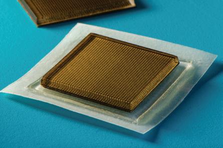
Stamp-sized ultrasound sticker developed to see inside the body while on the move
Childhood obesity
New clinical guideline from American Society for Metabolic and Bariatric Surgery calls for early treatment of obesity in children

Bahrain US$8, Egypt US$8, Iran US$8, Iraq US$8, Jordan US$8, Kuwait US$8, Lebanon US$8, Oman US$8, Qatar US$8, Saudi Arabia US$8, Syria US$8, UAE US$8, Yemen US$8 In the News • Cleveland Clinic Abu Dhabi signs MoU with UAE University’s College of Medicine and Health Sciences to promote research and education • Academic Health Association International designates Dubai’s MBRU as regional hub • Novo Genomics, Lenovo collaborate to
genomics research in KSA • WHO rolls out new holistic way to
early childhood development
accelerate
measure
Cancer diagnostics
www.MiddleEastHealth.com Serving the region for over 40 years March - April 2023
Prognosis
AI in Oncology
In this issue, one of our focus areas is Oncology, specifically at some new research which shows how artificial intelligence (AI) with infrared imaging helps improve colon cancer diagnostics.
In general, AI is beginning to have a major impact in the field of Oncology, from diagnosis to treatment and prognosis. With the help of AI, oncologists can better understand the disease, develop more accurate diagnostic tools, and create more targeted treatments.
One of the most significant contributions of AI in Oncology is in medical imaging. AI algorithms can analyze images from CT scans, MRIs, and other imaging tests to help detect cancer at earlier stages. AI-based software can also help physicians identify subtle changes in tumour size, shape, and location, making it easier to track the progression of the disease and plan treatments accordingly.
AI is also being used to develop more personalized cancer treatments. By analyzing large amounts of data from genomic testing and clinical trials, AI can identify patterns and predict how a particular patient will respond to a specific treatment. This information can help doctors tailor treatment plans to the individual patient, increasing the chances of success while reducing the risk of side effects.
AI is also helping to improve the accuracy of cancer diagnosis. By analyzing patient data, including medical history, family history, and genetic information, AI can help doctors identify patients who are at high risk for cancer, allowing for earlier screening and detection. Additionally, AI can help doctors differentiate between cancerous and non-cancerous cells, reducing the need for invasive biopsies.
Another area where AI is being used in Oncology is in drug development. By analyzing large amounts of data from clinical trials and scientific literature, AI algorithms can identify promising drug candidates and predict how they will interact with different types of cancer cells. This can help researchers develop more effective and targeted cancer treatments.
AI is transforming the field of Oncology, providing new tools and insights that are helping doctors diagnose, treat, and prevent cancer more effectively. As AI continues to evolve, it has the potential to revolutionize the way we approach cancer care, offering more personalized and precise treatments that improve patient outcomes and quality of life.
Callan Emery Editor editor@MiddleEastHealth.com
Publisher Michael Hurst Michael@MiddleEastHealth.com
Editor Callan Emery editor@MiddleEastHealth.com
Editorial Consultants
Dr Gamal Hammad, Dr Peter Moore, Harry Brewer
Middle East Editorial Office PO Box 72280, Dubai, UAE
Telephone: (+9714) 391 4775 editor@MiddleEastHealth.com
Marketing Manager
Foehn Sarkar
Telephone: (+9714) 391 4775 n Fax: (+9714) 391 4888 marketing@MiddleEastHealth.com
Subscription & Admin Manager
Savita Kapoor
Telephone: (+9714) 391 4775 n Fax: (+9714) 391 4888 Savita@MiddleEastHealth.com
Advertising Sales PO Box 72280, Dubai, UAE marketing@MiddleEastHealth.com
Americas, France Joy Sarkar P O Box 72280, Building No.2
2nd Floor, Dubai Media City
Dubai, United Arab Emirates
Tel: +971 4 391 4775 Fax: +971 4 391 4888 Joy@MiddleEastHealth.com
Japan
Mr Katsuhiro Ishii
Ace Media Service Inc
12-6, 4-chome, Adachi-ku, Tokyo 121-0824, Japan
Tel: +81-3-5691-3335 n Fax:+81-3-5691-3336
Email: amskatsu@dream.com
China
Miss Li Ying Medic Time Development Ltd, Flat 1907, Tower A, Haisong Building, Tairan 9th Road, Futian District, Shenzhen, China 518048
Tel: +86-755-239 812 21 n Fax: +86-755-239 812 33 Email: medic8@medictime.com
Taiwan
Larry Wang
Olympia Global Co Ltd
7F, No.35, Sec 3, Shenyang Rd, Taichung
Taiwan 40651 n P O Box: 46-283 Taichung Taiwan 40799
Tel: +886- (4)-22429845 n Fax:+886- (4)-23587689
Email: media.news@msa.hinet.net
Middle East Health is published by Hurst Advertising FZ LLC , Dubai Media City, License Number: 30309

UAE National Media Council - Approval Number: 2294781
Middle East Health online www.MiddleEastHealth.com

Middle East Health is printed by Atlas Printing Press. www.atlasgroupme.com

MIDDLE EAST HEALTHI 3 March - April 2023 Serving the region for 40 years


4 IMIDDLE EAST HEALTH contents NEWS 6 Middle East Monitor 10 Worldwide Monitor 14 The Laboratory Serving the region for 40 years March - April 2023 FOCUS EXPOS & CONFERENCES FINAL PAGES www.MiddleEastHealth.com 20 Oncology: Cleveland Clinic Abu Dhabi Oncology Institute launches 4th Angel Program for patient support 24 Oncology: Researchers use AI with infrared imaging to improve colon cancer diagnostics 25 Oncology: Cancer patients who don’t respond to immunotherapy lack important immune cells 26 Ultrasound: Stamp-sized ultrasound adhesives provide long-term imaging of diverse organs 30 Ultrasound: Ultrasound-guided surgery is quicker, more effective for treating ductal carcinoma in situ 31 Ultrasound: A combination of ultrasound and nanobubbles destroys cancerous tumours, eliminating the need for invasive treatments 32 Anaesthesia: Researchers identify what causes accidental awareness under anaesthesia 32 Anaesthesia: How does ketamine act as a ‘switch’ in the brain? 34 Anaesthesia: Neuron function altered by widely-used Propofol 38 Arab Health 2023 review 48 The Back Page 38 20 26 06 12

Image: Felice Frankel
middle east monitor

Update from around the region
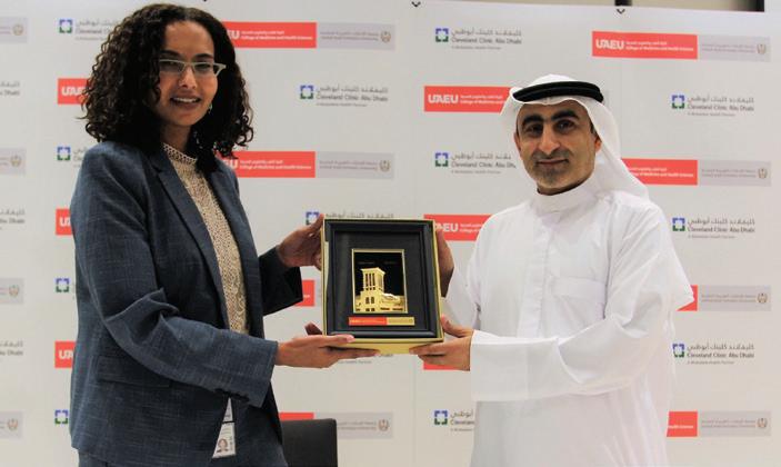
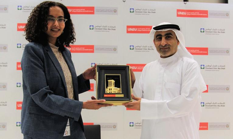
Cleveland Clinic Abu Dhabi signs MoU with UAE University’s College of Medicine and Health Sciences to promote research and education
Cleveland Clinic Abu Dhabi has signed a Memorandum of Understanding (MoU) with the College of Medicine and Health Sci ences - United Arab Emirates University. The agreement strengthens an alliance between the two parties, in research and education.
The MoU includes collaboration on re search projects, co-mentoring and place ment of students at Cleveland Clinic Abu Dhabi, as well as the creation of research and development partnerships. The educa tional arm of the MoU entails collaboration on educational projects at the undergradu ate, graduate, and postdoctoral levels. The MoU also includes partnering on the devel opment of jointly led events such as work shops, seminars and conferences.
Commenting on the MoU, Dr Sawsan AbdelRazig, Interim Chief Academic Of ficer at Cleveland Clinic Abu Dhabi said: “Innovation, research, and education are key areas that Cleveland Clinic Abu Dhabi focuses on. Partnering with one of the most recognized and respected academic institutions in the UAE puts us at the forefront to train the next generation of healthcare leaders and physicians, advance research, broaden our body of knowledge, and, ultimately, improve healthcare in the UAE. We recognize the importance of nurturing
‘homegrown’ talent to create sustainable health services in the UAE. This agreement serves as a mutually beneficial platform that will help us drive all of these objectives, which will, in turn, benefit the local communities and the wider region.”
Prof. Juma Alkaabi, Interim Dean from College of Medicine and Health Sciences
- United Arab Emirates University said: “The College of Medicine and Health Sciences strives to produce Emirati doctors educated to the highest international stan-
NAFFCO, one of the leading producers and suppliers of safety solutions in the GCC, presented the first-of-itskind electric ambulance made in the UAE, at the Arab Health 2023 exhibition. NAFFCO’s electric ambulance is outfitted with some of the most advanced communication technologies available, including hands-free, voiceactivated, wearable computing solutions that give medical professionals a comprehensive overview of medical conditions of the patients in the ambulance before reaching the hospital. The solutions allow for real-time communication with remote co-workers and medical professionals, in order to support medical training, rounding, proctoring, and specialist consultations. Additionally, the project represents a step towards the country’s zero emission goals, thereby driving sustainable operations in the sector.
dards. The collaboration with Cleveland Clinic Abu Dhabi will enhance student’s experience and also open up additional avenues for research. We are extremely proud to be aligned with one the most distinguished hospitals in the region. There is much that Cleveland Clinic Abu Dhabi and the College of Medicine and Health Sciences can achieve together by way of collaboration in research and education, and we look forward to working closely with the team.”

6 IMIDDLE EAST HEALTH
NAFFCO showcases first-of-its-kind electric ambulance made in UAE
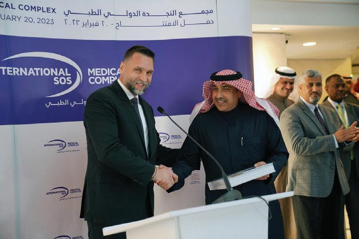
Al Rushaid Group, International SOS open International SOS Medical Complex in Khobar
A new medical facility, International SOS Medical Complex, has opened in Khobar, Kingdom of Saudi Arabia. The medical complex is a joint venture between International SOS and Al Rushaid Group (the operator). The medical facility features an extensive range of services, including: dental care, obstetrics, gynaecology, paediatrics, internal medicine, family medicine and ophthalmology. Additionally, it has a 24/7 emergency department and one of the largest medical laboratories to be hosted in a medical complex in the region.
The official opening of the International SOS Medical Complex on February 20
coincided with the Saudi Founding Day and showcased the companies’ commitment to enriching local communities and furthering Vision 2030 of the Kingdom of Saudi Arabia. The clinic features a team comprised of both expert local and international medical professionals.
The ceremony was attended by Sheikh Rasheed Al-Rushaid, Chairman of Al-Rushaid Group, and Laurent Sabourin, International SOS Group Managing Director.
Al-Rushaid said: “We are proud to introduce a new healthcare landmark in Khobar. Our Group is well known for bringing in worldwide industry leaders and
exchanging expertise to enrich our Saudi young talent and professionals. This is a step bringing us all towards achieving Vision 2030. Our commitment to our community will not stop here and we’ll continue expanding our footprint in the region and in the kingdom to always provide the highest quality of service and care.”
Reinforcing International SOS’ commitment to passion, expertise, respect and care, Sabourin, said: “We are not only bringing over 35 years of international medical leadership experience to Saudi Arabia; we are here to uphold these values and make a positive contribution to the community.”
• For more information, visit: https://my.internationalsos.com/InternationalSOSMedicalComplex
AMSA Healthcare Group launches new kidney care centre in Dubai with 13 haemodialysis units
AMSA Healthcare Group officially launched the AMSA Medical Centre and Kidney Care in Dubai on January 17, this year. The multi-specialty clinic and kidney care centre also includes an AMSA pharmacy and 13 haemodialysis units.
The medical centre has multiple consultation rooms dedicated to specialties in Nephrology, Cardiology, Nutrition, General Practice, and Internal Medicine. The availability of in-house pathology diagnostics allows for quicker assessment and intervention.
The 13 haemodialysis units are strategically placed to ensure patient comfort as well as privacy. There are an additional two private rooms and two Isolation rooms to cater to Hep B, Hep C, and Covid positive dialysis patients. The centre has 17 beds in total.
Academic Health Association International designates Dubai’s MBRU as regional hub
The Alliance of Academic Health Centers International (AAHCI) has designated Mohammed Bin Rashid University of Medicine and Health Sciences (MBRU) as the AAHCI Regional Office for The Middle East and North Africa Region (MENA) for a two-year period beginning July 1, 2023. The American University of Beirut Medical Center will remain the AAHCI MENA Regional Office host until June 2023.
Professor Alawi Alsheikh-Ali, AAHCI MENA Regional Ambassador and Chief Academic Officer of the Dubai Academic Health Corporation (DAHC), commented: “Academic health centres are ground-

ed in evidence that the integration of clinical practice, research, and education can accelerate the adoption of best prac tices in patient care. MBRU’s designa tion as the regional centre for academic health is a key milestone in our journey as Dubai’s First Academic Health System with a mission to impact lives and shape the future of health through the integra tion of care, learning, and discovery.”
MBRU has been a member of AAHCI since 2021 and hosted its latest regional meeting in October 2022 under the theme, Transforming Academic Health Centers: Innovation and Resilience. As the new AAHCI Regional Office, MBRU plans

MIDDLE EAST HEALTHI 7 s
Professor Alawi Alsheikh-Ali, AAHCI MENA Regional Ambassador and Chief Academic Officer of the Dubai Academic Health Corporation
to forge a coordinated approach aimed at fostering effective solutions the following areas: Evolving clinical care, transforming the patient experience, investment in biomedical research, resilience of academic health centres, adaptation in health professions and medical education, and partnerships and community outreach.

“Improvements in healthcare rarely oc-
cur in isolation. The challenges patients are facing in the region are being tackled by healthcare organizations across the globe. Every member of MENA AAHCI brings something valuable to the table. As the AAHCI Regional Office, we intend to leverage the strengths and expertise of each AAHCI member to expand knowledge and rapidly integrate medical ad-
vances into clinical practice and community outreach programmes,” Prof. Alawi said. “As Regional Ambassador, I’d like to officially invite healthcare organizations in the MENA region to join AAHCI. A well-coordinated, collaborative approach to solving today’s pressing challenges is essential to improve the health and vitality of the communities we serve.”
Novo Genomics, Lenovo collaborate to accelerate genomics research in KSA

Lenovo has formed a new partnership with Novo Genomics, one of Saudi Arabia’s leading biotech start-ups with a focus on localizing and applying the latest technologies in genomics and multi-omics. Novo Genomics leverages data through advanced analytics and data mining to promote health and wellness in the region. The partnership will also mark Novo Genomics as the first customer of the Lenovo Genomics Optimization and Analysis Tool (GOAST) in the EMEA region.
Genomics research is actively expanding in the Kingdom, supported by strong infrastructure for research with the establishment of dedicated research centres and institutions such as the King Abdul Aziz City for Science and Technology and King Abdullah International Medical Research Centre. The Kingdom has made significant investments in genomics research as a part of the country’s Vision 2030, with an aim to increase capacity for scientific research and innovation.
Lenovo, a leader in accelerating genomics analytics with its GOAST solution, enables research that is significantly faster than standard environments. This allows researchers to analyze a whole human genome in less than 53 minutes, and whole exomes in about a minute, enabling the processing of more genomes concurrently, finding answers faster, and making breakthroughs sooner. Lenovo is the only solutions provider who delivers this degree of performance in a cost-effective, open source, CPU-based solution. Lenovo ThinkSystem servers that run GOAST currently hold an incredible 196 world records for performance.
Commenting on the partnership, Alaa
Bawab, General Manager, Lenovo Infrastructure Solutions Group, MEA said “Lenovo is a pioneer in accelerating genomics analytics, which is evident by our GOAST tools which not only offer higher degrees of performance and reliability but also keeps it cost-effective. We are confident that with our tools, researchers in fields of life sciences, precision medicine and infectious diseases can understand data faster and more efficiently, making it the smarter way of decoding genomics. Our R&D groups, which have some of the industry’s best HPC engineers and world-class scientists, have ensured that all the supporting work is already done, which leaves users with optimized settings for high-performing results from the moment they setup.”
Dr Budoor Almansour, Chief Operating Officer and Co-founder said: “Novo Genomics is a Saudi biotech company focused on localizing and applying the technologies in genomics / multi-omics and leveraging data through advanced analytics and data mining to promote health and wellness in the region and beyond. Given the high prevalence of genetic diseases in the region, we are committed to genomics as a vital field to enrich the sector with qualified Saudi expertise. NOVO consists of a high-level team of bioinformatics scientists, consultants, and clinicians, who are empowered to put all the right strategies in place to transform the concept of health, wellness and quality of life for the benefit of the sector and society, not just for humans, but for animals and plants as well.”
8 IMIDDLE EAST HEALTH
Philips and Wipro partner to offer next-generation electronic medical record enterprise solution in KSA
Philips has entered into an agreement with Wipro Limited, a leading technology services and consulting company, to adapt Philips Electronic Medical Record (EMR) to provide a tailored EMR solution for the needs of healthcare providers in the Kingdom of Saudi Arabia. The agreement builds on the decadelong partnership between the two companies.
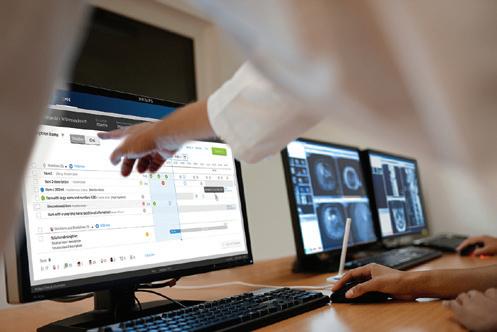
Philips EMR offers one integrated solution across all care settings through a single platform and database that enables centralized management of clinical, operational and administrative processes. The capabilities of the platform will be combined with Wipro’s expertise in professional service delivery, as well as established patient administration and revenue cycle management systems designed
for and delivered in the Kingdom.
The collaboration will help drive sustainable digital transformation of healthcare at a time when medical practices face pressure to operate at peak efficiency. It will leverage international clinical best practices while complying with local practices and regulations. With this offering, which may be extended to other markets in the future, healthcare providers will gain access to marketleading complementary technologies and services to ultimately deliver better health outcomes, improved patient and staff experience and lower the cost of care. It will particularly focus on supporting local clinical practice, configurable workflows,
Saudi German Health earns ACHSI accreditation across its medical facilities in KSA
Saudi German Health (SGH), a leading healthcare group in the MENA region, has received accreditation for its facilities by the Aus tralian Council on Healthcare Standards International (ACHSI), a globally-recognized organization that aims to ensure quality and safety standards in patient care. The accreditation reinforces SGH’s com mitment to uplift the standards of healthcare services and provide patients with cutting-edge and innovative solutions in Saudi Arabia in line with “Saudi Vision 2030” and the healthcare transformation programme.
As part of ACHSI cluster accreditation, which encompasses 13 accreditation certificates, SGH is the first healthcare provider in the Middle East to obtain Person-Centered Systems Standards accreditation and ACHSI dialysis certification in its medical facilities. SHG has also become the first in the Middle East to earn hemodialysis accreditation. Furthermore, the recently opened hospitals in Makkah and Al Jamea District, Jeddah, have earned ACHSI accreditation while Beverly Clinic in Jeddah and Abha clinic received ACHSI ambulatory accreditation.
Makarem Sobhi Batterjee, President and Vice Chairman, Saudi German Health, said: “The foundations of our organization are patient safety and ethical practice. The comfort and well-being of our patients and their families are extremely important to us at SGH, and we go above and beyond to provide the best possible clinical treatment. The accreditation will further strengthen our dedication to enhancing and delivering first-rate services to the community through our many cutting-edge healthcare treatments and solutions.”
By receiving this accreditation, SGH was able to add the expertise of the Australian healthcare system to its collection of global accreditations. SGH currently has more than 60 accreditation certificates from various national and international certification organizations..
data and service integrations, and clinical decision support in a bid to realize Saudi’s Health Sector Transformation ProgramVision 2030 <https://www.vision2030.gov. sa/v2030/vrps/hstp/>.

“As Philips, we have over 20 years’ experience in electronic medical records solutions, which forms the basis of this sophisticated system that standardizes and centralizes processes for enhanced efficiency and outcomes,” said Vincenzo Ventricelli, CEO of Philips Middle East, Türkiye & Africa. “Through this collaboration we will be able to offer our advanced Philips EMR solution to benefit healthcare
Dubai’s Marie Curie Cancer Institute
Prime Healthcare Group has established a strategic collaboration with Varian, a Siemens Healthineers company, with the acquisition of a Truebeam radiotherapy system and Bravos afterloader system for the Marie Curie Cancer Institute (MCCI) currently under development.
MCCI will be an integral part of Prime Healthcare Group’s US$122 million multi-specialty hospital, being built in Dubai’s Healthcare City district. The centre will provide a comprehensive cancer care system including medical oncology, surgical oncology and radiation oncology.
Announcing the strategic collaboration at Arab Health, 2023, Dr Jamil Ahmed, Managing Director of the Group said: “We are going to work with Varian, a Siemens Healthineers company, and extended the long-term collaboration with Siemens Healthineers in the region. Prime Health Care has acquired a TrueBeam radiotherapy system and BRAVOS afterloader system.”
This extended partnership comes on the back of an existing longterm relationship between Prime Healthcare Group and Siemens Healthineers, which entails the equipment and servicing of the radiology department of Prime Healthcare Group facilities in Dubai.

MIDDLE EAST HEALTHI 9
worldwide monitor Update from around the
globe
Genomics England deploys enterprise imaging technology in ground-breaking cancer data programme
Genomics England has completed installation of an enterprise imaging system that will help to support a world-pioneering initiative for cancer research. The programme is linking whole genome sequencing, pathology and radiology data, in what has been described as the world’s largest multimodal cancer research platform.
First announced in 2022 as a means to support new discoveries, Genomics England’s programme will help a wide range of researchers and scientists create a better understanding of cancer. It is hoped that this will lead to new treatments as well as supporting the development of cancer-targeting AI.
Genomics England has now deployed technology from medical imaging technology provider Sectra, that will play a central role in allowing the organisation to bring together underpinning data, so that researchers and developers from a wide variety of backgrounds can harness it in new ways.
In particular, the enterprise imaging system will allow Genomics England to incorporate NHS imaging data, whilst the Image Exchange Portal, a system used nationally in the NHS, will also allow it to transport images from participating NHS trusts.
This will mean diagnostic imaging data captured in the NHS, including radiology images such as x-rays, CT and MRI scans, and digital pathology images generated by NHS labora-
tories, can be linked with whole genome sequencing data from Genomics England.
To begin with 30 NHS trusts in England are providing data on solid tumours. This includes approximately 250,000 pathology images and 200,000 radiology scans, for 16,000 participants.
Once the radiology and pathology data in the system is matched with the genomics data, multi-modal data will be used by researchers to investigate and identify markers for cancer diagnostics and treatments.
Information will be kept highly secure with patient identifiable data removed for researchers outside of Genomics England, who will only have access to a Genomics England ID number, the age of the participant, and the name of the NHS site at which data was captured.
Dr Prabhu Arumugam, director of clinical data and imaging, and Caldicott Guardian for Genomics England, said: “This programme will push the boundaries of cancer research and how we work. It has the potential to transform clinical trials, change who can do research and development, and lead to the creation of new targeted treatments for cancer patients. The potential is vast.
“We will be able to understand mutations and when things go wrong in DNA, and importantly, whether that transpires into what clinicians see in medical imaging. We can also expose data in new ways to AI. All
of that can help to facilitate new drug discoveries, and better inform which patients might benefit from particular treatments.
“Innovative working with Sectra is an important part of our initiative. The imaging system is already a very recognisable interface in NHS clinical settings, but we are using it in new ways. It will help us to harness imaging that we can then match to our genomic data, whilst de-identifying data to ensure confidentiality. The resulting multi-modal dataset will enable important research, break down traditional barriers, and support a safe and secure but accessible cloud-based research environment, that means many more people than bioinformaticians can harness genomic, pathology and radiology data.”
Deployed in the Genomics England’s cloud environment, the new research platform will be easily accessible for users, through a secure, fast and reliable interface.
Cloud deployment will also provide the flexibility to scale the initiative as more users come on board, and as the programme potentially expands to support research for non-cancers in the future.
Sectra’s enterprise imaging system is widely used in the NHS, where it supports healthcare professionals in diagnosing patient illness. The system installation was completed in February 2023, after a contract was awarded earlier in 2022.
Global Fund provides US$867 million in additional funding for pandemic preparedness and response
Since December 2022, the Global Fund has awarded US$547 million in additional funding to 40 countries through its COVID-19 Response Mechanism (C19RM) and has now initiated the process to award a further $320 million, making a total of approximately $867 million.
These grants are focused on supporting both the immediate COVID-19 response and broader pandemic preparedness, while strengthening the underlying health systems. This includes investments in disease
surveillance, laboratory networks, community health worker networks and community-based organizations, medical oxygen and respiratory care systems, as well as the rollout of novel therapeutics to scale up test-and-treat programs in case of future COVID-19 surges. These investments reflect the deliberate rebalancing of C19RM from the immediate COVID-19 response towards strengthening key components of health systems to counter potential future variants and reinforce pandemic prepared-
ness. The total awarded for C19RM since 2020 now amounts to almost $5 billion.
The Global Fund’s C19RM follows a country-led, inclusive, and demand-driven approach to ensure funding goes where it is most needed. Uganda is one such example of how the Global Fund partnership is making a very substantial contribution to address gaps in disease surveillance and strengthen national laboratory systems to reinforce their capacity to detect COVID-19 and other pathogens.
10 IMIDDLE EAST HEALTH
s
“From the start of the pandemic, the Global Fund took a leading role in supporting low- and middle-income countries like Uganda to scale up testing for the new virus, leveraging 20 years of experience in procuring diagnostics and investing in laboratory capacities and disease surveillance. Most recently in 2022, our strong surveillance systems enabled Uganda to detect the Ebola outbreak in a timely manner, respond and eventually control the outbreak in a record time of 69 days,” said Dr Jane Aceng, Minister of Health of Uganda.
Elsewhere, with a US$30 million investment from the Global Fund, the government of Indonesia and Genome Science Initiative established a nationwide network of facilities able to conduct whole genome sequencing to strengthen early diagnosis and treatment of deadly diseases including tuberculosis, COVID-19, cancer, metabolic disorders, brain diseases and genetic disorders. This funding has benefited organizations like the Center for Environmental Health Engineering and Disease Control in Batam, Indonesia, which today are at the forefront of fighting disease and preparing for future health threats.
The funds are being used to purchase new instruments and train laboratory staff to help ensure facilities across the country are able to harness the new technology and transform the country’s health system.
“Prior to the initiative launching, laboratory workers in Batam had to send samples 1,100 km away to a laboratory in Jakarta to do genomic sequencing, and it would take more than two weeks to get results. Today, they are able to get this same result in less than five days, which is vital in deciding how to respond and manage the disease outbreak as early as possible. The initiative also enhances distributed laboratories capacities throughout the country for genomic surveillance and bioinformatics analysis,” said Budi Gunadi Sadikin, Minister of Health of Indonesia.
As part of the process to make the second wave of awards totalling approximately US$320 million, the Global Fund has invited countries to express an interest in having their funding requests considered for inclusion in a potential Global Fund proposal to the Pandemic Fund, which has recently announced a call for proposals for US$300 million in funding. Since there is a strong overlap in the priority areas of investment for both C19RM and the Pandemic Fund, the Global Fund is exploring how to help countries minimize any extra work and maximize the synergies between investments funded from the different sources.
The Fifth Global Ministerial Summit on Patient Safety closed in Montreux, Switzerland on 24 February, after endorsing the Montreux Charter on Patient Safety with recommended actions to address avoidable harm in health care. This was the first Global Ministerial Patient Safety Summit to take place after the COVID-19 pandemic, which has exposed the high risk of unsafe care to patients, health workers and the general public, and made visible a range of safety gaps across all core components of health systems. The Summit was hosted by the Swiss government. Unsafe care is among the leading causes of death and disability in the world. It is particularly acute in resource-constrained settings. In the years preceding the COVID-19 pandemic, 2.6 million people died every year due to safety lapses in hospitals in lower-income countries. Rich countries are not immune: nearly 15 per cent of hospital expenditure and activity in countries of the Organization for Economic Co-operation and Development could be attributed to treating safety failures.
It is estimated that more than half of cases of patient harm are preventable, by working together to create a safer healthcare system for all and to build a culture of safety that emphasizes continuous improvement, learning, and innovation.
Dr Tedros Adhanom Ghebreyesus, WHO Director-General, participated in the Summit with the host, the Swiss President Alain Berset. In his address to the ministerial segment, Dr Tedros urged health ministers to invest in patient safety as part of their commitment to universal health coverage and health security; to build a culture of safety and strengthen reporting and learning systems; to support health workforce and strengthen their capacity; to strengthen data systems; and to engage patients and families in their own care. Dr Tedros announced that the theme for World Patient Safety Day 2023 will be “Engaging patients for patient safety”.
In Montreux, delegations from more than 80 countries discussed the gaps and key challenges for the implementation of the World Health Assembly resolution (WHA72.6) “Global Action on Patient Safety” and the global roadmap for patient safety, the Global Patient Safety Action Plan 2021–2030: Towards eliminating avoidable harm in health care.
Despite progress to address patient safety challenges worldwide, concerted efforts are needed to ensure safety of patients and health and care workers, noted the delegations and stressed that lessons learned from the COVID-19 crisis hold huge potential to build safer and more resilient health systems.
The Montreux Charter on Patient Safety, endorsed at the Summit, reaffirms that patient harm in health care is an urgent public health issue, pertinent to countries of all income settings and geographies and therefore a shared global challenge. It identifies actions for countries to narrow implementation gaps in patient safety, including by treating patient safety as a global public health priority, building upon lessons learned from the COVID-19 pandemic, deepening partnerships, collaboration and mutual learning, and engaging patients and their families. The Charter also urged setting priorities for patient safety, including medication safety, safe surgery, infection prevention and control, and antimicrobial resistance.
The Summit in Montreux builds on the preceding Global Ministerial Summits on Patient Safety which have raised awareness about burden of avoidable patient harm in health care and fostered strategic approaches to strengthening Patient Safety, from London (2016), to Bonn (2017), to Tokyo (2018) and to Jeddah (2019).
The Sixth Summit will be held in Chile in 2024.
MIDDLE EAST HEALTHI 11
The Montreux Charter on Patient Safety galvanizes action to address avoidable harm in health care
The World Health Organization (WHO) has launched a new package of measures, the Global Scales for Early Development (GSED), to monitor the development of young children at population level up to three years of age.
The new GSED methodology allows for a comprehensive assessment of the develop ment of young children up to 36 months of age, capturing cognitive, socio-emotional, language and motor skills. The GSED pro vides a developmental score (D-score), a new common unit to measure development, to give an overall picture of children’s develop ment which can be tracked over time.

The GSED package will help countries, programmes and researchers gather and use data on early childhood development to better invest in services and support needed for young children and their families.
“The foundations of life-long health and well-being are formed during the vital first years of a child’s life,” said Dr Anshu Banerjee, WHO Assistant Director-General ad interim, Universal Health Coverage and Life Course and Director for Maternal, Newborn, Child and Adolescent Health and Ageing. “The GSED will help us make more informed decisions to better invest in nurturing care and support children’s right to reach their full potential”.
Countries have long required measures that are valid and reliable to monitor development among children from birth to 36 months but standardized and globally applicable population-based measures have been scarce.
Existing measures were either designed to monitor the development of children after 24 months or lacked sufficient consideration for the diversity of contexts in which children are raised. Other measures required extensive resources to be implemented.
The new methodology was developed based on a common dataset collected from 51 cohorts in 32 countries, of which 30
are low- or middle-income countries, by a multi-disciplinary team of global experts coordinated by WHO.

Investing in children and their families
The GSED provides open-access and consistent measures to monitor early childhood development across time and groups of children. It will help generate data that will inform policymakers and governments’ understanding of the barriers to children’s developmental progress in their countries. This will also enable them to target resources more effectively towards policies and interventions that will support the children most at risk of not reaching their developmental potential.
Global organizations can use the data for cross-country comparisons and trend analyses to inform interventions and funding allocation.
“Just like children are measured for height and weight to check that they are growing as best they can physically, the GSED will now allow for children’s development to be measured holistically” said Dr Dévora Kestel, WHO Director of Mental Health and Substance Use. “These new measures will help us ensure that no child is left behind, as set out in the Sustainable Development Goals”.
Implementing the Global Scales for Early Development
The GSED is delivered through open access, culturally neutral forms that are easy to use across the world, with minimal adaptation required beyond translation. They are relevant across different contexts, including in emergency situations as well as in large-scale data collection efforts.
To date, the GSED measures have been validated in Bangladesh, Pakistan, and United Republic of Tanzania (1248 children per country) and data collection is ongoing in Brazil, China, Côte d’Ivoire and the Netherlands.
The current package comprises the measures (Short form and Long form), related user manuals and item guides, translation and adaptation guide, scoring guide and technical report summarising the methodology and the results of its validation. A GSED App is also available. The GSED will continue to evolve and an expanded version of the package, including global norms and standards for child development, will be released following additional data collection.
Global Scales for Early Development
https://bit.ly/3SDUljb
12 IMIDDLE EAST HEALTH
WHO rolls out new holistic way to measure early childhood development
HUMAN DIAGNOSTICS
IVD experience that mattersReliable diagnostics worldwide
The Middle East as an important region for HUMAN’s international business
HUMAN is one of the global players in the in-vitro diagnostics industry. With its broad network of subsidiaries and long-standing distribution partners, the German medium-sized company with its headquarters in Wiesbaden (near Frankfurt) supports medical laboratories in more than 160 countries. For its customers of the Arab world, HUMAN also maintains a regional office in Sharjah, UAE.
Middle East and North Africa (MENA) is an important region for HUMAN’s international business. The major driving factor linked to the growth of the IVD industry in this region is the high burden of chronic diseases, especially infectious diseases, cancer, and diabetes. The UAE, for instance, has one of the world’s highest rates of diabetes, at about 18.7 percent, which is a huge medical, but also an economic burden.
With its Diabetes Dx products, HUMAN helps to ease and improve both an early diagnosis of diabetes, but also better diabetes monitoring and diabetes management. Regardless of whether this is to be done at the point of care or if the blood analysis is done in a specialized laboratory – HUMAN’s Diabetes Dx products fulfill every need with highest quality and precision in combination with rapid results and easy handling.
Reveal undiagnosed diabetics by WHO-recommended HbA1c testing
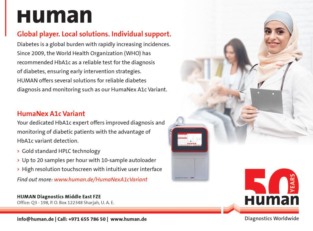
One of HUMAN‘s featured products at Medlab 2023 was HumaNex A1c Variant, an HPLC system for small and mediumscale measurement of glycated hemoglobin HbA1c. Since 2009, the World Health Organization (WHO) has recommended HbA1c as a reliable test for the diagnosis of diabetes, ensuring early intervention strategies.
MIDDLE EAST HEALTHI 13
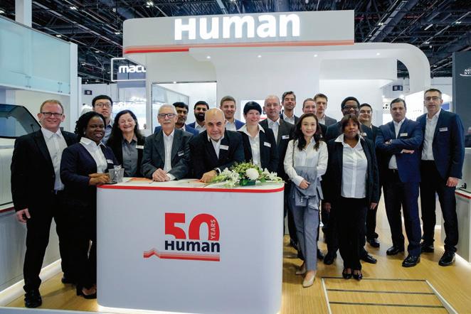
Sales & service staff members of the HUMAN family at Medlab Middle East 2023 in Dubai
the laboratory
Medical research news from around the world
New study a major step forward in understanding autism
Results of a new study led by Roberto Araya, a Canadian neuroscientist, biophysicist and researcher at the CHU Sainte-Justine Research Centre, in Montreal, show that in Fragile X syndrome (FXS), the most common cause of autism, sensory signals from the outside world are integrated differently, causing them to be underrepresented by cortical pyramidal neurons in the brain.
This phenomenon could provide important clues to the underlying cause of the symptoms of this syndrome. The research team’s work not only provides insight into the mechanism at the cellular level, but also opens the door to new targets for therapeutic strategies.
The study was published on January 3 in the journal Proceedings of the National Academy of Sciences [1] .
Autism is characterized by a wide range of symptoms that may stem from differences in brain development. With advanced imaging tools and the genetic manipulation of neurons, the team of researchers at the CHU Sainte-Justine Research Center was able to observe the functioning of individual neurons – specifically pyramidal neurons of cortical
layer 5 – one of the main information output neurons of the cortex (the thin layer of tissue found on the surface of the brain).
The researchers found a difference in how sensory signals are processed in these neurons.
“Previous work has suggested that FXS and autism spectrum disorders are characterized by a hyperexcitable cortex, which is considered to be the main contributor to the hypersensitivity to sensory stimuli observed in autistic individuals,” said Araya, also a professor in the Department of Neurosciences at Université de Montréal.
“To our surprise, our experimental results challenge this generalized view that there is a global hypersensitivity in the neocortex associated with FXS. They show that the integration of sensory signals in cortical neurons is underrepresented in a murine model of FXS,” added Diana E. Michell, first co-author of the study.
A protein, FMRP, that is absent in the brains of people with FXS modulates the activity of a type of potassium channel in the brain. According to the research group’s work, it is the absence of this pro-
tein that alters the way sensory inputs are combined, causing them to be underrepresented by the signals coming out of the cortical pyramidal neurons in the brain.
Soledad Miranda-Rottmann, also first coauthor of the study, attempted to rectify the situation with genetic and molecular biology techniques. “Even in the absence of the FMRP protein, which has several functions in the brain, we were able to demonstrate how the representation of sensory signals can be restored in cortical neurons by reducing the expression of a single molecule,” she said.
“This finding opens the door to new strategies to offer support to those with FXS and possibly other autism spectrum disorders to correctly perceive sensory signals from the outside world at the level of pyramidal neurons in the cortex,” explained Araya.
“Even if the over-representation of internal brain signals causing hyperactivity is not addressed, the correct representation of sensory signals may be sufficient to allow better processing of signals from the outside world and of learning that is better suited to decision making and engagement in action.”
Reference
doi: https://doi.org/10.1073/pnas.2208963120
Cleveland Clinic-led trial shows first drug designed for statin-intolerant patients reduces serious cardiovascular events
Findings from a clinical trial led by global health system Cleveland Clinic have shown that the use of bempedoic acid (a-
tein (LDL) cholesterol and reduced heartcent Late Breaking Science Session at the American College of Cardiology’s 72nd Annual Scientific Session in New Orleans New
Elevated LDL cholesterol can accumulate -
es and raising the risk of heart attack or stroke. Statins are the standard first-line treatment for the prevention of cardiovascular disease and work by lowering cholesterol levels in the blood. Statins have been shown in many studies to reduce the risk of stroke, heart attack and death when administered to patients, both with a history of cardiovascular disease and without. However, some patients struggle with adverse side effects like muscle pain, headaches or weakness that prevent them from using statins at recommended doses.
Bempedoic acid is approved by the U.S. Food and Drug Administration as an additional treatment to help lower cholesterol in pa-
1. Diana E. Mitchell, Soledad Miranda-Rottmann, et. al. Altered integration of excitatory inputs onto the basal dendrites of layer 5 pyramidal neurons in a mouse model of Fragile X syndrome. PNAS, 3 January 2023.
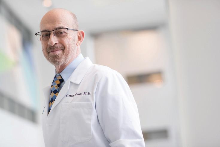
Steven E. Nissen, MD, chief academic officer of the Heart Vascular & Thoracic Institute at Cleveland Clinic s
tients with certain conditions who have high cholesterol despite maximally tolerated statin therapy. The trial, called CLEAR Outcomes, is the first to explore whether bempedoic acid could reduce cardiovascular outcomes.
The study enrolled 13,970 statin-intolerant patients between December 2016 and August 2019 at 1,250 sites in 32 countries. To participate, patients and their physicians were required to confirm that the patient could not tolerate statins. All participants had LDLc levels of 100 mg/dL or higher and either a previous cardiac event or other risk factors for heart disease. Participants were randomly assigned to take 180 mg of bempedoic acid or a placebo daily and followed for an average
of over 3 years. The primary outcome studied was a composite of cardiovascular death, heart attack, stroke or coronary revascularizations.
The primary outcome was reduced 13% with bempedoic acid treatment. When different types of cardiac events were analyzed separately, bempedoic acid was found to reduce heart attacks by 23% and coronary revascularizations by 19%.
“Until now, there have not been any drugs designed specifically for statin-intolerant patients,” said the study’s lead author Steven E. Nissen, MD, chief academic officer of the Heart Vascular & Thoracic Institute at Cleveland Clinic. “While statins remain the cornerstone of risk reduction in patients with
elevated LDL cholesterol, this is a major step forward for a population who need statins but suffer troublesome side-effects.”
Bempedoic acid differs from statins by not activating until it reaches the liver. This limits the drug’s effects on muscle, or other tissues or organs, reducing the likelihood of side effects reported with statins.
Researchers note that the 20-25% reduction in LDL cholesterol reported for bempedoic acid is less than the 40-50% reductions typically achieved with statins.
“Overall, these results reveal bempedoic acid can still make a significant difference in the risk of serious cardiac events for patients who cannot tolerate statins,” said Dr Nissen.
Reference
1. doi: https://doi.org/10.1056/NEJMoa2215024
New American Society for Metabolic and Bariatric Surgery clinical guideline calls for early treatment of obesity in children and teens
The American Society for Metabolic and Bariatric Surgery (ASMBS) fully supports the new “Clinical Practice Guideline for the Evaluation and Treatment of Children and Adolescents With Obesity” [1] issued by the American Academy of Pediatrics (AAP) calling for earlier and more intensive treatment of obesity in children and teens. Published in the journal Pediatrics in February, this is the first comprehensive guideline on obesity in 15 years from the AAP, the largest professional association of paediatricians in the U.S.
According to AAP, more evidence exists than ever that obesity treatment is safe and effective while there is “no evidence to support either watchful waiting or unnecessary delay of appropriate treatment of children with obesity”.
According to data from the U.S. Centers for Disease Control and Prevention (CDC) [2], about 14.7 million children and teens or nearly 20% of the younger population in the United States have obesity, a number that rises dramatically in the adult population. The CDC reports 42.4% of American adults have obesity with 9.2% having severe obesity.
The AAP Guideline “recommends early evaluation and treatment at the highest in-
tensity level that is appropriate and available”. Treatment options may include exercise, nutrition support, behavioural or lifestyle interventions, and pharmacotherapy as early as age 12. It also recommends that patients 13 years and older with severe obesity be referred to comprehensive and multidisciplinary centres that provide paediatric-focused metabolic and bariatric surgical care.
This strong endorsement comes after the publication of Pediatric Metabolic and Bariatric Surgery, Evidence, Barriers, and Best Practices [3], a 2019 AAP policy statement that called for better access to bariatric surgery for teens with severe obesity .
“These are significant recommendations from the American Academy of Pediatrics that provide further recognition and understanding that diet and exercise alone are insufficient solutions for obesity,” said Teresa LaMasters, MD, President, ASMBS. “Younger patients need not get any sicker or gain more weight or wait until adulthood to access the most proven and effective treatments available. As with many serious diseases including obesity, early diagnosis and treatment leads to better outcomes.”
In its own paediatric guidelines issued in 2018 [4], the ASMBS stated that “children
who suffer from obesity are at a significant disadvantage if they are denied metabolic and bariatric surgery”, generally considered the most effective long-term treatment for severe obesity that has been shown to improve quality of life and longevity, as well as produce significant weight loss and prevent, improve or resolve related diseases including type 2 diabetes, high blood pressure, heart disease, stroke and certain cancers.
“It is our hope paediatricians, health systems, community partners, payers, and policy makers recognize the significance and urgency of treating obesity early in the disease process and advance its availability and accessibility to all children and adolescents who need it,” said Marc P. Michalsky, MD, MBA, FAAP, co-author the AAP Guideline and paediatric surgeon and surgical director at the Center for Healthy Weight and Nutrition at Nationwide Children’s Hospital in Ohio.
• References:
1. https://doi.org/10.1542/peds.2022-060640
2. www.cdc.gov/nchs/products/databriefs/ db360.htm
3. https://doi.org/10.1542/peds.2019-3223
4. https://doi.org/10.1016/j.soard.2018.03.019
16 IMIDDLE EAST HEALTH
Music beats beeps: Researchers find redesigned medical alarms can better alert staff and improve patient experience
Changing the tune of hospital medical devices could improve public health, according to researchers at McMaster University and Vanderbilt University.
“By simply changing the sounds in medical devices, we can improve the quality of healthcare delivery and even save lives,” said Michael Schutz, co-author and professor of music cognition and percussion at McMaster.

The research builds on previous studies on sound design in medical devices, focussing on the sound’s shape. Researchers compared industry-standard flat beeps against alarm tones that rise and fall gradually like musical notes and how the different tones affect speech recognition.
“This is the first time we’ve shown musical alarms work well for speech comprehension while also reducing annoyance. It’s hugely important for future sound design in medical settings. Patients in hospitals generate hundreds of beeps and alerts per day and how these devices sound has not been a priority beyond being loud and alerting to staff,” says Michael Schutz, coauthor of the study and professor of music cognition and percussion at McMaster.
Study participants also rated the detectability of flat tones and percussive alarms while performing a speech recognition
task designed to approximate real-world demand on healthcare workers. Respon dents found that the percussive tones were detectable and did not interfere with speech comprehension.
In a separate experiment, respondents rated how annoying they found the tones, finding musical tones to be less bother some than flat tones.
“Healthcare settings are a horrible cacophony of sound, we’re barraged by auditory alarms that are loud, annoying, not informative, and often false or non-actionable,” says Dr Joseph Schlesinger, coauthor and associate professor of anaesthesiology at Vanderbilt University. “There’s also the sounds of conversations and other equipment. Imagine you’re a patient being woken up. You can end up developing sleep deprivation or ICU delirium which can lead to long-term cognitive impairment.”
Auditory alarms play a crucial role in healthcare because they alert staff faster than speech or visual warnings, while allowing them to keep their eyes focussed on other tasks. Schutz says most efforts to cut down on noise pollution in hospitals have centred around reducing alarms rather than changing the sounds.
“Making the sounds less disruptive is a
management procedures and protocols industry wide. Think of it like playing the same song on different instruments. You still recognize the tune,” says Schutz. “By simply changing the sounds we can make alarms that allow for better staff communication and reduce recovery times. This can even save lives.”
Schutz is a professional musician and directs McMaster’s percussion ensemble, which gives him a unique perspective when researching hospital soundscapes at McMaster’s MAPLE (Music, Acoustics, Perception & Learning) Lab.
“Think about your own experience. We choose to listen to music and voluntarily subject ourselves to thousands of notes daily, so it’s possible to structure sound in a way that’s not so annoying while messages still get through. There are other ways to make a sound pop out from the background than just making it loud and maximally annoying.”
• The study is published in the January 17, 2023 issue of The British Journal of Anaesthesia. doi: https://doi.org/10.1016/j.bja.2022.10.045
The Society of Thoracic Surgeons (STS) has developed and launched a new risk calculator to estimate the risk of mitral valve repair for patients with mitral valve prolapse and degenerative primary mitral regurgitation, or primary MR.
STS’s Risk Calculator for Surgical Repair of Primary Mitral Regurgitation
enables providers to enter a patient’s demographics, risk factors such as smoking and diabetes, lab results, and details about their heart disease and surgical history. The calculator then produces a result that provides statistically robust objective estimates of the patient’s individual risk for death or surgical complications.
“Risk estimation for surgical intervention is an essential component of a heart team’s shared decision-making process,” said STS President Thomas MacGillivray, MD. “This important new STS Risk Calculator will make a significant difference for patients with mitral valve prolapse and regurgitation as it will give their cardiolo-
MIDDLE EAST HEALTHI 17
Michael Schutz
McMaster University
s
New online tool helps clinicians calculate patient risk associated with mitral valve repair surgery
gists and surgeons an objective method to help determine the optimal course of treatment tailored to the individual.”
The STS National Database accounts for over 97% of all heart operations performed in the United States. The new calculator utilizes contemporary and procedure-specific data from the STS Adult Cardiac Surgery Database that are most representative of the surgeries being performed today.
“Recently published studies demonstrated that in the vast majority of patients with primary MR due to mitral valve prolapse, the risk of mortality associated with mitral valve repair surgery is less than 1%. These new data, specific for these patients, has informed the new STS risk calculator,” Dr MacGillivray said.
STS is a global leader in risk model development as a means to improve quality and patient care. Risk calculators, such as
this new tool for primary MR, help inform multidisciplinary medical providers as they choose the optimal care for their patients.
The STS National Database <https:// www.sts.org/registries/sts-national-database> is one of largest clinical registries in healthcare, including more than 8 million records and over 97% of cardiothoracic operations performed in the United States.
Getting tested for levels of HDL (the good) and LDL (the bad) cholesterol is part of the annual physical exam. But emerging research is showing that these standard tests may not be the most accurate way to test for heart disease risk.
Instead, emerging data suggest that testing for levels of Apolipoprotein B-100 (ApoB), a protein that carries fat molecules, including LDL cholesterol – the so-called “bad cholesterol” – around the body, may be a more accurate risk predictor of atherosclerotic cardiovascular disease, which occurs when cholesterol plaque builds up, hardens, and creates narrowing inside the arteries.
In a new study presented at the recent 2023 American College of Cardiology annual Scientific Sessions in New Orleans, researchers from Intermountain Health found that ApoB testing may help identify patients who may still be at increased risk for a cardiovascular event, despite having normal LDL cholesterol levels.
“Testing for ApoB doesn’t tell you how much cholesterol a patient has, but instead it measures the number of particles that carry it,” said Jeffrey L. Anderson, Intermountain Health cardiologist and principal investigator of the study, noting that ApoB testing is still fairly uncommon, but on the rise.
“While it’s still not a commonly or-
dered test, we found that it’s both being used more often, and it could lead to a more accurate way to test for lipoproteinrelated risk than how we do it now,” Dr Anderson added. “For example, some people have normal LDL cholesterol levels but still have a large number of particles due to an abundance of small, dense LDL particles.”
ApoB levels measure atherogeneic particle numbers, and an increasing number of studies indicate that particle numbers beat cholesterol levels as risk predictors of disease.
In the retrospective study, Intermountain Health researchers examined all patients’ electronic health records from 2010 to February 2022.
They found that Apo B testing increased from 29 cases in 2010 to 131 in 2021. They also found that ApoB levels positively correlated with LDL cholesterol, but that the ApoB/LDL cholesterol ratio increased as LDL cholesterol decreased, suggesting the presence of an excessive number of atherogenic small, dense LDL particles – those particles with smaller amounts of LDL cholesterol per particle.
A better assessment of particle numbers is why Dr Anderson suggests that ApoB may be better at evaluating risk, especially for patients with normal LDL
cholesterol levels, including those with metabolic syndrome, such as diabetes or prediabetes or low HDL and high triglyceride levels.
“Data suggest that these particle numbers increase risk to a greater extent than just cholesterol levels alone,” Dr Anderson said. “ApoB could help us identify a population of patients with normal or even low LDL numbers but who are at higher risk and should be more aggressively treated,” he said.
However, Dr Anderson doesn’t expect ApoB to eclipse standard HDL and LDL testing anytime soon.
ApoB testing is slightly more expensive, and it’s not yet ingrained in the healthcare system in nearly as firmly a way, but it should increasingly be considered a valuable tool for clinicians to refine cardiovascular risk, especially in these specific patient groups.
18 IMIDDLE EAST HEALTH
Testing for ApoB protein may be a more accurate marker for heart disease risk than testing for cholesterol alone
Testing for ApoB doesn’t tell you how much cholesterol a patient has, but instead it measures the number of particles that carry it.
Cleveland Clinic Abu Dhabi Oncology Institute launches 4th Angel Program for patient support
Cleveland Clinic Abu Dhabi Oncology Institute has launched the 4th Angel Program, a personalized peer support initiative, aimed at empowering those affected by cancer and its treatment.
The 4th Angel Program idea came from a U.S. figure skating Olympic gold medalist Scott Hamilton who recovered from cancer. He revealed that three angels had helped him through his journey. Scott’s oncologist at Cleveland Clinic was his first angel; his oncology nurse was the second; and his family and friends were his third.
However, what he felt was missing was a fourth angel – someone who had been in his shoes and who understood what he was going through and how he was feeling. And so, the 4th Angel Program was created – bringing patients together with survivors of the same cancer type.
Following its huge success and positive impact on well-being, this program has been extended from Cleveland Clinic and brought to the UAE in what is its first international expansion.
Shared experience
Dr Stephen Grobmyer, Chair of the Oncology Institute at Cleveland Clinic Abu Dhabi, explained: “What Scott went through was something that many also experience, that feeling of being alone and of being afraid of what the world has in store for them post-diagnosis. Not all angels have wings. Some angels are people who have had their world turned upside down by cancer, either by battling the disease themselves or caring for a loved one, and now want to share their experience with someone about to go through the same journey. This runs at the very heart of the personalized peer support initiative.”
Cleveland Clinic Abu Dhabi’s 4th Angels are empathetic cancer patients who want to give back and can provide one-onone support to cancer patients with similar experiences. They are trained volunteers with firsthand experience of cancer, who

can answer difficult questions and provide helpful, positive, and much-needed strategies learned from their own experiences.
Despite its very recent launch, the Cleveland Clinic Abu Dhabi 4th Angel Program, which was pioneered with breast cancer patients and survivors – as breast cancer is the most prevalent cancer in the UAE – has already attracted 14 volunteers, who are in the process of being trained by oncology nurses and other staff from the Oncology Institute and Cleveland Clinic Abu Dhabi.
Compassion and empathy
Zeina Kassem, Nursing Director Oncology Institute, Cleveland Clinic Abu Dhabi said: “No doubt, by volunteering their time and sharing experiences, recovered cancer patients, and their family and friends hold the key to the success of this incredible program. This is coupled with the vast experience and expertise, as well as the compassionate and empathetic care of our oncology nurses who are dedicated to understand the journey of each cancer patient and play a vital role in making it seamless and comfortable. They support the 4th Angel program by educating pa-
tients, providing support to their caregivers, and matching them with suitable 4th Angels.”
Anyone diagnosed with cancer is eligible to be matched with an ‘Angel’. When an Angel is requested for, the patient will be matched based on similar experiences. The Angel then contacts the patient to offer one-on-one support via phone, email or face-to-face meetings.
Dr Grobmyer concluded: “We know that a cancer diagnosis for you or a loved one is a challenging time and can be a lifechanging event. The program empowers patients and their families via education and guidance for their journey through cancer treatment and survivorship, providing awareness, hope and a much-needed helping hand. At our Oncology Institute, we offer expert, specialized treatment at every step of the journey, ensuring you receive the most appropriate and compassionate care you need. The 4th Angel Program is an extension of the compassionate care we provide. It aims to promote and support world-class research and quality care that will hopefully, one day lead to a cure for cancer.”
• To request a 4th Angel, or if you would like to become a 4th Angel yourself, send an email to: 4thangel@clevelandclinicabudhabi.ae or call 80082223/ 025019205 to request your patient mentor today. The program is free of charge.
20 IMIDDLE EAST HEALTH
Oncology

Almana Group of Hospitals
World Cancer Day 2023: Almana Group of Hospitals
At its core, the annual World Cancer Day aims to prevent millions of deaths each year by raising awareness and education about cancer under the slogan “close the care gap”, so more people have access to better cancer prevention, diagnosis, and treatment.
With more than 27,0001 new cancer cases recorded in Saudi Arabia in 2020 alone, there has never been a more pressing time to ensure all those who need it receive the most specialized and advanced cancer care possible.
To meet this growing need, Almana Group of Hospitals, the first private medical centre established in the Eastern Province of Saudi Arabia and one of the oldest and largest medical groups in the Kingdom, has now opened a new dedicated Oncology Center in Dammam, which provides access to the latest cancer care treatment to thousands more patients across the Kingdom.
With over 73 years of providing exceptional medical care, Almana has not only cemented their expertise in oncology in the region but continues to make an impact to provide the best possible care for its patients.

The hospital group has expanded its already successful oncology offering to create a holistic space where patients can receive
individualized and tailored treatment in a centralized and fully-fledged unit.
Almana recognizes that, more than any other disease, cancer is highly individualized. Each patient is on their own unique journey and deserves to be treated with the utmost compassion and care. Providing this humancantered care is central to Almana’s DNA.
70 specialised medical oncologists
With every diagnosis different from the last, treating cancer needs to have a high degree of multifactorial diagnosis from specialists who have a long-established successful track record of treating cancer. No matter what a patient faces, Almana’s Oncology Center team of 70 specialised medical oncologist experts have likely faced it before and know the most effective way to treat it.
When it comes to treatment, Almana’s team understands the complexity of cancer. That’s why they’ve developed new state-of-the-art treatment methods precision-fit for specific types of cancer to provide personalized treatments to achieve the best outcomes.
Fully equipped with unique imageguided radiotherapy and precision therapy, the six new dedicated oncology
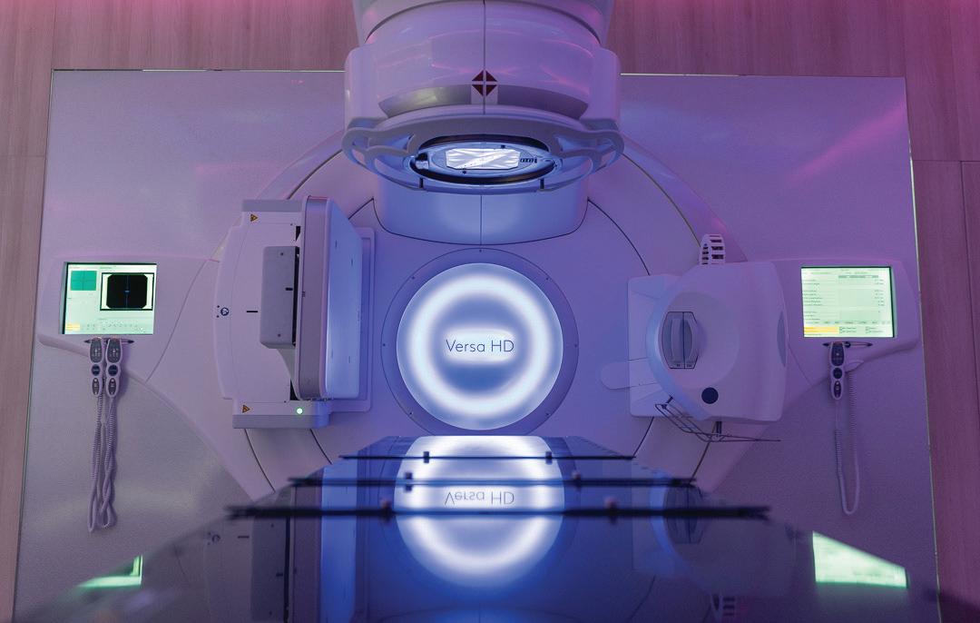
clinics within the centre specialize in treating a range of cancer conditions and offer chemotherapy, cancer diagnostics, screening, therapeutic services, surgical services as well as palliative care, and much more.
As well as providing exceptional treatment for patients, Almana focuses on preventive cancer care measures, such as free year-round breast cancer screenings at all hospital branches in Dammam, Khobar, Ahsa, Jubail, and Rakah.
Known for being pioneers in healthcare in the region, the hospital group has continuously advanced their services to meet the growing needs of their communities and support the Saudi 2030 vision to transform healthcare in the Kingdom. Dammam’s new oncology centre is the latest addition to their existing footprint of eight hospitals and clinics spread over Khobar, Dammam, Jubail, Ahsa, and Rakah.
• The Oncology Center is located at Dammam and is open 6 days a week from 8:00 am to 8:00 pm.
• For more information on the Center please visit: https://www.almanahospital.com.sa/ or call +966 92 00 33 440.
References
1. https://gco.iarc.fr/today/data/factsheets/populations/682-saudi-arabia-fact-sheets.pdf
22 IMIDDLE EAST HEALTH
opens first dedicated oncology centre in KSA’s Eastern Province in a collective effort to “close the cancer care gap”

Oncology
Researchers use AI with infrared imaging to improve colon cancer diagnostics
The immense progress in the field of therapy options over the past several years has significantly improved the chances of cure for patients with colon cancer. However, these new approaches, such as immunotherapies, require precise diagnosis so that they can be specifically tailored to the individual. Researchers at the Centre for Protein Diagnostics PRODI at Ruhr University Bochum, Germany, are using artificial intelligence in combination with infrared imaging to optimally tailor colon cancer therapy to individual patients. The label-free and automatable method can complement existing pathological analyses. The team led by Professor Klaus Gerwert reports in the “European Journal of Cancer” in January 2023.
Deep insights into human tissue within one hour
The PRODI team has been developing a new digital imaging method over the past few years: the so-called label-free infrared (IR) imaging measures the genomic and proteomic composition of the examined tissue, i.e. it provides molecular information based on the infrared spectra. This information is decoded with the help of artificial intelligence and displayed as false-colour images. To do this, the researchers use image analysis methods from the field of deep learning AI.
In cooperation with clinical partners, the PRODI team was able to show that the use of deep neural networks makes it possible to reliably determine the so-called microsatellite status, a prognostically and therapeutically relevant parameter, in colon
automated process and enables a spatially resolved differential classification of the tumour within one hour.
Indication of the effectiveness of therapies
In classical diagnostics, microsatellite status is determined either by complex immunostaining of various proteins or by DNA analysis.
“15 to 20 per cent of colon cancer patients show microsatellite instability in the tumour tissue,” says Professor Andrea Tannapfel, head of the Institute of Pathology at Ruhr University. “This instability is a positive biomarker indicating that immunotherapy will be effective.”
With the ever-improving therapy options, the fast and uncomplicated determination of such biomarkers is also becoming more and more important. Based on IR microscopic data, neuronal networks were modified, optimised, and trained at PRODI to establish label-free diagnostics. Unlike immunostaining, this approach does not require dyes and is significantly faster than DNA analysis.
“We were able to show that the accuracy of IR imaging for determining microsatellite status comes close to the most common method used in the clinic, immunostaining,” says Stephanie Schörner, PhD student. “Through constant further development and optimisation of the method, we expect a further increase in accuracy,” adds Dr Frederik Großerüschkamp.
long-standing, intensive cooperation between the Institute of Pathology at Ruhr University (Prof. Tannapfel), the Clinic for Haematology and Oncology at the St. Josef Hospital, Clinical Centre of Ruhr University (Professor Anke ReinacherSchick) and the Centre for Protein Diagnostics (Prof. Gerwert).
The PRODI researchers were able to access the ColoPredict Plus 2.0 molecular registry, a non-interventional, multicentre registry study for patients with early-stage colorectal cancer, to develop this diagnostic approach.
“The ColoPredict registry also enables a more targeted therapy for patients through the targeted analysis of biomarkers. Thus, the registry recently serves as a study platform for precision oncology approaches,” says Prof. Reinacher-Schick.
In addition to providing tissue samples, the registry offers a sound database of prognostically and therapeutically relevant baseline characteristics.
“In such a project, it is of immense importance to be able to draw on an excellent cohort and pathological expertise,” emphasises Prof. Gerwert.
“Our work on the classification of microsatellite status in colon cancer patients is based on one of the largest cohorts we have published to date and clearly demonstrates the potential for use in translational cancer research,” says Prof. Tannapfel.
Reference:
https://doi.org/10.1016/j.ejca.2022.12.026
24 IMIDDLE EAST HEALTH
Klaus Gerwert, Stephanie Schörner, Frederik Großerueschkamp, et. al. Fast and label-free automated detection of microsatellite status in early colon cancer using artificial intelligence integrated infrared imaging. European Journal of Cancer, 28 January 2023. doi:
© RUB, Marquard
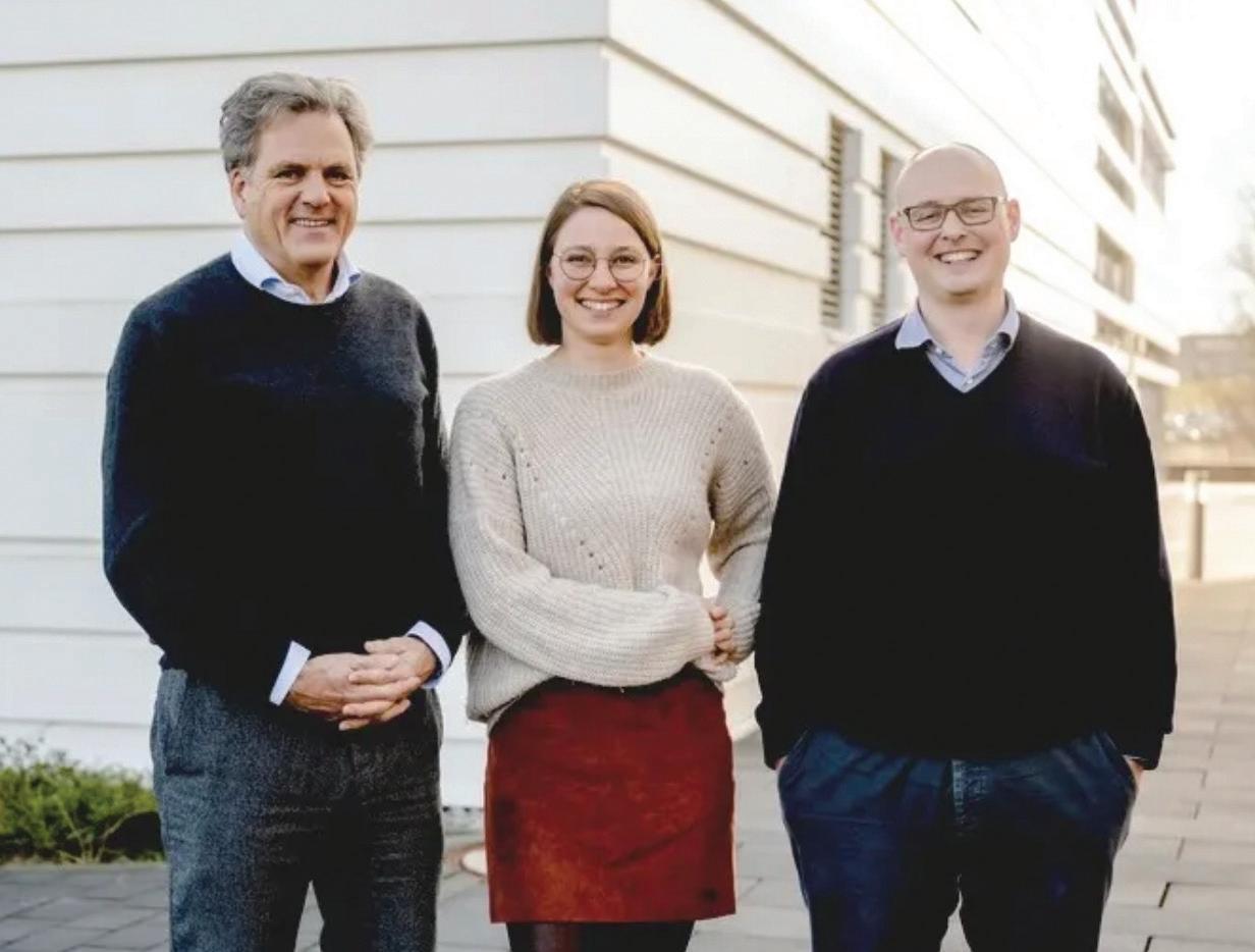
(from left) Klaus Gerwert, Stephanie Schörner and Frederik Großerüschkamp are using artificial intelligence to improve the diagnosis of colon cancer.
Cancer patients who don’t respond to immunotherapy lack important immune cells
Immunotherapy has transformed cancer care. In advanced melanoma, for example, the most fatal form of skin cancer, the five-year survival rate has risen from less than 10% to more than 50% since immunotherapy was introduced in 2011. Still, only about half of melanoma patients respond to immunotherapy, and those who do not respond face a difficult future.
Researchers at Washington University School of Medicine in St. Louis have discovered that the difference between people who do and do not respond to immunotherapy may have to do with an immune cell known as CD5+ dendritic cells because they bear the protein CD5 on their outer surfaces. Their research showed that people with a variety of kinds of cancers, including melanoma, lived longer if they had more CD5+ dendritic cells in their tumours, and that mice that lacked CD5 on their dendritic cells were unable to respond well to immunotherapy.
The findings, published 17 February 2023 in the journal Science, suggest that a supplementary therapy designed to increase the number or activity of CD5+ dendritic cells potentially could extend the lifesaving benefits of immunotherapy to more cancer patients.
“Immunotherapy has revolutionized the field of cancer therapy, but there are a lot of patients with cancer who don’t benefit from it,” said senior author Eynav Klechevsky, PhD, an assistant professor of pathology & immunology and a researcher at Siteman Cancer Center at Barnes-Jewish Hospital and Washington University School of Medicine. “Part of the reason some people do not respond well to some forms of immunotherapy is because this population of dendritic cells is reduced dramatically. We’re developing some novel immunebased approaches to boost the activation of these CD5-expressing dendritic cells with a goal of helping more patients respond to immunotherapy.”
The immune system defends the body against cancer by activating immune cells known as T cells to recognize and kill tumour cells. In response, tumour cells manipulate the immune checkpoint system — a safeguard that prevents T cells from mistakenly attacking healthy cells — to hoodwink
T cells into leaving them alone. Immune checkpoint blockade therapy works by thwarting tumour cells’ manipulations, thereby freeing T cells to recognize and destroy tumours. But even with therapy, some people’s T cells are unable to do their job effectively.
Klechevsky and colleagues – including first author Mingyu He, PhD, a staff scientist, and co-author Kate Roussak, MD, a postdoctoral researcher – suspected that people who don’t respond to immunotherapy may have a problem with their dendritic cells. If T cells are the players on a soccer field, dendritic cells are the coaches who get the players pumped up for the game and give them instructions. Without dendritic cells, T cells are subdued and aimless.
By analyzing data in The Cancer Genome Atlas – a public database with information on 20,000 tumours representing 33 cancer types – Klechevsky and colleagues discovered that patients with types of skin, lung, bone and soft tissue, breast and cervical cancers fared better if they had higher levels of CD5+ dendritic cells in their tumours.
Further experiments with human cells and mice showed that CD5+ dendritic cells are required for effective T cell activity against tumours. CD5+ dendritic cells from people powerfully induced T cells to activate and multiply. Mice with tumours responded only weakly to immunotherapy and failed to reject the tumours if they lacked CD5 on their dendritic cells.
The findings suggest that the amount of CD5+ dendritic cells inside tumours could be used to help doctors assess which patients are most likely to benefit from immunotherapy. They also suggest that increasing the numbers or the activity of such dendritic cells potentially could help more people benefit from immunotherapy. As part of this study, the researchers discovered that the immune protein IL-6 increases the amounts of CD5+ dendritic cells.
“We still don’t completely understand how immunotherapies work,” Klechevsky said. “This study indicates that there is more we can do to increase the efficacy of these treatments. I’m confident that if we can find ways to harness these cells or expand these cells in patients, we can help more people.”
Reference:
MIDDLE EAST HEALTHI 25
M, Roussak K, Klechevsky E, et. al. CD5 expression
Feb. 17, 2023.
He
by dendritic cells directs T cell immunity and sustains immunotherapy responses. Science.
doi: https://doi.org/10.1126/science.abg2752
Stamp-sized ultrasound adhesives provide long-term imaging of diverse organs
Ultrasound imaging is a safe and noninvasive window into the body’s workings, providing clinicians with live images of a patient’s internal organs. To capture these images, trained technicians manipulate ultrasound wands and probes to direct sound waves into the body. These waves reflect back out to produce high-resolution images of a patient’s heart, lungs, and other deep organs.
Currently, ultrasound imaging requires
bulky and specialized equipment available only in hospitals and doctor’s offices. But a new design by MIT engineers might make the technology as wearable and accessible as buying Band-Aids at the pharmacy.
In a paper published in Science, the engineers present the design for a new ultrasound sticker – a stamp-sized device that sticks to skin and can provide continuous ultrasound imaging of internal organs for 48 hours.
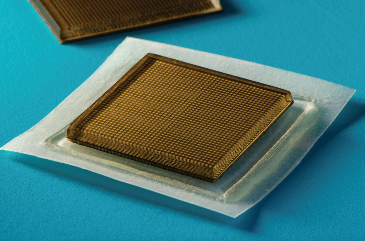
The researchers applied the stickers to volunteers and showed the devices produced live, high-resolution images of major blood vessels and deeper organs such as the heart, lungs, and stomach. The stickers maintained a strong adhesion and captured changes in underlying organs as volunteers performed various activities, including sitting, standing, jogging, and biking.
The current design requires connecting the stickers to instruments that translate
26 IMIDDLE EAST HEALTH
Ultrasound
MIT engineers designed an adhesive patch that produces ultrasound images of the body. The stamp-sized device sticks to skin and can provide continuous ultrasound imaging of internal organs for 48 hours.
Image: Felice Frankel

the reflected sound waves into images. The researchers point out that even in their current form, the stickers could have immediate applications: For instance, the devices could be applied to patients in the hospital, similar to heart-monitoring EKG stickers, and could continuously image internal organs without requiring a technician to hold a probe in place for long periods of time.
If the devices can be made to operate wirelessly – a goal the team is currently working toward – the ultrasound stickers could be made into wearable imaging products that patients could take home from a doctor’s office or even buy at a pharmacy.
“We envision a few patches adhered to different locations on the body, and the patches would communicate with your smartphone, where AI algorithms would analyze the images on demand,” said the study’s senior author, Xuanhe Zhao, professor of mechanical engineering and civil and environmental engineering at MIT. “We believe we’ve opened a new era of wearable imaging: With a few patches on your body, you could see your internal organs.”
A sticky issue
To image with ultrasound, a technician first applies a liquid gel to a patient’s skin, which acts to transmit ultrasound waves. A probe, or transducer, is then pressed against the gel, sending sound waves into the body that echo off internal structures and back to the probe, where the echoed signals are translated into visual images.
For patients who require long periods of imaging, some hospitals offer probes affixed to robotic arms that can hold a transducer in place without tiring, but the liquid ultrasound gel flows away and dries out over time, interrupting long-term imaging.
In recent years, researchers have explored designs for stretchable ultrasound probes that would provide portable, lowprofile imaging of internal organs. These designs gave a flexible array of tiny ultrasound transducers, the idea being that such a device would stretch and conform with a patient’s body.
But these experimental designs have
produced low-resolution images, in part due to their stretch: In moving with the body, transducers shift location relative to each other, distorting the resulting image.
“Wearable ultrasound imaging tool would have huge potential in the future of clinical diagnosis. However, the resolution and imaging duration of existing ultrasound patches is relatively low, and they cannot image deep organs,” says Chonghe Wang, who is an MIT graduate student.
How is it made?
The MIT team’s new ultrasound sticker produces higher resolution images over a longer duration by pairing a stretchy adhesive layer with a rigid array of transducers. “This combination enables the device to conform to the skin while maintaining the relative location of transducers to generate clearer and more precise images.” Wang says.
The device’s adhesive layer is made from two thin layers of elastomer that encapsulate a middle layer of solid hydrogel, a mostly water-based material that easily transmits sound waves. Unlike traditional ultrasound gels, the MIT team’s hydrogel is elastic and stretchy.
“The elastomer prevents dehydration of hydrogel,” says Chen, an MIT postdoc. “Only when hydrogel is highly hydrated can acoustic waves penetrate effectively and give high-resolution imaging of internal organs.”
The bottom elastomer layer is designed to stick to skin, while the top layer adheres to a rigid array of transducers that the team also designed and fabricated. The entire ultrasound sticker measures about 2 square centimetres across, and 3 millimetres thick – about the area of a postage stamp.
Observations
The researchers ran the ultrasound sticker through a battery of tests with healthy volunteers, who wore the stickers on various parts of their bodies, including the neck, chest, abdomen, and arms. The stickers stayed attached to their skin, and produced clear images of underlying structures for up to 48 hours. During this time, volunteers
performed a variety of activities in the lab, from sitting and standing, to jogging, biking, and lifting weights.
From the stickers’ images, the team was able to observe the changing diameter of major blood vessels when seated versus standing. The stickers also captured details of deeper organs, such as how the heart changes shape as it exerts during exercise. The researchers were also able to watch the stomach distend, then shrink back as volunteers drank then later passed juice out of their system. And as some volunteers lifted weights, the team could detect bright patterns in underlying muscles, signalling temporary microdamage.
“With imaging, we might be able to capture the moment in a workout before overuse, and stop before muscles become sore,” says Chen. “We do not know when that moment might be yet, but now we can provide imaging data that experts can interpret.”
The team is working to make the stickers function wirelessly. They are also developing software algorithms based on artificial intelligence that can better interpret and diagnose the stickers’ images. Then, Zhao envisions ultrasound stickers could be packaged and purchased by patients and consumers, and used not only to monitor various internal organs, but also the progression of tumours, as well as the development of foetuses in the womb.
“We imagine we could have a box of stickers, each designed to image a different location of the body,” Zhao says. “We believe this represents a breakthrough in wearable devices and medical imaging.”
28 IMIDDLE EAST HEALTH
We believe we’ve opened a new era of wearable imaging: With a few patches on your body, you could see your internal organs.
• doi: https://doi.org/10.1126/science.abo2542 Ultrasound

MIDDLE EAST HEALTHI 29
surgery
for treating ductal carcinoma in situ
Using ultrasound to guide surgery for patients with ductal car cinoma in situ (DCIS) gives better results than the standard technique of using a wire inserted into the breast, according to research presented at the 13th European Breast Cancer Con ference.
The technique, known as intraoperative ultrasound, or IOUS, enables surgeons to remove a smaller quantity of breast tissue, while still removing all the of DCIS tissue. Using IOUS improves the chance of having no cancer cells at the outer edge of the tissue that was removed (positive margins), reducing the risk of patients needing a second operation.
Because IOUS negates the need for inserting a guide wire, this technique could also reduce pain and preoperative stress for patients and save time for medical staff.
The research was carried out by a team at Clinica Univer sidad de Navarra in Madrid, Spain, and was presented by Dr Antonio J. Esgueva. He said: “DCIS is a common form of early breast cancer that can develop into a more serious, invasive cancer. To ensure it does not progress, patients are usually of fered surgery. Because DCIS does not usually create lumps in the breast, we need a good technique to guide surgery and make it as accurate as possible.”
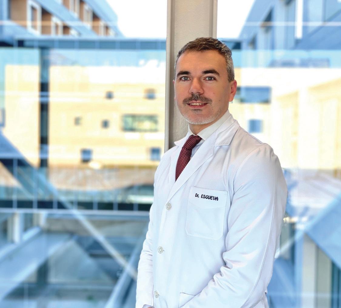
The research involved 108 people who were diagnosed with DCIS and treated at the Clinica Universidad de Navarra be tween February 2018 and December 2021. Forty-one were treated with IOUS-guided surgery while 67 were treated with surgery guided by wire localisation (WL).
Following each operation, the tissue removed was analysed to see how much was removed and whether there were ‘positive margins’. This means that DCIS cells were found at the edge of the tissue removed, suggesting some DCIS cells could have been left behind and patients would probably need a second operation.
Among those treated using WL, seven (10.4%) had positive margins and needed a second operation while in those treated using IOUS, there were only two patients (4.8%) with positive margins who needed a second operation.
The patients have been followed up for a year and half so far and cancer has only recurred in one (who was treated with WL).
Antonio Esgueva
the very best oncological surgery in terms of removing any trace of DCIS but also removing as little of the breast tissue as possible in order to have the best cosmetic result possible. At the same time, we also want to improve patients’ experience during treatment by using less invasive techniques and reducing their anxiety. Our research suggests that using intraoperative ultrasound, a quicker and less invasive technique, is effective for guiding DCIS surgery.”
The researchers plan to continue gathering information about patients having surgery for DCIS in the hopes of seeing long-term benefits of using IOUS.
30 IMIDDLE EAST HEALTH
Ultrasound-guided
is quicker, more effective
Ultrasound
Dr
Researchers at Tel Aviv University have developed a new technology that makes it possible to destroy cancerous tumours in a targeted manner, via a combination of ultrasound and the injection of nanobubbles into the bloodstream. According to the research team, unlike invasive treatment methods or the injection of microbubbles into the tumour itself, this latest technology enables the destruction of the tumour in a non-invasive manner.
The study was conducted under the leadership of doctoral student Mike Bismuth from the lab of Dr Tali Ilovitsh at Tel Aviv University’s Department of Biomedical Engineering, in collaboration with Dr Dov Hershkovitz of the Department of Pathology. Prof. Agata Exner from Case Western Reserve University in Cleveland also participated in the study. The study was published in the journal Nanoscale.
Dr Tali Ilovitsh noted: “Our new technology makes it possible, in a relatively simple way, to inject nanobubbles into the bloodstream, which then congregate in the area of the cancerous tumour. After that, using a low-frequency ultrasound, we explode the nanobubbles, and thereby the tumour.”
The researchers explain that today, the prevalent method of cancer treatment is surgical removal of the tumour, in combination with complementary treatments such as chemotherapy and immunotherapy. Using therapeutic ultrasound to destroy the cancerous tumour is a non-invasive alternative to surgery.
This method has both advantages and
disadvantages. On the one hand, it allows for localized and focused treatment; the use of high-intensity ultrasound can produce thermal or mechanical effects by delivering powerful acoustic energy to a focal point with high spatial-temporal precision. This method has been used to effectively treat solid tumours deep within in the body. Moreover, it makes it possible to treat patients who are unfit for tumour resection surgery. The disadvantage, however, is that the heat and high intensity of the ultrasound waves may damage the tissues near the tumour.
Overcoming off-target toxicity
In the current study, Dr Ilovitsh and her team sought to overcome this problem. In the experiment, which used an animal model, the researchers were able to destroy the tumour by injecting nanobubbles into the bloodstream (as opposed to what has been until now, which is the local injection of microbubbles into the tumour itself), in combination with low-frequency ultrasound waves, with minimal offtarget effects.
Dr Ilovitsh explained: “The combination of nanobubbles and low frequency ultrasound waves provides a more specific targeting of the area of the tumour, and reduces off-target toxicity. Applying the low frequency to the nanobubbles causes their extreme swelling and explosion, even at low pressures. This makes it possible to perform the mechanical destruction of the tumours at low-pressure thresholds. Our method has the advantages of ultrasound, in that it is safe, cost-effective, and clini-
cally available, and in addition, the use of nanobubbles facilitates the targeting of tumours because they can be observed with the help of ultrasound imaging.”
Dr Ilovitsh adds that the use of lowfrequency ultrasound also increases the depth of penetration, minimizes distortion and attenuation, and enlarges the focal point. “This can help in the treatment of tumours that are located deep within the body, and in addition facilitate the treatment of larger tumour volumes. The experiment was conducted in a breast cancer tumour mouse model, but it is likely that the treatment will also be effective with other types of tumours, and in the future, also in humans.”
Keren Primor Cohen, CEO of Ramot noted: “Ramot - Tel Aviv University Tech Transfer Company, applied for several patents to protect this technology and its application. We believe in the commercial potential of this breakthrough technology in cancer treatment, and we are in contact with several leading companies in Israel and abroad to promote it.”
• doi: https://doi.org/10.1039/D2NR01367C
MIDDLE EAST HEALTHI 31
A combination of ultrasound and nanobubbles destroys cancerous tumours, eliminating the need for invasive treatments
Our method has the advantages of ultrasound, in that it is safe, cost-effective, and clinically available.
Anaesthesia
Researchers identify what causes accidental awareness under anaesthesia
Brain structures which could predict an individual’s predisposition to accidental awareness under anaesthetic have been identified for the first time by neuroscientists in Trinity College Dublin.
The findings, published in the journal Human Brain Mapping [1], could help identify individuals who may require higher than average doses of anaesthetic.
Although anaesthesia has been used in clinical medicine for over 150, scientists do not fully understand why its effect on people is so varied. One in four patients presumed to be unconscious during general anaesthesia may in fact have subjective experiences, such as dreaming, and in very rare cases (0.05–0.2%) individuals become accidentally aware during a medical procedure.
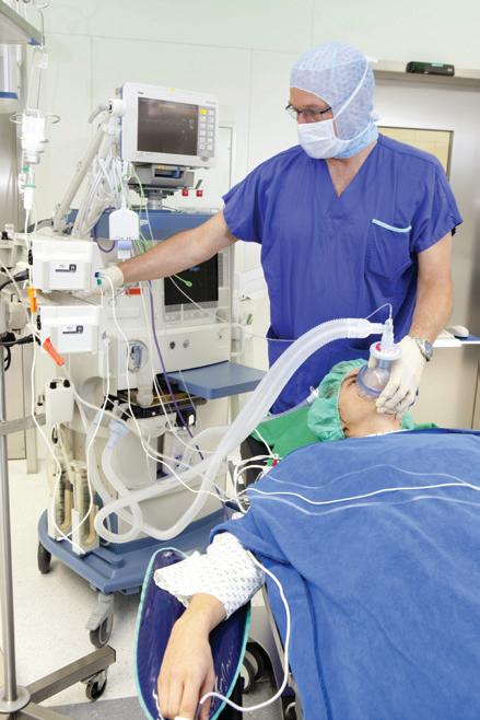
The research found that one in three participants were unaffected by moderate propofol sedation in their response times, thus
thwarting a key aim of anaesthesia – the suppression of behavioural responsiveness.
The research also showed, for the first time, that the participants who were resistant to anaesthesia had fundamental differences in the function and structures of the fronto-parietal regions of the brain to those who remained fully unconscious. Crucially, these brain differences could be predicted prior to sedation.
Commenting on the research, Lorina Naci, Associate Professor of Psychology, Trinity and lead author on the paper said: “The detection of a person’s responsiveness to anaesthesia prior to sedation has important implications for patient safety and wellbeing. Our results highlight new markers for improving the monitoring of awareness during clinical anaesthesia. Although rare, accidental awareness during an operation can be very traumatic and lead to
as post-traumatic stress disorder, as well as clinical depression or phobias.
“Our results suggest that individuals with larger grey matter volume in the frontal regions and stronger functional connectivity within fronto-parietal brain networks, may require higher doses of propofol to become nonresponsive compared to individuals with weaker connectivity and smaller grey matter volume in these regions.”
The research, conducted in Ireland and Canada, investigated 17 healthy individuals who were sedated with propofol, the most common clinical anaesthetic agent. The participants’ response time to detect a simple sound was measured when they were awake and as they became sedated. Brain activity of 25 participants as they listened to a simple story in both states was also measured.
Reference:
1. Responsiveness variability during anaesthesia relates to inherent differences in brain structure and function of the fronto-parietal networks. Deng F, Taylor N, Owen AM, Cusack R, Naci L. Human Brain Mapping. doi: https://doi.org/10.1002/hbm.26199
How does ketamine act as ‘switch’ in the brain?
Ketamine, an established anaesthetic and increasingly popular antidepressant, dramatically reorganizes activity in the brain, as if a switch had been flipped on its active circuits, according to a new study by Penn Medicine researchers. In a Nature Neuroscience paper <https://doi.org/10.1038/s41593-022-012035>, the team described starkly changed neuronal activity patterns in the cerebral cortex of animal models after ketamine administration – observing normally active neurons that were silenced and another set that were normally quiet suddenly springing to action.
This ketamine-induced activity switch in key brain regions tied to depression may impact our understanding of ketamine’s treatment effects and future research in the field of neuropsychiatry.
“Our surprising results reveal two distinct populations of cortical neurons, one engaged in normal awake brain function, the other linked to the ketamine-induced brain state,” said the co-lead and co-senior author Joseph Cichon, MD, PhD, an assistant professor of Anesthesiology and Critical Care and Neuroscience in the Perelman School
of Medicine at the University of Pennsylvania. “It’s possible that this new network induced by ketamine enables dreams, hypnosis, or some type of unconscious state. And if that is determined to be true, this could also signal that it is the place where ketamine’s therapeutic effects take place.”
Ketamine’s signature characteristics
Anaesthesiologists routinely deliver aesthetic drugs before surgeries to reversibly alter activity in the brain so that it enters its unconscious state. Since its synthesis in
s
32 IMIDDLE EAST HEALTH
New markers in brain structures and function could help identify people who require a higher than average dose of anaesthetic
Anaesthesia
the 1960s, ketamine has been a mainstay in anaesthesia practice because of its reliable physiological effects and safety profile. One of ketamine’s signature characteristics is that it maintains some activity states across the surface of the brain (the cortex). This contrasts with most anaesthetics, which work by totally suppressing brain activity. It is these preserved neuronal activities that are thought to be important for ketamine’s antidepressant effects in key brain areas related to depression. But, to date, how ketamine exerts these clinical effects remains mysterious.
In their new study, the researchers analyzed mouse behaviours before and after they were administered ketamine, comparing them to control mice who received placebo saline. One key observation was that those given ketamine, within minutes of injection, exhibited behavioural changes consistent with what is seen in humans on the drug, including reduced mobility, impaired responses to sensory stimuli, which are collectively termed “dissociation”.
“We were hoping to pinpoint exactly what parts of the brain circuit ketamine affects when it’s administered so that we might open the door to better study of it and, down the road, more beneficial therapeutic use of it,” said co-lead and co-senior author Alex Proekt, MD, PhD, an associate professor of Anesthesiology and Critical Care at Penn.
Two-photon microscopy was used to image cortical brain tissue before and after ketamine treatment. By following individual neurons and their activity, they found that ketamine turned on silent cells and turned off previously active neurons.
The neuronal activity observed was traced to ketamine’s ability to block the activity of synaptic receptors called NMDA receptors and ion channels called HCN channels. The researchers found that they could recreate ketamine’s effects without the medications by simply inhibiting these specific receptors and channels in the cortex. The scientists showed that ketamine weakens several sets of inhibitory cortical neurons that
normally suppress other neurons. This allowed the normally quiet neurons, the ones usually being suppressed when ketamine wasn’t present, to become active.
The study showed that this dropout in inhibition was necessary for the activity switch in excitatory neurons – the neurons forming communication highways, and the main target of commonly prescribed antidepressant medications. More work will need to be undertaken to determine whether the ketamine-driven effects in excitatory and inhibitory neurons are the ones behind ketamine’s rapid antidepressant effects.
“While our study directly pertains to basic neuroscience, it does point at the greater potential of ketamine as a quick-acting antidepressant, among other applications,” said co-author Max Kelz, MD, PhD, the Distinguished Professor and Vice Chair of Research of Anesthesiology & Critical Care. “Further research is needed to fully explore this, but the neuronal switch we found also underlies dissociated, hallucinatory states caused by some psychiatric Hillnesses.”
Neuron function altered by widely-used Propofol
By Katie Malatino
Propofol is the most commonly used drug to induce general anaesthesia. Despite its frequent clinical application, it is poorly understood how propofol causes anaesthesia.
In a new study published in Molecular Biology of the Cell <https://doi.org/10.1091/
mbc.E22-07-0276>, a team of Rensselaer Polytechnic Institute researchers identified a previously unknown propofol effect in neurons. The study found that propofol exposure impacted the process by which neurons transport proteins, biomolecules that perform most cellular functions, to the cell surface.
Almost all animal cells, including human cells, are highly compartmentalized and rely on efficient movement of protein material between compartments. Proteins are moved from their site of synthesis to the location at which they perform their function in small carriers called “vesicles.” This transport must be efficient and highly specific to maintain cellular organization and function.
vin Bentley, an assistant professor in the Department of Biological Sciences, whose laboratory studies vesicle transport in neurons. Neurons are particularly reliant on vesicle transport because axons – which are often organized in nerve bundles – can span distances of up to 1 meter in humans. Errors in vesicle transport have been linked to neurodevelopmental and neurodegenerative diseases such as Alzheimer’s and Parkinson’s.
This new study found that propofol affects a family of proteins called kinesins. Kinesins are small “motor proteins” that move vesicles on tiny filaments called microtubules.
Dr Bentley’s team observed that vesicle movement of two prominent kinesins, Kinesin-1 and Kinesin-3, was substantially reduced in cells exposed to propofol. The team then showed that propofol-induced transport delays led to a significant decrease of protein delivery to axons.
Overall, the research contributes significantly to our understanding of how propofol works. Most studies that address the anaesthetic mechanism of propofol have been focused on its interaction with an ion channel called the GABAA receptor, which inhibits neurotransmission when activated.
This new study demonstrates that vesicle transport is an additional mechanism that may be important for propofol’s anaesthetic effect. Discovery of this new propofol effect has important applications for human health and may lead to the development of better anaesthetic drugs.
Need more story here
The research team was led by Dr Mar-
“The mechanism by which propofol works is not fully understood,” Bentley said. “What we discovered was unexpected: propofol altered the trafficking of vesicles in live neurons.”
“By using state-of-the-art live cell imaging technologies, Dr Bentley’s team has furthered our understanding of the mechanism of action of a widely used drug that is already impacting human health on a daily basis,” said Curt M. Breneman, Dean of the School of Science. “Dr Bentley’s research may pave the way for the development of related compounds that use these same mechanisms to target debilitating neurodegenerative diseases.”
34 IMIDDLE EAST HEALTH

Becton Dickinson
Target-Controlled Infusion anaesthesia: New more universal models
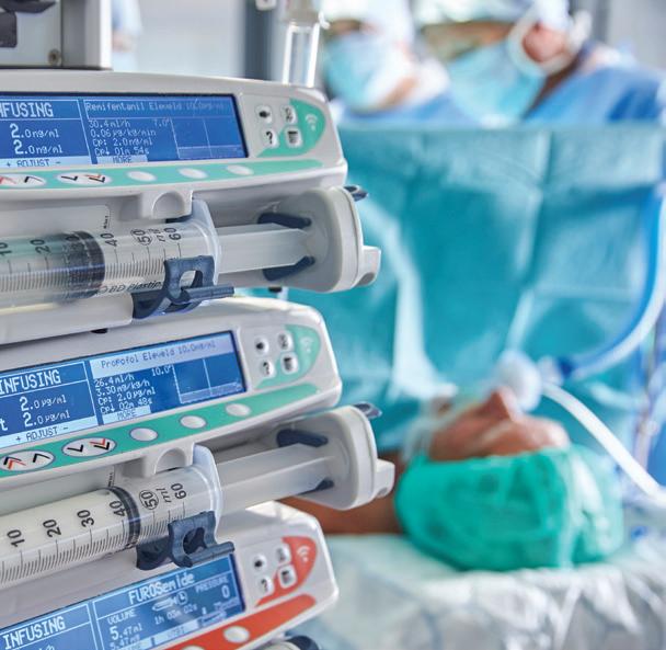 By James Waterson, RN, M.Med.Ed. MHE. Becton Dickinson. Medical Affairs Manager, Middle East & Africa
In simple terms Target-Controlled Infusion (TCI) means that instead of setting a dose-rate on the pump, the pump is programmed to target a required plasma concentration or effect-site concentration. A TCI pump automatically calculates how much drug is needed during induction and maintenance to maintain the desired effect-site or plasma concentration.
A TCI algorithm (the ‘target’ and plan on which the pump relies to deliver appropriate induction and maintenance rates to maintain anaesthesia without overdosing the patient) is based on pharmacokinetic (PK) and pharmodynamic (PD) models and on Absorption, Distribution, Metabolism, and Excretion of medications by the body.
For example, the effect-site concentration of Propofol required to produce loss of consciousness is about 3 to 6 mcg/ml, depending on the patients’ demographics. Patients waking from anaesthesia generally have a blood concentration of around 1- 2 mcg/ml, although this is dependent on other drugs given during anaesthesia.
Adequate analgesia with Remifentanil is generally achieved with 3-6 ng/ml. A Remifentanil infusion of 0.25-0.5 mcg/kg/ min in an ‘average’ man – 70 kg, 170 cm, 40 years old – produces a blood concentration of around 6ng/ml after 25 minutes.
PK models are based on body compartments
Conventionally the body compart-
ment that the drug is injected into is V1 (plasma/blood), the next compartment is the ‘vessel-rich’ or ‘fast re-distribution’ compartment and is characterized as V2 (heart, liver, etc.). The final compartment, which is anatomically ‘vessel-poor’ and ‘slow’ in terms of re-distribution, is V3 (fatty tissue).
Drug distribution and the metabolism/ elimination of each drug in each compartment is also part of each TCI model, as is the pharmacodynamics of the time taken between the plasma and effect-site effect.
Computer simulations and mathematical modelling of infusion schemes based on the above theories of compartments and clearances give models for both Target Plasma Concentration (Cpt) and Target Effect Concentration (Cet) and these can be incorporated into specialist infusion pumps.
The Marsh model for Propofol requires only age and weight to be programmed in the pump. The Schnider model is an alternative model for Propofol and has advantages in elderly patients as it is based on a lean body mass (LBM) calculation for each patient. Elderly patients receive a lower induction and maintenance dose, which can assist with hemodynamic stability.
The Remifentanil Minto model uses age, height, gender and weight, and determines LBM for its calculations.
TCI pumps deliver the infusion at a constantly altering rate, but it is useful to think of this one infusion as being a meanaverage of three continually calculated infusion rates: a constant rate to replace drug elimination and two exponentially de-
creasing infusions to match drug removed from central compartments to other peripheral compartments of distribution.
Key features of an ideal TCI infusion system or pump are:
• Critical information such as decrement time, current Cet or Cpt and respective targets, current dose rate and concentration and type of agent being infused can be displayed at the same time on one screen.
• Patient parameters used during the setting-up of infusions appear on one screen to avoid the need for shuttling through multiple screens to check vital information.
• An Induction Time adjustable from seconds to minutes to allow for a gentle induction for patients with cardiovascular conditions or established hypotension.
Obese patients have previously presented a problem for ‘classic’ TCI, and the physiological differences between paediatrics and adults had required separate models for children.
Now, however, we have the Eleveld model for both Propofol and Remifentanil, and the Kim-Obara-Egan Remifentanil model which are much more universal and can potentially allow TCI in age ranges from 6 months to 99 years of age, and from 2.5 to 215 kg.
TCI, with its emphasis on evidencebased anaesthesia, and new near-universal patient models seems primed to change our approach to the management of all patients receiving sedatives and analgesic agents.
36 IMIDDLE EAST HEALTH
Abdul Latif Jameel Health to exclusively
distribute iSono Health’s ATUSA 3D breast scanner in Global South
Abdul Latif Jameel Health, part of inter national diversified family business Abdul Latif Jameel, announced a new distribu tion agreement with iSono Health, a med ical technology company in San Francisco, USA, with the vision to transform breast care with automated imaging and artificial intelligence (AI).
The partnership, announced on the first day of Arab Health 2023, will see Abdul Latif Jameel Health become the exclu sive distributor of iSono Health’s ATUSA scanner in the Global South, making it available to hundreds of millions of wom en in an initial 31 countries covering the Middle East and North Africa, Africa, South Asia, and Southeast Asia.
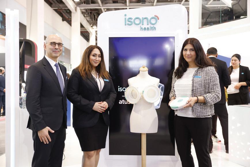
The female-founded iSono Health is transforming breast imaging with a first-ofits-kind, compact automated whole breast ultrasound system featuring a unique wear able accessory and an intuitive, intelligent software for automated image acquisition and analysis. The patented and FDAcleared ATUSA system is a compact ultrasound scanner that captures 3D images through automated scanning of the whole breast in just two minutes, independent of operator expertise. The device connects to laptop or tablet for real-time image acquisition and 3D visualization; the data is transferred to a secure cloud for storage. The ATUSA system is designed to seamlessly integrate with machine learning models that will give physicians a comprehensive set of tools for decision making and patient management.
Maryam Ziaei, PhD, Founder and CEO, iSono Health, said: “This new partnership is a significant milestone in our history and an important step forward in making our ATUSA scanner accessible to millions more women across the world. Working with Abdul Latif Jameel Health will empower so many more women to access the healthcare they need, to improve patient
experiences and to bring peace of mind.
“We have been able to develop a scanner which takes two minutes to scan and makes breast imaging painless and convenient. We’re very much looking forward to bringing this technology to the region and making a lasting, sustainable impact.”
The agreement will see Abdul Latif Jameel Health distribute the ATUSA scanner across the Middle East and North Africa as well as African markets including South Africa, Kenya and Nigeria, in addition to South Asia covering India, Pakistan, Bangladesh, Bhutan, and Nepal, and Southeast Asian territories of Indonesia, Malaysia, Singapore, Philippines, Thailand and Brunei.
Last year, Abdul Latif Jameel Health announced the creation of its new international commercial ecosystem, helping to accelerate access to modern medical care and drive health inclusivity across
the Global South. The network of confirmed partners will cover key markets across a wide geographic area and will see on-the-ground distribution of Abdul Latif Jameel Health approved products into hospitals, clinics and medical institutions to fast-track health inclusion for those who need it most.
Abdul Latif Jameel Health was created as a response to the ongoing global disparity in access to modern medical care, focusing on accelerating healthcare inclusion across the Global South. Reflecting the Jameel Family’s long-established commitment to innovating for a better future, Abdul Latif Jameel Health works in the commercial environment to address tangible real-world needs today, for a better tomorrow. It works with partners from around the world to open and grow new markets for distribution of existing solutions and investing in the future of MedTech.
38 IMIDDLE EAST HEALTH
Arab Health 2023 Review
Akram Bouchenaki, CEO Abdul Latif Jameel Health, Maryam Ziaei and Shadi Saberi, co-founders iSono Health at Arab Health 2023.
Dunlee unveils oncology bundles
Dunlee unveiled its new oncology bundles at Arab Health 2023. The bundles combine components that have been tested and verified to work together so clinicians can offer state-of-the-art onboard Cone Beam CT (CBCT). As cancer cases rise globally, an estimated 60% of new patients will need some form of radiation therapy, but only 25% of those patients will receive treatment, either because the equipment is not available or because treatment is too costly. It is estimated that by 2030, if every patient who needs radiotherapy has access to it, almost one million more lives will be saved every year worldwide.
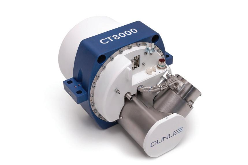
Image guidance has been a major breakthrough in efforts to make radiation therapy more precise, because it enables physicians to take images immediately before treatment while the patient is in the treatment position.
The oncology bundles include:
• X-ray tubes, which customers rely on Dunlee products’ small focal spot size to enable high resolution imaging

• X-ray flat panel detectors, providing excellent soft tissue visualization to identify tumour borders, a large field of view, fast frame speed of up to 150 frames-persecond, sharp images, and minimal motion artifacts
• Anti-scatter grids, which significantly improve soft tissue visibility and uniformity without the need to increase imaging dose
• Cables, including all modifications customers need for efficient integration into their linear accelerator
Dunlee offers two types of bundles:
• DA1094 X-ray Tube Bundle, which consists of a flat panel detector,
DA 1094 X-ray tube, anti-scatter grid and high voltage cables
• Grid and Detector Bundle, which combines a state-of-the-art antiscatter grid with the Dunlee XD-300 Detector for excellent soft tissue visualization and fast operation
• Dunlee Solutions+, Dunlee’s service portfolio, which supports customers in every step of product development, from system definition through operation
LMB Technology
As imaging departments continue to face lofty demands to maintain efficient operations, reduce costs and provide a positive patient experience, Dunlee’s unique ‘CoolGlide’ LMB technology performs just as well as the OEM alternative, and helps alleviate increased pressures for radiologists, including tube longevity, the wait time associated with tube cooling and a quieter, smoother exam.
Dunlee’s CoolGlide Liquid Metal Bearings are designed and manufactured in Hamburg, Germany, based on knowledge gained from over 30 years of LMB technology development and over 100,000 LMB units sold worldwide. The invention of Liquid Metal Bearing Technology by the Hamburg R&D team in 1989 had an enormous impact on performance and ease-ofuse, becoming the gold standard for CT and Image Guided Therapy systems.
CoolGlide LMB technology features benefits to address the following:
• Throughput: A cooling channel within the bearing shaft directs cooling fluid to the anode. This design provides much higher heat conduction and cool-
ing capacity than traditional ball bearing tubes.
• Workflow: The anode rotates continuously and provides constant high speed through the entire working day without stopping, speeding up workflow and facilitating productivity. There is no preparation time necessary between every scan, as there is for ball bearing tubes.
• Reliability: The bearing shaft and sleeve glide on a liquid film. The liquid is pumped to the interior of the bearing by spiral grooves. As there is no direct contact during operation, there‘s virtually no wear.
• Noise: As there is no direct contact , there is no audible bearing noise during operation.
Dunlee’s new oncology bundles combine components that have been tested and verified to work together so clinicians can offer state-of-the-art onboard Cone Beam CT (CBCT)
MIDDLE EAST HEALTHI 39
The Xpert bundle with CT8000 is the newest and most advanced product bundle in Dunlee’s components portfolio. It has been developed and designed to fit into high-end CT systems.
Middle East Health speaks to Nerio Alessandri, Founder & CEO, Technogym about the wellness trend and their new Biostrength REV smart fitness training product.

Middle East Health: At the recent Arab Health exhibition in Dubai, Technogym launched Biostrength REV smart fitness training equipment. The training equipment is designed for the sport, fitness and health for wellness sectors. Before we look at this new equipment in more detail, can you comment on the health for wellness sector, who does this include? Is there increasing demand from this sector for your equipment, and if so, why do you think this demand is growing?
n Nerio Alessandri: Health is now more than ever the number one priority for people. In reality, non-communicable diseases are the leading cause of death worldwide, and this depends on people’s lifestyles. There is an increased awareness of the benefit of physical exercise. People are more informed, but also at an institutional level, governments and local authorities are understanding that investing in health and prevention is a great tool to reduce healthcare costs that will be unsustainable going forward since the population is ageing.
That said, health is a huge trend and this is clearly revealed in the fitness sector. More and more fitness clubs are integrating rehabilitation and prevention programmes within their training formats.
In this arena, Technogym has a strategic advantage thanks to its unique digital ecosystem that allows professionals to prescribe precise exercises or training programmes based on the needs of each individual user and at the same time the user can keep track of their movement and improvement.

MEH: These are smart devices and presumably relatively expensive. Do you think this form of wellness is a luxury reserved for those who have the time and money to afford to use these advanced fitness training devices? Can you explain why?
n NA: Technogym has always invested
in the health sector and in specific smart solutions for rehabilitation and prevention in order to support operators (both fitness clubs and medical centres) in offering peo ple different fitness training formats and programmes. That’s why we believe this is not a luxury product since people can have access to them in a wide variety of for mats. Technogym supplies its solutions to thousands of rehabilitation centres, fitness clubs, medical centres and even commu nity centres all over the world. So, there are plenty of ways people can access this.
MEH: The Biostrength REV is Techno gym’s latest offering among their range of fitness training equipment. According to a recent press release, it incorporates a new technology called BIODRIVE, which is based on aerospace technol ogy. Can you tell us a bit about the Bio strength REV, how does it work and what is unique about this equipment compared to other fitness training devices?
n NA: The new Biostrength REV prod ucts are the fastest and most efficient path to recover and improve performance. Two new additions – Leg Press REV and Leg Extension REV – add to Biostrength’s unique technology with a specific focus on rehabilitation and recovery. The sensors placed on the equipment enable it to detect and collect the necessary data to achieve the goal more safely and effectively
MEH: What is BIODRIVE and why has this been incorporated in Biostrength REV?
n NA: The patented Biodrive system, which uses aerospace technology, offers six types of resistance, which can improve the effectiveness of the exercise depending on the goal to achieve. The user is guided through every aspect of the workout to achieve maximum results in a safe and effective way. Biodrive recognises
market or for those fitness clubs offering products and services for rehabilitation and recovery. When it comes to home fitness, we have a full range of equipment integrated with the ecosystem and Technogym Live platform, which includes an extensive ondemand library of video content for fitness, such as live classes with incredible trainers, one-to-one cardio or strength training sessions, athletic training routines and more.
Biostrength REV is just one example of how innovation is always leading Technogym growth. Launched for the first time in 1992, the Technogym REV was the first isokinetic machine in the world to feature advanced technology and testing that enabled operators to carry out more in-depth analyses and diagnostics, thus advancing performance and speeding up recovery.
40 IMIDDLE EAST HEALTH
Biostrength REV – advanced, smart fitness training for rehabilitation and recovery
Technogym

Philips launches MR 7700 3T MRI
Philips launched the MR 7700 3T MRI at Arab Heath as part of its effort to showcase how its innovations support clinical teams in making informed decisions. The company says the MR 7700 reaches new levels of precision in both anatomical and functional imaging to support confident diagnosis for patients. It combines neuroscience capabilities, unsurpassed gradients, with an 35% faster scan time (compared to Ingenia Elition X with Vega HP gradients ), streamlined workflow, and delivers greater patient comfort.
Philips notes that now more than ever it is important to ease the burdens on clinical teams and ensure that hospital resources are put to their most productive use. Their portfolio of integrated workflow solutions supports this aim by connecting workflows, across the radiology enterprise to help enhance the efficiency and work
experience of radiologists, technologists and administrators. These include its next-generation Advanced Visualization Workspace platform with AI-enabled algorithms and workflows, as well as Philips’ AI-enabled PACS, which provides automatic analysis of medical data and extraction of relevant information to generate meaningful – and actionable – insights.
In addition, a variety of time saving features are included across Philips’ entire portfolio including AI-driven technologies in the MR5300 with BlueSeal Magnet, automating the most complex clinical and operational tasks, resulting in patient set-up in under a minute and the reduction of change-over time between exams by up to 30%.
Additionally, Philips is leveraging its AI expertise with Philips SmartSpeed to deliver fast high-quality imaging for a
wider range of patients, at imaging speeds nearly three times faster than parallel scanning, including for patients who are in pain, or struggle to hold their breath. This reduced examination time helps support higher throughput, while the time gained through these efficiencies can be used to scan more patients and reduce the cost per scan, or even to add unplanned patients to the schedule and to reduce staff overtime.
Philips points out that their technology is designed to be scalable and futureproof. This includes solutions such as Philips Capsule, which offers vendor neutral device integration that can capture streaming clinical data from nearly any patient device, and deliver contextually rich information, decision support, clinical surveillance, retrospective analytics and more.
Siemens Healthineers showcases a comprehensive portfolio of products
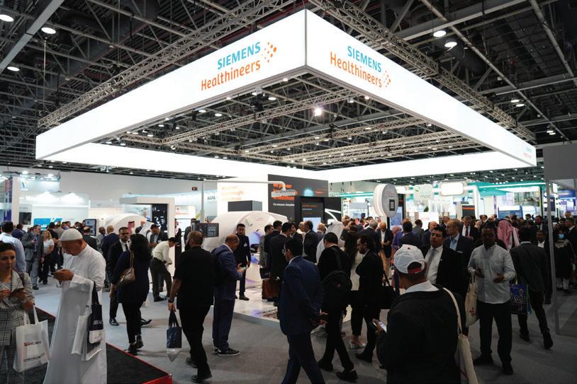
Siemens Healthineers showcased a com prehensive portfolio of products at Arab Health. One of the highlights was the world’s first photon-counting CT, the NAEOTOM Alpha with Quantum Tech nology, representing a total reinvention of computed tomography, offering high-reso lution images at minimal dose with spectral information in every scan and improved contrast at lower noise.
The company also exhibited the MAG NETOM Free.Max, with its 80 cm bore, which sets a new paradigm in patient com fort and expands access to MRI.
Also on the show floor was their MOBI LETT Impact, an economical high-quality, mobile digital X-ray machine that can be used at the patient’s bedside, enabling fast and undisrupted workflow.
Siemens Healthineers also introduced the ACUSON Juniper 2.5 ultrasound sys tem, which enhances the user experience while addressing clinical challenges across a variety of clinical applications.
They also showcased their Sequoia ultrasound system powered by BioAcoustic technology, which helps to deliver
effective clinical insights by reducing the effects of ultrasound variability between users, patients, and technology.
42 IMIDDLE EAST HEALTH
Arab Health 2023 Review

Proximie introduces PxLens to share a first-person perspective of open surgeries
Proximie, health tech company, launched their PxLens. These head-mounted smart glasses with embedded software system connects to Proximie’s cloud-based OR telepresence software, enabling surgeons to share a first-person perspective of open surgeries and minimally invasive procedures.
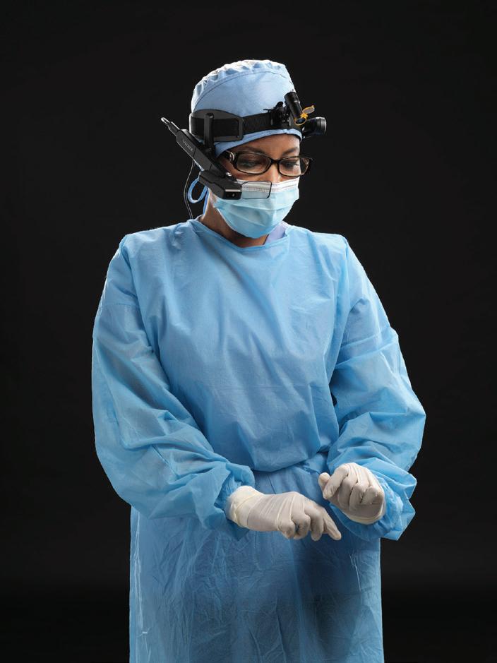
The PxLens is ideal for use in any procedural context. They are equipped with a 4K camera which facilitates hi-resolution video quality. The company reports that the new PxLens solution has been used in the US and UK in late 2022 in more than 20 successful procedures, ranging from colorectal, otolaryngol-
ogy, orthopaedics and urology. This enables live collaboration between surgeons beyond OR, and recorded sessions become post-procedural learning resources for those in training.
Jonathan Noël, MBBS, FRCS, Consultant Urological Surgeon, Guy’s & St Thomas’ NHS Foundation Trust said: “The first-person view and vantage point is a new way of learning. There are opportunities for training with live mentorship and retrospective case view. I think it will be useful for endoscopic and open cases.”
44 IMIDDLE EAST HEALTH Arab Health 2023 Review
Medlab Middle East puts focus on sustainability
Medlab Middle East 2023, held from 6-9 February 2023 at the Dubai World Trade Centre, put the focus on sustainability with the theme: ‘Paving the way for sustainability in laboratory medicine’.
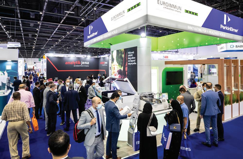
During a panel discussion by global leaders in the field who spoke at the ‘Sustainability in the clinical lab conference’ it was noted that the sustainability strategies of clinical laboratories are crucial to reducing the environmental burden of the healthcare sector.
Speaking at the conference, Dr Rana Nabulsi, Head of Operations and QualityPathology and Genetics at Dubai Health Authority (DHA), said: “Climate change is a big responsibility for us all. It is a global issue that we all need to devote our attention, commitment, and leadership.
“2023 is mandated by the UAE Government to be the ‘year of sustainability’,
and Dubai will host COP 28 in November this year. This crisis is going beyond national borders, and we have to build leadership at the top to keep our planet safe, save resources for future generations, improve biodiversity and reduce carbon emissions.”
Other speakers at the conference included Dr Bernard Gouget, Assistant Professor, Fédération Hospitalière de France, who led a session on ‘green’ labs and how to get started. He said: “Sustainability is now becoming a way of life as global concern for the environment grows. Medical labs are an important part of hospitals and healthcare systems, so we must be engaged in this dynamic area.
“It is essential to distinguish between ‘green’ and ‘sustainable’. Green practices are, of course, very important for the environment, but sustainability addresses the long-
term picture for future generations,” he added.
Gouget noted that there are many ways in which a lab can address sustainability. Following international guidelines from bodies such as the International Organisation for Standardisation (ISO) was outlined as one of the main strategies at the session.
The two preferred tools for labs are ISO 26000-2010 and ISO 14001-2015. ISO 26000-2010 provides social responsibility guidelines such as transparency, ethical behaviour, human rights and respect for stakeholder interests. ISO 14001-2015 focuses on Environmental Management Systems (EMS). EMS adoption also offers potential commercial advantages by reducing the risk of government-imposed fines, lowering energy consumption, improving resource efficiency and optimising waste management processes.
46 IMIDDLE EAST HEALTH
Medlab Middle East 2023 review
ChatGPT scores passing mark on the US Medical Licensing Exam
ChatGPT, the new artificial intelligence system making waves round the world, can score at or around the approximately 60 percent passing threshold for the United States Medical Licensing Exam (USMLE), with responses that make coherent, internal sense and contain frequent insights, according to a study published February 9, 2023 in the open-access journal PLOS Digital Health.
ChatGPT, known as a large language model (LLM), is designed to generate human-like writing by predicting upcoming word sequences. Unlike most chatbots, ChatGPT cannot search the internet. Instead, it generates text using word relationships predicted by its internal processes.
The researchers tested ChatGPT’s performance on the USMLE, a highly standardized and regulated series of three exams (Steps 1, 2CK, and 3) required for medical licensure in the United States. Taken by medical students and physiciansin-training, the USMLE assesses knowledge spanning most medical disciplines, ranging from biochemistry, to diagnostic reasoning, to bioethics.
After screening to remove image-based questions, the authors tested the software
on 350 of the 376 public questions available from the June 2022 USMLE release.
After indeterminate responses were removed, ChatGPT scored between 52.4% and 75.0% across the three USMLE exams. The passing threshold each year is approximately 60%. ChatGPT also demonstrated 94.6% concordance across all its responses and produced at least one significant insight (something that was new, non-obvious, and clinically valid) for 88.9% of its responses. Notably, ChatGPT exceeded the performance of PubMedGPT, a counterpart model trained exclusively on biomedical domain literature, which scored 50.8% on an older dataset of USMLE-style questions.
While the relatively small input size restricted the depth and range of analyses, the authors note their findings provide a
glimpse of ChatGPT’s potential to enhance medical education, and eventually, clinical practice. For example, they add, clinicians at AnsibleHealth already use ChatGPT to rewrite jargon-heavy reports for easier patient comprehension.
“Reaching the passing score for this notoriously difficult expert exam, and doing so without any human reinforcement, marks a notable milestone in clinical AI maturation,” say the authors of the study

Author Dr Tiffany Kung added that ChatGPT’s role in this research went beyond being the study subject: “ChatGPT contributed substantially to the writing of [our] manuscript... We interacted with ChatGPT much like a colleague, asking it to synthesize, simplify, and offer counterpoints to drafts in progress...All of the co-authors valued ChatGPT’s input.”
• The journal article is freely available in PLOS Digital Health: https://doi.org/10.1371/journal.pdig.0000198
48 IMIDDLE EAST HEALTH The Back Page



 By James Waterson, RN, M.Med.Ed. MHE. Becton Dickinson. Medical Affairs Manager, Middle East & Africa


